
Chone veleronis Banse, 1972
|
publication ID |
https://doi.org/ 10.1080/00222930701250912 |
|
persistent identifier |
https://treatment.plazi.org/id/03B1879F-300D-5C78-FE24-E2D4C586FD09 |
|
treatment provided by |
Felipe |
|
scientific name |
Chone veleronis Banse, 1972 |
| status |
|
Chone veleronis Banse, 1972 View in CoL
( Figure 17 View Figure 17 )
Chone veleronis Banse 1972, p 477 View in CoL –479, Figure 7 View Figure 7 .
Material examined
Type material. California [ LACM-AHF POLY 0460 , 46 paratypes, Velero] .
Non-type material. California [ LACM-AHF], Oceanside, A-4 # 3, 11 December 1982 (4). Dawson, T-2-80 # 1 (1). [ CSD-MBL], Point Loma, Sta. A 10, 32 ° 39.509N, 117 ° 16.139W, 14 January 1986, 47 m (2) GoogleMaps ; Sta. A 11, 32 ° 39.989N, 117 ° 16.279W, 28 January 1986, 49 m (12). [ PC-RR], City of San Diego , PLOO GoogleMaps Survey, A-10, 0.76 m, 8 July 1986, 113 (2). [ LACM-AHF], 003209, 2120-52 (1). [ LACM-AHF], 003216, 4845-57 (1). [ LACM-AHF], ORCOSAN, NB-30, 57 m, 25 July 1975 (1). [ LACM-AHF], BLM, BFI , 22662 (1); 22912 (1); 23039 (1); 23040 (1); 23189 (1); 23189 (1); 23195 (1); 23201 (1); 23205 (1); 23911 (1); 24380 (1); 81504 (1); 81507 (1); 81509 (1); 81514 (1); 81520 (2);
81521 (1); 81901 (1); 81902 (5); 81903 (4); 81904 (3); 81906 (1); 81908 (1); 81919 (6); 81920 (8); 81921 (2); 81922 (4); 81923a (5); 81923b (5); 81924 (3); 81925 (4); 81927 (1); 81927 (2); 83502 (1); 86201 (1); 86701 (4).
Description (based on paratypes)
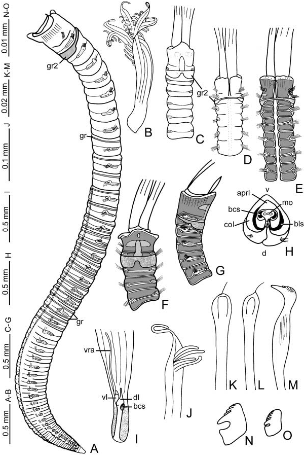
Colour in preserved material, body shape, and size. Body and base of the branchial crown cream coloured. Glandular ridge brown or orange. Glandular ridges yellow in segments from anterior part of abdomen ( Figure 17A View Figure 17 ). Body depressed for all its length, with a ventral compression along last 14 abdominal segments. Body length 7–9 mm, width 0.35– 0.5 mm. Tubes unknown.
Colour in fresh material. Base of the branchial crown cream coloured. Mid-dorsal collar margins red/orange, colour extending towards the dorsal lips. Radioles cream coloured in the basal half, then two distal red bands occupying the length of three pinnules, colour extending into pinnules. One pair of dorsal pinnular appendages with white spots on the basal half. Flanges and body wall transparent. Orange oocytes in thorax and anterior abdomen. Glandular ridge on chaetiger 2, and anterior abdominal ridges white [ LACM, AHF, Santa Monica Bay, SMB-LACSD, Sta. 8D17, LH 04-438].
Branchial lobes and branchial crown. Insertion of the branchial lobes exposed beyond collar laterally ( Figure 17A, G View Figure 17 ). Branchial crown length 3–4 mm. Base of the branchial crown long, about one-quarter the length of branchial crown ( Figure 17C–F View Figure 17 ). Radioles: five to six pairs. Radioles with median pinnules twice as long as distal pinnules. Radiolar tips long ( Figure 17J View Figure 17 ). The palmate membrane extends to three-quarters the length of branchial crown (almost reaching the radiolar tips). Lateral flanges broad ( Figure 17J View Figure 17 ). Dorsal lips triangular, elongate, erect, three times longer than wide, without mid-rib (dl) ( Figure 17I View Figure 17 ). Ventral lips rounded (vl) and about one-half the length of dorsal lips ( Figure 17I View Figure 17 ). Ventral radiolar appendages: one pair occupying one-half the radiole length (vra) ( Figure 17I View Figure 17 ). Dorsal pockets not developed.
Peristomium . Base of the peristomium composed of two lateral horns of basal central skeleton (bcs) surrounding the mouth (mo); anterior peristomial ring lobe (lo) not exposed beyond collar, distally entire, triangular ( Figure 17H View Figure 17 ). The base of the posterior peristomial ring collar is supported by basal lateral skeleton (bls). Posterior peristomial ring collar: dorsal, ventral, and lateral margins entire ( Figure 17A, C–G View Figure 17 ); ventral margin higher than dorsal ( Figure 17A, G View Figure 17 ); entire length of mid-dorsal collar margins forms a narrow gap ( Figure 17D, E View Figure 17 ). Ventral shield of collar swollen, extending to the second thoracic segment, horseshoe-shaped, three times longer than wide ( Figure 17C, F View Figure 17 ). Ratio of posterior peristomial ring collar length versus chaetiger 2 length, in lateral view: 2:1.
Thorax. Chaetiger 1: two groups of elongate, narrowly hooded chaetae. Chaetigers 2–8: notopodia—two irregular rows of elongate, narrowly hooded chaetae; one anterior row with bayonet chaetae; two posterior rows with symmetrical; paleate chaetae with medium-sized mucro ( Figure 17K, L View Figure 17 ); neuropodia—one row of acicular uncini with the main fang surmounted by four rows of teeth equal in size, occupying half the length of main fang ( Figure 17M View Figure 17 ). Glandular ridge on chaetiger 2 (gr2): ventrally, the glandular ridge is sunglasses-shaped, extending to the first half of the third segment; dorsally, inverted Ushaped. The ridge is located near to the superior margin of second segment dorsally and ventrally, on the sides it is located near to the inferior margin of the second segment, under noto- and neurochaetae ( Figure 17A, C–G View Figure 17 ).
Abdomen. Abdominal segments: 33–35. Anterior segments: one glandular ridge in most anterior segments, then two ridges per segment (gr) ( Figure 17A View Figure 17 ); two transverse rows of elongate, narrowly hooded chaetae; uncini with the main fang surmounted by four regular rows of teeth in frontal view, equal in size, occupying half the length of main fang, main fang not extending beyond breast, breast rectangular ( Figure 17N View Figure 17 ). Posterior segments: very elongate, narrowly hooded chaetae; modified uncini with the main fang surmounted by six to seven regular vertical rows of teeth equal in size ( Figure 17O View Figure 17 ), occupying threequarters the length of main fang, main fang not extending beyond breast, breast rectangular. Pygidium with triangular posterior margin ( Figure 17A View Figure 17 ).
Gametes. Oocytes in thorax and anterior abdomen [ LACM-AHF, LH 04-438].
Methyl green staining. The epidermis is completely glandular and stains uniformly in thorax and abdomen, dorsally and ventrally, except the anterior margin of collar ( Figure 17E–G View Figure 17 ). Posterior end lost colour quickly, but pygidium stayed darker for several days.
Remarks
Chone veleronis is unique among Chone species in having a very long base of the branchial crown, and the glandular ridge of chaetiger 2 being sunglasses-shaped ventrally (extending to the first half of the third segment) and inverted U-shaped dorsally.
No known copyright restrictions apply. See Agosti, D., Egloff, W., 2009. Taxonomic information exchange and copyright: the Plazi approach. BMC Research Notes 2009, 2:53 for further explanation.
|
Kingdom |
|
|
Phylum |
|
|
Class |
|
|
Order |
|
|
Family |
|
|
Genus |
Chone veleronis Banse, 1972
| Tovar-Hernández, María Ana 2007 |
Chone veleronis
| Banse K 1972: 477 |