
Cissococcus braini, 2011
|
publication ID |
https://doi.org/ 10.11646/zootaxa.2996.1.1 |
|
persistent identifier |
https://treatment.plazi.org/id/03FAA75F-FF9F-FFE1-FF00-060BDDF2E952 |
|
treatment provided by |
Felipe |
|
scientific name |
Cissococcus braini |
| status |
sp. nov. |
CISSOCOCCUS BRAINI Hodgson & Millar , sp. n.
( Figs 1H–J View FIGURE 1 , 13-15 View FIGURE 13 View FIGURE 14 View FIGURE 15 )
Cissococus fulleri Cockerell, Brain, 1918: 133–134 [misidentification as C. fulleri ].
Type material. Holotype ad ♀: SOUTH AFRICA, Eastern Cape, Boesmansriviermond , 33°40'S 26°39'E, 26.xi.2005, in gall of Rhoicissus digitata (Vitaceae) , P.J. Gullan ( SANC: HC 6970 View Materials ): 1/1 ad ♀. GoogleMaps
Paratypes: As for holotype: 1 3 rd -instar ♀ on holotype slide + 1 3rd-instar ♀; also 1 ad ♀ mounted by T. Kondo 2006 as DNA voucher TK0236 ( BME); Eastern Cape, Gxulu River , ca 20 km SW of East London, 33°07'S 27°44’E, 4.xii.2005, on Rhoicissus digitata, P.J. Gullan ( SANC: HC 6971 View Materials ): 1/1 ad ♀ GoogleMaps ; KwaZulu-Natal, Natal Coast, July 1916, "Type locality", C.K. Brain ( SANC: CKB 40 View Materials ):1/1 ad ♀ ; KwaZulu-Natal, Natal, Scottburgh, on Cissus cuneifolia (see below under “ Comment ” below), 5.vii.1915, C. Fuller ( SANC: CKB 40 View Materials ): 12/8ad ♀ +
many 1 st instar nymphs; KwaZulu-Natal, Natal, Mtabetabe (misspelling of Mtubatuba), 28º25’S 32º13’E, 13.viii.1970, P. Insley ( SANC: HC 4429): 1/1 ad ♀. KwaZulu-Natal, Natal, Durban, July 1935, on wild grape, H.K. Munro ( BMNH, 1958.229 & 1958.579) 4/4 ad ♀ in poor condition (these specimens are those illustrated in fig. 34 in Hodgson (1994) as the non-type and used for the main drawing). Also “Co-type”, C. fulleri , South Africa, on Cissus cuneifolia ( BMNH 1925.174): 1/1adf (poor). Also 3 slides in USDA, each with 1 specimen, variously labelled: slide 1: left label: Cissococcus / fulleri Ckll / Natal Coast. / C.F., July, 1916; right label (under cover slip): Type / locality. / 40. / C.K.B. Also slide 2: left label: Cissococcus / fulleri Ckll. ? Natal Coast. C.F. / anal armature / shows well C.K.B.; right label under cover slip: Type locality. / 40. / C.K.B. Also slide 3: left label: Cissococcus / fulleri Ckll. / Natal Coast. / C.F. July, 1916; right label under cover slip: Type / locality. / 40. / C.K.B. Also 2 slides in USDA, both labelled: Cissococcus fulleri / Ckll / Durban Bot. Garden / Natal / Jan. 1920 / Brain #[727] (USDA): 1 slide with 2 pupae, other with 3 1 st instar nymphs + 1 2 nd -instar male nymph.
Comment. Although Brain (1918) listed the host plant as Cissus cuneifolia , which today is R. tridentata , it is possible that he misidentified the plant species since the two kinds of vines can look similar ( Moore, 1984). Brain’s description of the gall of purported C. fulleri probably was made from the material with the collection data C.K.B. 40. These slides consist of 9 adult females on 9 slides and numerous crawlers on 4 slides. Apparently, this is a composite sample of two collections, with inconsistent label data. Slide C.K.B. 40:1 is labelled in C.K. Brain’s handwriting “ Cissococcus fulleri Ckll – Natal Coast – C.F. July 1916 [no host plant recorded] – Type locality – 40. C.K.B.”. Slides C.K.B. 40: 2–13 each has a new label in De Lotto’s handwriting with “C.K.B. No. 40 – S. Afr.: Natal Scottburgh – 5.vii.1915 – ex.: Cissus cuneifolia – coll.: C. Fuller”, and an old locality label on the reverse side of each slide in E.K. Hartwig’s handwriting with “Natal Coast near Durban – [no date] – C. Fuller – on native vine – CKB 40”. On the basis of the present study, it seems clear that all of this material actually refers to C. braini — and indeed, Brain’s description of the gall, repeated below, describes well that of this species.
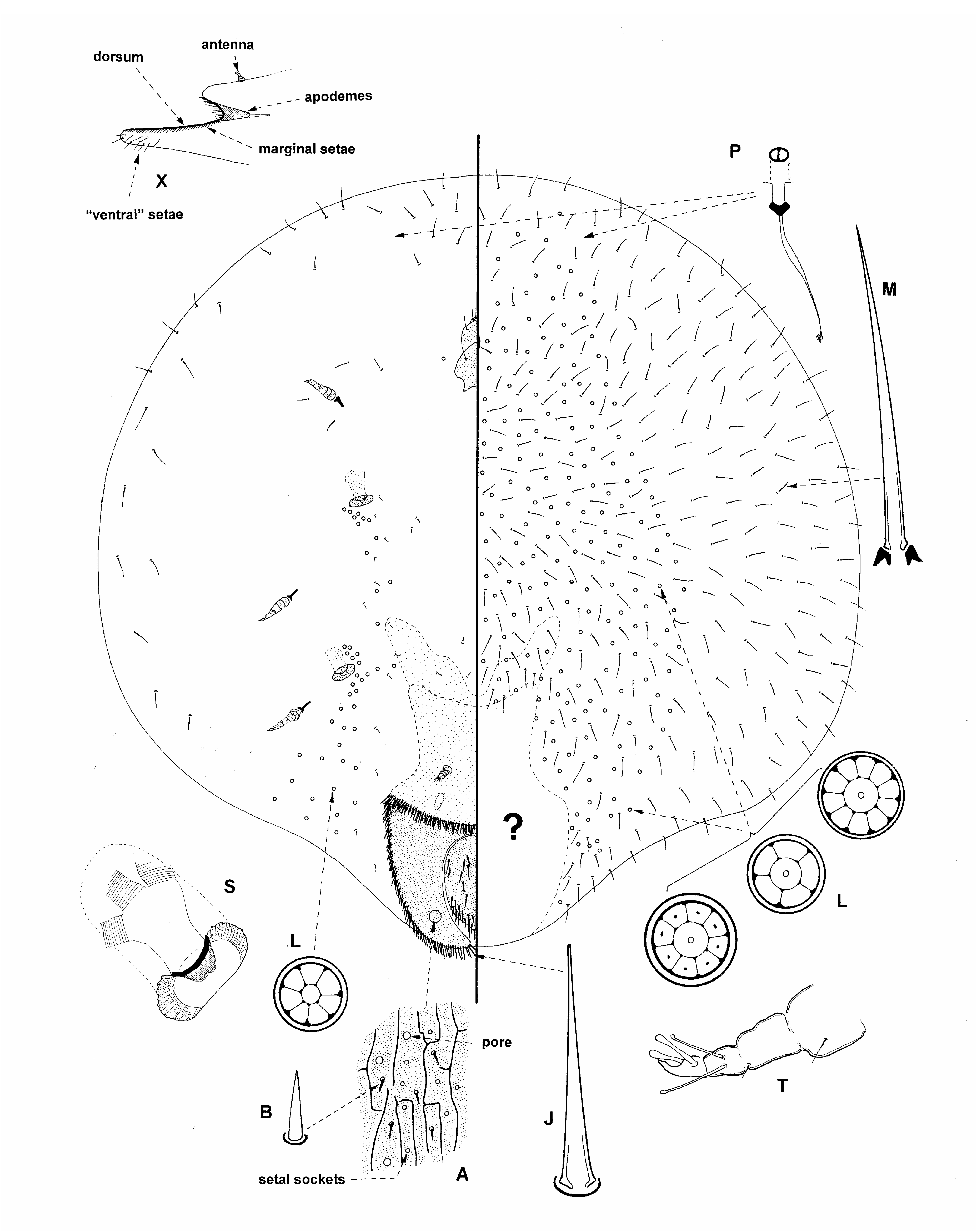
ADULT FEMALE ( Fig. 13 View FIGURE 13 ). Described mainly from 3 specimens in good condition plus reference to several others.
Unmounted material. The female induces a tilted, conical to ovoid or pear-shaped gall ( Fig. 1H–J View FIGURE 1 ), 8–13 mm long, 7–8 mm high and of similar width, with apex truncate; orifice leading to gall chamber minute, circular to elongate, maximum dimension 0.3–0.5 mm, at bottom of a small outer chamber or concavity at apex of gall. Males are not known but presumably are free-living as for C. fulleri . The following description of the female and its gall is from Brain (1918: 133–134): "Insect causing large galls on the stems, tendrils or leaf-stalks of the host-plant. The galls are normally solitary, but in some cases six to eight are clustered together and are then distorted. The normal gall averages 12 mm. long, is broad pear-shaped, almost as broad as long, broadly rounded at the base and slightly tapering to the end where the orifice is situated. The galls are usually fixed by one side, so that the long axis of the gall is parallel with the stem or tendril to which it is attached. The galls apparently grow very rapidly from June to August….” “The twigs and leaves are green, but the galls brown, and where a number are clustered together the intermediate stem is decidedly reddish. The stem beneath the normal galls remains green, but the broad attachment is half green and half brown. The outside of the gall is not smooth, but is much wrinkled and appears bark-like. The orifice of the gall is conical, the thin outer edge being brown and hard in texture, the inside appearing softer and green. At the base of the wide opening there is a minute circular pore. When opened in the fresh state the walls of the galls are hard and woody, from 1 mm. to 1.5 mm. in thickness, with the outer bark loosely attached.” ”The adult female at the time the eggs are developing fills approximately half the space of the gall, being very convex, with the blunt caudal extremity fitting tightly into the conical portion near the aperture. The dorsal surface is distinctly segmented and is flat with prominent ridges.” “The colour of the body is delicate flesh-pink, slightly obscured by a thin layer of powdery secretion.".
Mounted material. See generic diagnosis. Body approximately round and apparently highly convex. Radius from 2.0 to at least 5 mm.
Dorsum. Length of dorsum 445–665 µm; greatest width 500–700 µm. Derm sclerotised throughout as per generic diagnosis, except with a longitudinal fissured pattern that appears to run into area of apodeme. Dorsum bent upwards and over about 1/3rd from anterior margin (i.e. becoming ⊃-shaped), so that anterior margin lies almost over anal plates. Derm with many pale pores each about 3–4 µm wide. Dorsal setae hard to discern but each rather spinose and 8 µm long but those near posterior margin longer, up to about 30 µm long; frequent throughout; when setae not visible, setal sockets appear similar to small pores, each about 1 µm wide. Anal plates together oval, each 290–340 µm long; together 170–190 µm wide; dorsal surface of each plate with about 15–40 spinose setae, mainly restricted to posterior half but with a few more anteriorly; each seta 25–35 µm long, but with 1 or 2 near apex 45–65 µm long. Eyespots indistinct, hidden under folded-over dorsum, size unclear.
Margin. Marginal setae very similar to those on dorsal surface of anal plates, each mainly sharply spinose and only rarely curved, 25–50 µm long, in more or less a single line along margin, bases almost touching, with 60–75 on each side from anal cleft to shoulder of folded dorsum, and with 25–40 on each half of folded-over anterior margin.
Venter. Becoming enormously swollen in mature specimens as described under generic diagnosis. Microducts frequent throughout, each about 2.0–2.5 µm wide. Loculate pores abundant medially on lower venter, apparently only rarely appearing to be segmentally arranged (frequency of loculate pores and number of loculi somewhat variable between collections); each with mainly 5–8 loculi, but a few with 10 loculi, and each 6–8 µm wide; no long inner ductule arising from central loculus detected. Similar loculate pores also associated with each spiracle on upper venter as per generic diagnosis. Setae all setose; setae on lower venter abundant and long, each 50–85 µm long (up to 100 µm long on Brain's no. 40); setae on upper venter infrequent and short, mostly 16–25 µm long.
Antennae distorted; located dorsad to large apodeme and often not observable. Clypeolabral shield 135–185 µm long; labium with 4 pairs of setae. Spiracles quite large, width of peritremes: anterior 75–85 µm, posterior 80–95 µm. Legs as in generic diagnosis.
Comment. The morphology of the adult females of Cissococcus species is very similar apart from the structure of the dorsum. It appears that the dorsum of the type species, C. fulleri , is almost flat (at least, it comes to lie flat on the slide once mounted) whereas the dorsum of the new species, C. braini , is rather larger and perhaps C-shaped, so that, during the mounting process, the dorsum is bent on itself and becomes ⊃-shaped. The consequence of this is that the actual dorsal surface of the anterior end of the dorsum (with the eyespots) faces ventrally and is more or less hidden on C. braini . Other differences between the adult females of the two species are (character states on C. fulleri in parentheses): (i) setae on dorsal surface of anal plates relatively few and not covering more than about half surface (numerous, covering almost entire surface); (ii) spiracles rather larger, smallest about 75 µm wide (smaller, largest 65 µm wide), and (iii) setae on lower venter very long, mostly more than 65 µm long (shorter, mostly 50 µm or less).
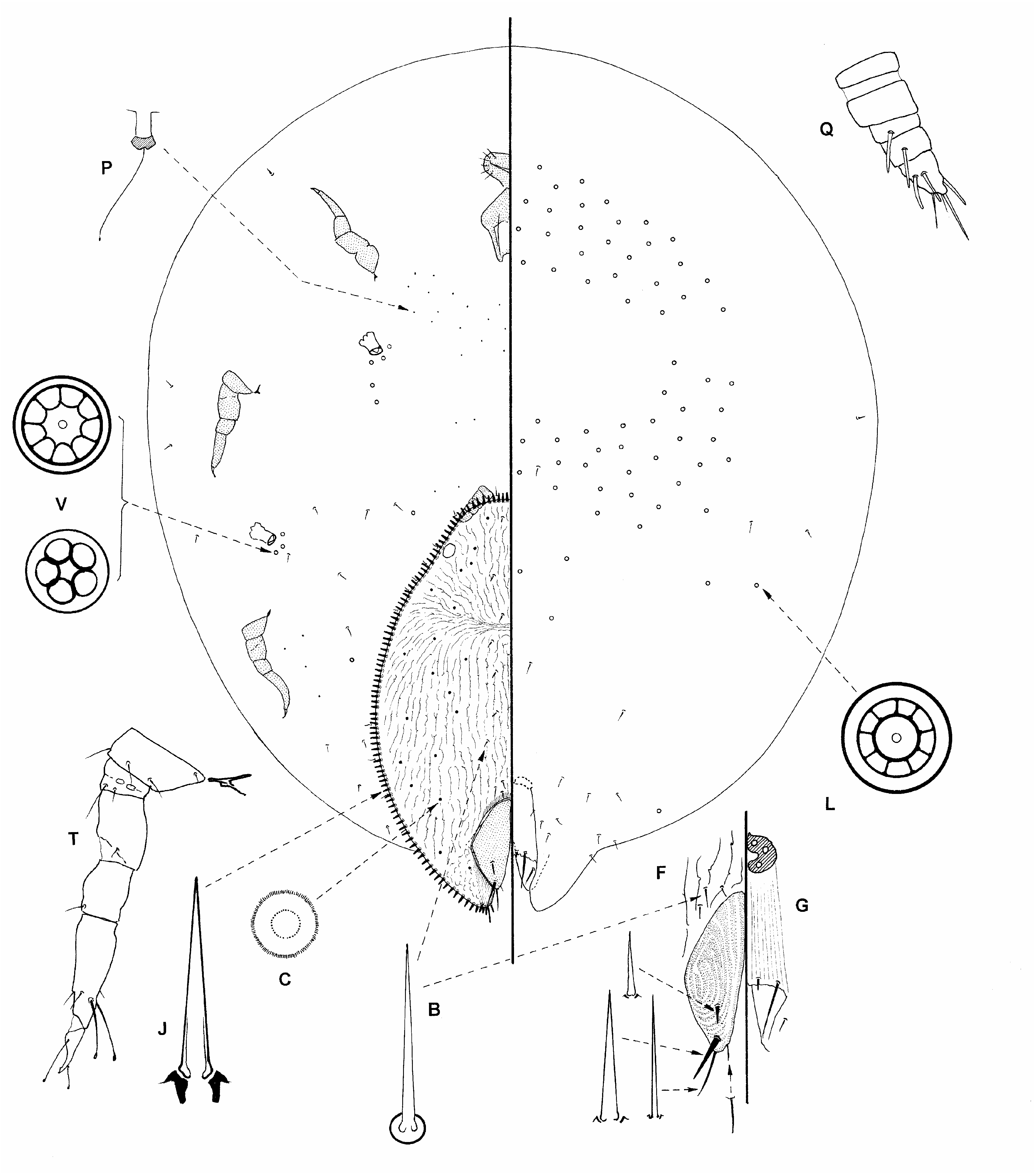
THIRD-INSTAR FEMALE ( Fig. 14 View FIGURE 14 ). Described from a single specimen in good condition.
Mounted material. See generic diagnosis. Body approximately round and apparently highly convex. Total length 1.49 mm; 1.39 mm wide.
Dorsum. Length 0.73 mm, width 0.48 mm. Derm barely sclerotised throughout, with a longitudinal fissured pattern converging on apodeme; a barely sclerotised bar present just anterior to anal plates; derm on each side of anal cleft also slightly more heavily sclerotised. Dorsum lying flat, with a clear indication of an apodeme arising about 1/4 th length of dorsum from anterior end. Derm with sparse dorsal pores, more lateral pores each 4.5–5.0 µm wide; more medial pores appearing smaller, each 3.5–4.0 µm wide; apparently randomly distributed. Dorsal setae easily discernible, in two submedial lines, with 1 pair on head (each about 7.5 µm long), 1 pair on prothorax (anterior to apodeme), 3 pairs posterior to apodeme plus 3 or 4 pairs just anterior to anal plates (each about 6.5–13 µm long); totaling 8 or 9 per side. Anal plates together oval, each 165 µm long; together about 140 µm wide, with longitudinal ridges; dorsal surface of each plate with 3 or 4 spinose setae, anterior setae (present on one plate only) 16 µm long, posterior pair 35–38 µm, apical 35 µm and inner margin 10–12 µm long. Anal fold with perhaps two pairs of setae along anterior margin, longest 38–40 µm long (but these could be preanal setae). Anal ring with 3 pairs of setae; anal tube 165 µm long. Eyespots distinct, near margin, each oval, 21 x 25 µm wide.
Margin. Marginal setae sharply spinose and only rarely curved, 15–25 µm long, mainly in a single line along margin, bases almost touching, with 36 between eyespots anteriorly and 85–90 on each side between eyespots and anal cleft; with a small group of about 4 setae on each anal lobe, longest up to 28 µm long.
Venter. Clearly enormously swollen as described under generic diagnosis; antennae located on upper venter just anterior to dorsum on flattened, slide-mounted specimens; legs placed near edges of mounted body. Microducts minute, each about 1.5 µm long, with a short outer ductule about 1.5–2.0 µm long and a long filamentous inner ductule; mainly detected in a band anterior to mouthparts, but occasional elsewhere. Loculate pores abundant medially on lower venter, appearing to be segmentally arranged, most abundant on pro- and mesothorax, many fewer on metathorax and on abdomen; each with mainly 6–9 (range 5–10) loculi, and each 6–8 µm wide. Similar loculate pores, but slightly smaller and with fewer loculi, also present in elongate groups of 3–5 associated with each spiracle on upper venter, as per generic diagnosis. Setae all setose and short on lower venter, length of each about 2x width of loculate pores; setae on upper venter similar but sparser. Submarginal setae sparse, with perhaps 10 on each side.
Antennae 5 segmented, located just anterior to anterior margin of dorsum, each about 75 µm long; apical stiff seta 33–35 µm long. Clypeolabral shield 120 µm long; number of labial setae uncertain. Spiracles: width of peritremes 30–32 µm. Legs as in generic diagnosis, almost always distorted; length of metathoracic leg (µm): coxa 60–62, trochanter + femur 55–60, with 1 long and 1 short seta on ventral surface; tibia 33, and tarsus 56–65; claw 23–27; claw with a distinct denticle; claw digitules probably similar, extending beyond claw.
Comment. The third-instar female of C. braini differs from that of C. fulleri in having: (i) abundant loculate pores medially on the thorax, (ii) many more marginal setae, and (iii) antennae only 5 segmented rather than 6 segmented. It differs from adult female C. braini (data for adult female in brackets) as follows: (i) dorsum of third instar about half total body length (much less than half total body length); (ii) ventral loculate pores frequent only medially on thoracic segments (abundant throughout most of venter); (iii) setae on lower venter few and fairly short (abundant and long), and (iv) dorsum with 8 or 9 pairs of setae (frequent throughout).
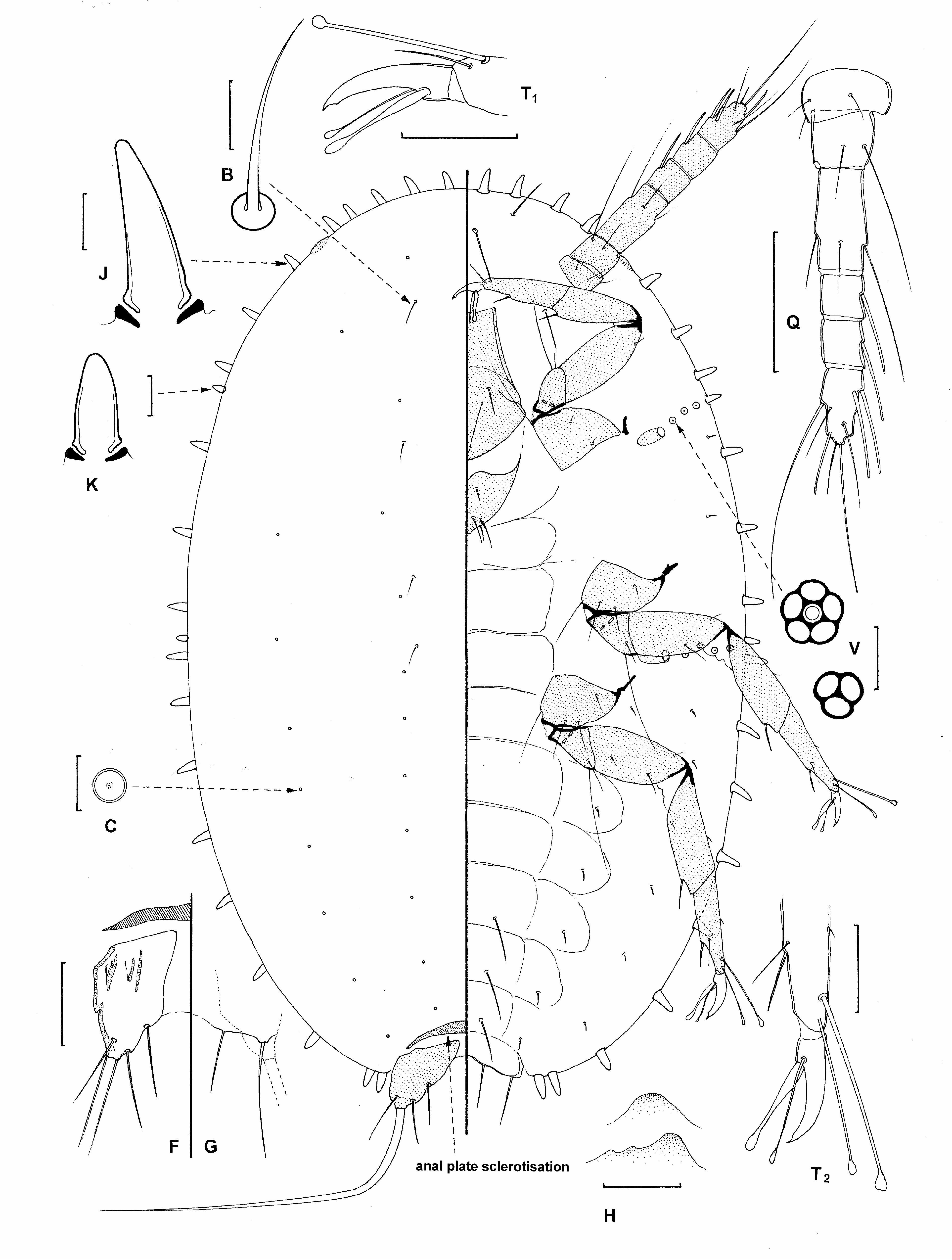
FIRST-INSTAR NYMPH (sex not determined) ( Fig. 15 View FIGURE 15 ).
Illustrated from about 3 specimens in fair to good condition.
The morphology of the 1 st -instar nymphs of C. braini appears to be indistinguishable from those of C. fulleri except that the claw digitules on C. braini appeared to be more dissimilar than those on C. fulleri .

| SANC |
Agricultural Research Council-Plant Protection Research Institute |
No known copyright restrictions apply. See Agosti, D., Egloff, W., 2009. Taxonomic information exchange and copyright: the Plazi approach. BMC Research Notes 2009, 2:53 for further explanation.
|
Kingdom |
|
|
Phylum |
|
|
Class |
|
|
Order |
|
|
Family |
|
|
Genus |
Cissococcus braini
| Hodgson, C. J., Millar, I. M. & Gullan, P. J. 2011 |
Cissococus fulleri Cockerell, Brain, 1918: 133–134
| Brain, C. K. 1918: 134 |