
Congocepheus thailandae, Fernandez & Theron & Leiva & Jordaan, 2018
|
publication ID |
https://doi.org/ 10.11646/zootaxa.4504.3.4 |
|
publication LSID |
lsid:zoobank.org:pub:5D91D1A9-413B-4704-A78E-4402FC449943 |
|
DOI |
https://doi.org/10.5281/zenodo.5997003 |
|
persistent identifier |
https://treatment.plazi.org/id/0389B40B-FFD8-781A-FF0A-FA7CFA25FB0F |
|
treatment provided by |
Plazi |
|
scientific name |
Congocepheus thailandae |
| status |
sp. nov. |
Congocepheus thailandae sp. nov.
( Figures 42–67 View FIGURES 42–48 View FIGURES 49–51 View FIGURES 52–60 View FIGURES 61–70 )
Etymology. The specific epithet “thailandae” derives from Thailand, country of origin of type material.
Material examined. Holotype . Female. “ THAILAND N°286. NE Bangkok Khao Yai Nai Park.759–859 mt. 26/XI /– 3/XII/85. Leg. LOBL”; material deposited in the Collection of the Natural History Museum of Geneva (NHMG), Switzerland; preserved in 70% ethanol. Paratype. One adult female, same locality and date as Holotype; deposited in Collection of MHNG; preserved in 46 % ethanol. Material studied with SEM: three specimens, not deposited .
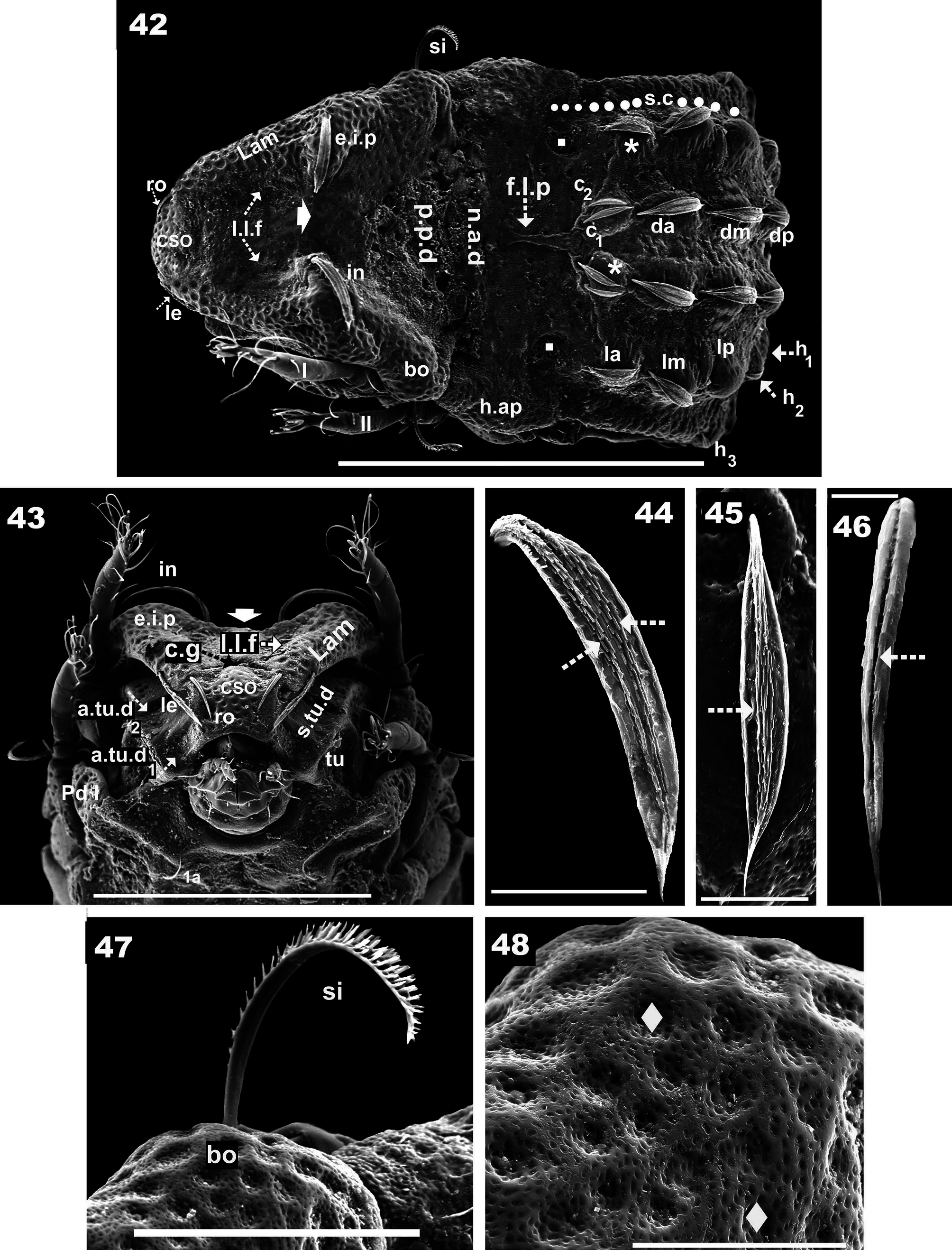
Diagnosis. Prodorsum. Large, triangular, central zone depressed or flat; setae in large, curved, situated on elevated interlamellar process, directing externally; ro setae curved, directing anteriorly; lamelar setae inserted far from apical lamellar zone. Anterior zone of lamellae welded to lateral prodorsal zone, lacking lamellar tip. Sensillus uncinate. Bothridial ring smooth; low lamellar furrow present, only slightly developed. Cornea superior of naso, with semicircular groove in posterior zone.
Notogaster. Fingerlike projection large, extending forward. Fourteen pairs of setae: c 1, c 2, da, dm, dp, la, lm, lp, h 1, h 2, situated on promontories; h 3, p 1, p 2, p 3, situated marginally on notogaster, all setae directing backwards; setae c 1, c 2 parallel and located side by side; integument with rugous elevated ridges.
Ventral region. Epimeral, central zones depressed, marginal region elevated. Epimera 3–4 fused; epimeral chaetotaxy 1-1-3-3; four pairs of genital setae in a simple line; ag setae situated on rounded elevated structure.
Description. Measurements: SEM 556 (460–602) x 260 (242–273) (measurements on three specimens). Light microscopy: 580 (560–582) x 273 (256–281).
Shape: Elongate oval ( Figure 42 View FIGURES 42–48 ).
Colour: Specimens without cerotegument, light brown to brown, observed in reflected light.
Cerotegument. Thin grainy layer (ce) ( Figure 69 View FIGURES 61–70 , indicated by 5), mainly on posterior zone of notogaster ( Figure 57 View FIGURES 52–60 ) and epimeral zone ( Figure 61 View FIGURES 61–70 ), and around ag setal insertion ( Figure 68 View FIGURES 61–70 ); rest of body and legs absent; cerotegumental layer may have been affected by the long period in conservation liquid.
Integument. Foveate-foveolate ( Figure 48 View FIGURES 42–48 , indicated byw): prodorsum: lamellar zone up to l.l.f ( Figures 42, 43 View FIGURES 42–48 ); anterior of prodorsum around CSO ( Figure 43 View FIGURES 42–48 ); bothridial and p.p.d zones ( Figures 42, 47 View FIGURES 42–48 ); notogaster: h.ap ( Figure 42 View FIGURES 42–48 ); Pd I, Pd II and Tu ( Figures 43 View FIGURES 42–48 , 52 View FIGURES 52–60 ).
Circular-ovoid fovea: notogastral zone: n.a.d and both sides of f.l.p ( Figure 42 View FIGURES 42–48 ).Ventral region: subcapitular zone h setae ( Figure 58 View FIGURES 52–60 ); epimeral zone (Figures 61,62,63); around genital opening ( Figure 56 View FIGURES 52–60 ); aggenital zone, but not very close to ag setae ( Figure 68 View FIGURES 61–70 ); lateral to ventral dep ( Figure 61 View FIGURES 61–70 ). Legs: femurs I, II and trochanter, femur III, IV ( Figure 52 View FIGURES 52–60 ).
Rugous with elevated ridges: lateral notogastral margin, zone of setae p 1, p 2, p 3, h 3, up to h.ap level ( Figure 57 View FIGURES 52–60 indicated by ●), in this zone, combined with circular-ovoid fovea; marginal ventral region; anal zone ( Figure 61 View FIGURES 61–70 ).
Pusticulate ( Figures 69, 70 View FIGURES 61–70 indicated by¿): zone epimeral surrounding setae 4c ( Figure 63 View FIGURES 61–70 ); zone near ag setae ( Figure 68 View FIGURES 61–70 ).
Setation. Lanceolate elongate with: smooth surface: ag setae ( Figure 68 View FIGURES 61–70 ); medial ridge: notogastral c 1, c 2, da, dm, dp, la, lm, lp ( Figures 42, 45 View FIGURES 42–48 ) ad setae ( Figure 67 View FIGURES 61–70 ); large basal, and thin anterior zone, medial ridge several lines: notogastral setae: h 3, p 3, p 2, p 1 ( Figures 53, 57, 60 View FIGURES 52–60 ); rugous surface: epimeral setae 1a, 2a, 3a, 3b, 4a, 4b ( Figures 62, 63, 65 View FIGURES 61–70 ); thin medial furrow: ro setae ( Figure 46 View FIGURES 42–48 ); two parallel lines with small barbs: in setae ( Figure 44 View FIGURES 42–48 ); barbate on both sides: le setae ( Figure 54 View FIGURES 52–60 ).
Filiform, irregular margin: epimeral setae, 3c, 4c ( Figures 64 View FIGURES 61–70 ). Simple: subcapitular (a, m, h) ( Figures 58, 59 View FIGURES 52–60 ), ge ( Figure 56 View FIGURES 52–60 ), an ( Figure 66 View FIGURES 61–70 ).
Prodorsum. Triangular (dorsal view) ( Figure 42 View FIGURES 42–48 ); convex in lateral view ( Figure 49 View FIGURES 49–51 ); rostral margin rounded (Figures 43,51); e.i.p elevated ( Figure 52 View FIGURES 52–60 ), central depressed zone flat (Figures 42,43 indicated by¿); large curving setae in, situated on e.i.p, directing externally ( Figures 42, 43 View FIGURES 42–48 ); ro setae clearly visible, curved, directing anteriorly, apical tips far from one another ( Figure 43 View FIGURES 42–48 ); le setae inserted laterally to lam, far from apical lamellar zone; setal insertion level more or less same level as ro setal insertion ( Figures 43 View FIGURES 42–48 ); anterior zone of lamellae welded to lateral prodorsal zone, lamellar tip absent ( Figure 54 View FIGURES 52–60 ).
Sensillus (si) uncinate ( Figure 47 View FIGURES 42–48 ). Bothridial ring (bo.ri) smooth, well defined, with bothridial tooth; l.l.f present, slightly developed ( Figures 42, 43 View FIGURES 42–48 , 51 View FIGURES 49–51 ), CSO present between ro setae ( Figures 43 View FIGURES 42–48 , 51 View FIGURES 49–51 ); poorly developed semicircular groove (c.g) in posterior zone ( Figure 43 View FIGURES 42–48 ).
Notogaster. Shape: in dorsal view anterior polyhedral and posterior oval ( Figure 42 View FIGURES 42–48 ); in lateral view convex ( Figures 49 View FIGURES 49–51 , 52 View FIGURES 52–60 ); d.sj narrow, slightly curving, well delimited; notogastral anterior depression (n.a.d) clearly visible ( Figure 42 View FIGURES 42–48 ).
Notogaster with promontories ( Figure 49 View FIGURES 49–51 ); large, forward extending finger-like projection (f.l.p) in front of promontory zone ( Figure 42 View FIGURES 42–48 ). Lateral to f.l.p, at level of origin of lateral promontories, depressed semi-circular area ( Figure 42 View FIGURES 42–48 indicated by n). Fourteen pairs of setae: c 1, c 2, da, dm, dp, la, lm, lp, h 1, h 2, h 3, p 1, p 2, p 3. Four longitudinally aligned promontories separated by depressed zone ( Figures 42 View FIGURES 42–48 , 49 View FIGURES 49–51 ); setae c 1, c 2, da, dm, dp, situated on aligned central promontories; la, lm, lp, h 1, h 2 situated on aligned lateral promontories ( Figures 42 View FIGURES 42–48 , 49 View FIGURES 49–51 , 52 View FIGURES 52–60 ); h 3, p 1, p 2, p 3, situated marginally notogastral zone ( Figures 42 View FIGURES 42–48 , 49 View FIGURES 49–51 , 52, 57 View FIGURES 52–60 ); all setae directing backwards; setae c 1, c 2 situated side by side, parallel ( Figure 42 View FIGURES 42–48 ); polyhedral h.ap large, easily observed ( Figures 42 View FIGURES 42–48 , 49 View FIGURES 49–51 , 52 View FIGURES 52–60 ); excavated Vshaped depression present, but angle of observation needs to be changed to permit clear view ( Figure 52 View FIGURES 52–60 indicated by 5).
Circumgastric depression (s.c) easily discernible ( Figures 42 View FIGURES 42–48 , 52, 57 View FIGURES 52–60 indicated by l), originating slightly posterior to h.ap, with the following trajectory: lateral to la, lm, lp, h 1, h 2, and internally to h 3, p 1, p 2, p 3 ( Figures 42 View FIGURES 42–48 , 52 View FIGURES 52–60 ); in the posterior notogastral zone the trajectory is partially obscured by rugous elevated cuticular ridges ( Figure 57 View FIGURES 52–60 ).
Lateral region. Dorsolaterally, lamellae clearly discernible, anterior zone welded to lateral notogastral zone, lamellar tip absent ( Figure 54 View FIGURES 52–60 ). Tu large lamina, curving margin ( Figure 43 View FIGURES 42–48 , 49 View FIGURES 49–51 , 52 View FIGURES 52–60 ). Deep s.tu.d running between and parallel to lamellae and Tu; anteriorly a large pocket depression is observed (a.tu.d); followed by deep depression, p.tu.d ( Figure 49 View FIGURES 49–51 , 52 View FIGURES 52–60 ). Pd I, conspicuous extended lamina, rounded apex, particular microsculpture (See Integument). Pd II medium sized, ovoid lamina; dis hardly discernible ( Figures 49 View FIGURES 49–51 , 52 View FIGURES 52–60 ).
Bothridia cup-shaped; bothridial opening directing slightly downwards; smooth bo.ri incomplete, with bo.to clearly discernible ( Figure 49 View FIGURES 49–51 ).
Long, extending polyhedral humeral apophysis, rounded apex, basally undulated, anterior tip overlapping posterior bothridial part ( Figures 49 View FIGURES 49–51 , 52 View FIGURES 52–60 ). Series of large dep discernible behind leg IV ( Figures 49 View FIGURES 49–51 , 52 View FIGURES 52–60 ). Posterior notogastral zone ( Figure 57 View FIGURES 52–60 ) with clearly visible rugous elevated cuticular ridges.
Ventral region. Epimera: central zones depressed, marginal elevated zones ( Figure 61 View FIGURES 61–70 ). Epimera 3–4 fused, small; apo.1, apo.2, apo.sj and apo.3 well discernible ( Figure 50 View FIGURES 49–51 ).
Epimeral chaetotaxy 1-1-3-3 (See Remarks); dis clearly observed as triangular protuberance with rounded apex ( Figure 61 View FIGURES 61–70 ).
Genital plates small relative to anal plates; four pairs of genital setae in a simple line ( Figure 50 View FIGURES 49–51 , 56 View FIGURES 52–60 ); all setae more or less equal length ( Figure 56 View FIGURES 52–60 ); ag setae far from genital opening, situated laterally and posterior to genital opening, placed on elevated rounded structure ( Figures 50 View FIGURES 49–51 , 61 View FIGURES 61–70 indicated by l); elevated structure covered by cerotegumental layer. Three pairs of adanal setae; ad 3 situated far from ag setae ( Figure 50 View FIGURES 49–51 ). Anal plate rectangular to polyhedral, small sharp tip ( Figure 50 View FIGURES 49–51 ); two pairs of anal setae. Lyrifissures not discernible. Ovoid paired depressions situated between anal and genital openings; ovoid-elongate depressions situated laterally to genital and anal openings ( Figures 50 View FIGURES 49–51 , 61 View FIGURES 61–70 )
Legs. Setal formulae I(1-3-2-3-16 -1) (1-2-2); II(1-4-3-3-15 -1) (1-1-2); III(2-3-1-2-14 -1) (1-1-0); IV(1-2-2-3- 13 -1) (0-1-0). Large femoral groove (Femur III).
Remarks. Epimeral chaetoxy 1-1-3-3 was observed in five specimens studied. However, due to the lengthy preservation period, the authors are uncertain if the setae have degraded during this time. There is a possibility that they exist, especially as in some instances, vestiges of setal insertions were observed. Two noteworthy aspects are: a) the large variation in shape of notogastral setae, where setae h 3, p 1, p 2, p 3 are very different to the other notogastral setae; b) the different shapes of epimeral setae, where 3b, 4b are very different compared to 3c and 4c.


| MHNG |
Museum d'Histoire Naturelle |
No known copyright restrictions apply. See Agosti, D., Egloff, W., 2009. Taxonomic information exchange and copyright: the Plazi approach. BMC Research Notes 2009, 2:53 for further explanation.