
Diplotrema tabascensis, Fragoso & Rojas, 2019
|
publication ID |
https://doi.org/ 10.11646/zootaxa.4688.4.2 |
|
publication LSID |
lsid:zoobank.org:pub:FE1F719B-D97F-4FCB-80D5-E5E2974DE80E |
|
persistent identifier |
https://treatment.plazi.org/id/C97C0EE2-B44F-4C1D-ABED-E5C1102C149D |
|
taxon LSID |
lsid:zoobank.org:act:C97C0EE2-B44F-4C1D-ABED-E5C1102C149D |
|
treatment provided by |
Plazi |
|
scientific name |
Diplotrema tabascensis |
| status |
sp. nov. |
Diplotrema tabascensis View in CoL sp. nov.
( Figures 5 View FIGURE 5 , 6 View FIGURE 6 , 7 View FIGURE 7 )
urn:lsid:zoobank.org:act:C97C0EE2-B44F-4C1D-ABED-E5C1102C149D
Localities and material. Mexico, Tabasco, Highway 186, municipality of Centro, 11.5 km from Villahermosa on the road to Macuspana , forest reserve in the proximities of Villahermosa airport; tropical subdeciduous forest with abundant palm trees, inside and below decaying palm logs; 18°0’1.98’’N, 92°48’32.83’’W, 22 m asl, two clitellate adults (one fragmented), two semi-adults with genital marks (GM) and two posterior fragments, 12/19/1991, C. Fragoso and P. Rojas. ( Fig. 10 View FIGURE 10 ) GoogleMaps .
Holotype. Clitellate adult IEOL 119 .
Paratypes. One fragmented clitellate adult (head of 9.7 mm length): IEOL 120 ; two entire semi-adults with GM: IEOL 121 , IEOL 122 .
Description. External. Length 35, 35, 39 (holotype) mm (average 36.3, n=3). Width after clitellum 1.22–1.33 mm (average=1.27, n=4), holotype 1.33 mm. Number of segments 113 (holotype), 121, 123, (average=119, n=3). Secondary annulation clearly seen after clitellum, characterized by two or three presetal and postsetal furrows. Middle segments quadrangular, 1.1–1.5 times wider than long; last segments rectangular, 5.7–8 times wider than long. Pigment absent. Prostomium open epilobous. Setae eight per segment, visible from 2; closely paired throughout ( Fig. 5A,B View FIGURE 5 )). Setal formula (averages) (aa:ab:bc:cd:dd) at 10 (n=4): 8.5:1:8.6:0.9:26.4 and 0.9 dd =1/2 C; at 30 (n=4): 7.6:1:7.7:1.1:26.7 and 0.98 dd =1/2 C; ten segments before anus (n=5): 6.1:1:5.2:1.1:16 and 0.8 dd =1/2 C.
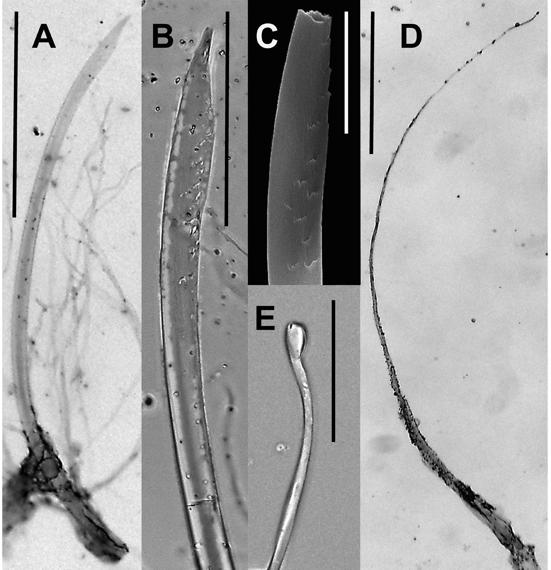
Hair-like penial setae (a and b) in 17 and 19, thin (width at base 11 μm) and long (2.5, 2.9 mm) ( Fig. 7D View FIGURE 7 ); they are delicate, smooth, without ornamentation, curved in some of their extension, and with a blunt apex ( Fig. 7E View FIGURE 7 ). Externally these setae were not visible; internally setae are contained within long follicles ( Fig. 6A View FIGURE 6 ) attached to the lateral-dorsal body wall and that probably perform as retractor tissue. Follicles of 17 and 19 run independently until segment 21, where they are fixed together by connective tissue; joined follicles extending 5–6 segments backwards, reaching segments 25 or 26. Genital setae a and b present in segment 16, within clearly visible follicles extending freely in the coelom; follicles larger than anterior and posterior follicles of somatic setae ( Fig. 6A View FIGURE 6 ). Genital setae slightly curved ( Fig. 7A View FIGURE 7 ), with scarce and irregular ornamentation limited to the distal part ( Fig. 7C View FIGURE 7 ), with a slight subapical dilatation, and with pointed tip ( Fig. 7B View FIGURE 7 ); length: 629 μm; width: 17.5, 18 μm.
Clitellum ring-shaped in 1/2 13, 14–17 (4.5 segments), of vermilion color, interrupted at BB in 16 and 17 by a genital mark and the prostatic pores, respectively ( Fig. 5A,C View FIGURE 5 ). Dorsal pores present, first functional pore in 9/10 (2 ind.); in 8/9 visible but closed. Nephropores in CD. Paired spermathecal pores in A of 7/8 and 8/9, the posterior ones tending to be larger ( Fig. 5B,D View FIGURE 5 ); in one individual slightly shifted into 8 and 9, respectively ( Fig. 5D View FIGURE 5 ). Female pores in 14, presetal in A. Two pairs of prostatic pores in B of segments 17 and 19, joined by slightly straight or bracket shaped seminal grooves which run in A ( Fig. 5A,B,C View FIGURE 5 ). Male pores in the equator of 18, within seminal grooves, but not seen externally; internally confirmed by discharge of male gonoduct. One large, transversely elliptical genital mark in segment 16 in BB, with a central transversal fissure in AA ( Fig. 5A,B,C View FIGURE 5 ); genital setae visible inside the fissure or above it. Body wall of region AA in segments 15 (one ind. Fig. 5A View FIGURE 5 ) or 17 (two individuals, Fig. 5C View FIGURE 5 ) slightly thickened.
Internal. Septa 6/7 thin and membranous; 7/8, 12/13 and 13/14 slightly thicker; following septa more muscular: 8/9<9/10=10/11>11/12; from 14/15 backwards membranous; 7/8–13/14 funnel shaped. Septum 5/6 only seen in holotype; thus the single spherical large gizzard is placed in segment 5 (one ind.) or 6 (three ind.). Gizzard dimensions (length by width): 0.47 by 0.52 mm (holotype), 0.75 by 0.75 mm, and 0.80 by 0.73 mm. Esophagus tubular, without dilatations; some internal lamellae present in segments 9–12; internal walls thicker in segments 10, 11, 12, 13. Intestine starting in 14/15. Laminar, dorsal typhlosole starting very small as a ribbon in 16, gradually increasing in size until reaching maximal size in segment 18 or 22; from segment 42 on, gradually diminishing size, ending as a thin cord.
Dorsal vessel single, visible throughout the intestine; in the esophagus visible in segments 8–13. Supra-esophageal vessel visible in segments 9–12. Lateral hearts in 9, 10; latero-esophageal hearts in 11 and 12. Ventral vessel present. Subneural trunk absent. Nephridial system holoic, avesiculate, exonephric and stomate, clearly parietal from segment 11, 12 backwards ( Fig. 6A View FIGURE 6 ); in one individual also parietal in 4 and 5. Nephrostomes and nephropores not seen. Male apparatus holandric. Testes and iridiscent male funnels in 10 and 11. Iridiscent male gonoduct double (from segment 14), straight, superficial, running along inner face of body wall in or slightly outside AB of segments 13–18, entering body wall in 17 or the equator of 18. Two pairs of acinous seminal vesicles in 11 and 12; the posterior one larger, covering entire dorsal portion of the esophagus. Paired coiled tubular prostates in 17 and 19, extending one to three segments backwards; conspicuous muscular ducts transversely or obliquely placed in the respective segments; glandular regions 2–4 times thicker and 2–3 times longer than muscular duct ( Fig. 6A View FIGURE 6 ).
Paired female funnels and shrub-fan ovaries in 13. Ovules large, clearly seen; ventral female funnels at both sides of mid-ventral line, close to 13/ 14 in AB. Small acinous glands in 14, joined to both sides of the esophagus, probably ovisacs. Two pairs of spermathecae in 8 and 9, opening in 7/8 and 8/9, respectively; ampulla joining duct at right angle; diverticulum joining duct along the same axis ( Fig. 6B,C View FIGURE 6 ); duct enlarged, almost the same size as ampulla and diverticulum. Ovoid ampulla with a fold; diverticulum with its distal region dilated, curved 180° and full of iridescent sperm. Length of ampulla almost equal (1.1 times) to that of diverticulum, or slightly smaller (0.83). Maximal total length of spermatheca: 0.87 mm (paratype IEOL 120; Fig. 6C View FIGURE 6 ), 0.91 mm (holotype; Fig. 6B View FIGURE 6 ).
Etymology. The name of the species refers to the Mexican state where all individuals were found.
Remarks. By the shape of the spermathecae, D. tabascensis sp. nov. is related to the group of species (D. jen- niferae, D. murchiei , D. oxcutzcabensis , D. chajulensis , and D. oaxacana sp. nov.) with a transversally connected ampulla, sharing with D. murchiei , D. jenniferae and D. oxcutzcabensis a similar spermathecae shape; from these species, however, it is separated by the pattern of genital marks (midventral in 16 in D. tabascensis sp. nov. vs. midventral in 17–19, 17 or 19 in D. murchiei , midventral in 6–9, 17–20 and paired in 17 and 19 in D. jenniferae and absent in D. oxcutzcabensis ), the presence of genital setae (present vs. absent in the three species) and the position of seminal vesicles [11 and 12 vs. 12 in D. murchiei and 9 and 11 in D. jenniferae and D. oxcutzcabensis ( Fragoso & Rojas 2018) ]. The new species shares with D. chajulensis and D. oaxacana sp. nov. the position of seminal vesicles [segments 11 and 12, a feature present also in two other gizzard-less species from northeast Mexico: D. albida and D. mexicana ( Gates, 1967) ] and the long, haired-like penial setae; from both species it is separated by the shape of the spermathecae. Apart from the gizzard, D. tabascensis sp. nov. differs from D. albida and D. mexicana in the pattern of the genital marks (mid-ventral elliptical in 16 vs. paired swellings in 16/17 and 19/ 20 in D. albida , and absent in D. mexicana ) and in the genital setae, present in segment 16 (absent in both species), a character that further separates this new species from all other neotropical Diplotrema species.


No known copyright restrictions apply. See Agosti, D., Egloff, W., 2009. Taxonomic information exchange and copyright: the Plazi approach. BMC Research Notes 2009, 2:53 for further explanation.
|
Kingdom |
|
|
Phylum |
|
|
Class |
|
|
Order |
|
|
Family |
|
|
Genus |