
Melaenini
|
publication ID |
https://doi.org/ 10.11646/zootaxa.1099.1.1 |
|
publication LSID |
lsid:zoobank.org:pub:CA0A0ED3-D49D-4198-A409-407EF86A164E |
|
persistent identifier |
https://treatment.plazi.org/id/227887BE-FFF0-FFA8-250D-F6E9FCCAFCBD |
|
treatment provided by |
Felipe |
|
scientific name |
Melaenini |
| status |
|
Tribe Melaenini
Scaritides (in part) Dejean 1831: 471, 478–483. Ditomides (in part) Lacordaire 1854: 166–167. Coscinides Chaudoir 1876: 115–118. Nomiini (in part) Horn 1881: 130–131. Cosciniinae Bates 1892: 296.—Bedel, 1895: 15.—1897: 109. Granigerini Bedel 1900: 24.—Sloane 1923: 244–247.—Andrewes 1930: XI.—Burgeon 1935: 192. Granigerina Iakobson 1905 200. Siagonini (in part) Peringuey 1908: 276.—Erwin 1979: 592.—1984: 381. Ditomini (in part) Peringuey 1926: 607. Cymbionotini Andrewes 1933: 3 .—1935: 18.—van Emden 1936: 46.—Burgeon 1937:
402.—Kryzhanovskij 1976 (cited by Ball 1979: 93).—Erwin and Sims 1984: 355.—Erwin
1985: 468, 469.—Madge 1989: 462.—Erwin 1991: 30.—Bousquet and Larochelle 1993:
29.—Kryzhanovskij et al. 1995: 61.—Liebherr and Will 1998: 137, 150.—Lorenz 1998b: 149. Melaenini Csiki 1933: 1650 .—Andrewes 1935: 26.—van Emden 1936: 46.—Erwin 1979:
590.1985: 468–469.—Erwin and Sims 1984: 355.—Erwin 1991: 50.—Bousquet and Laro
chelle 1993: 29.—Liebherr and Will 1998: 150.—RoigJuñent 1998: 352, Fig. 10 View FIGURE 10 .—2000: 2,
13, Figs. 14 View FIGURE 14 and 15 View FIGURE 15 , respectively. Melaeninae Alluaud 1934: 30 .—Basilewsky 1953: 14.—Madge 1989: 465.—Lorenz 1998b: 149. Melaenitae (subfamily) Jeannel 1941: 291–292. Cymbionotidae Jeannel 1946: 97.—Antoine 1955: 64. Cymbionotinae Basilewsky 1953: 13. Melaenitae (supertribe) Erwin 1984: 374.1985: 468, 469.—Erwin and Sims 1984: 355.—Erwin
1991: 30.—Liebherr and Will 1998: 137. Melaenidae Deuve 1993: 146 . Trechinae (in part) Lawrence and Newton 1995: 812.
Notes about synonymy. The suprageneric names Scaritides (Dejean 1831) and Ditomides (Lacordaire 1854), with which the melaenines were associated in the first part of the 19 th Century, referred to groups now recognized as paraphyletic or polyphyletic. These taxa were systematically disassembled until each comprised only the genera presently included in the Scaritini and Ditomina , respectively.
The names Coscinides and Granigerini (and derivatives depending upon formally accepted rankdesignating suffixes) were invalid as applied to melaenines: Coscinides, because Coscinia Dejean 1831 was a junior homonym of Coscinia Hübner 1822 ; and the type species of Graniger Motschulsky 1864 ( G. algirinus Motschulsky 1864 ) proved to be a ditomine (tribe Harpalini ) (see International Code of Zoological Nomenclature, 1999, page 46, article 39). Two names were proposed independently, in the same year, for the melaenines: Cymbionotini (Andrewes 1933) , with type genus Cymbionotum Baudi di Selve the valid generic name; and Melaenini (Csiki 1933) , with type genus the name Melaenus Dejean, 1831 . Both Andrewes (1935) and Csiki (1933) concluded that Cymbionotum and Melaenus were tribally distinct from one another, an opinion accepted by most subsequent authors, although Erwin (1979 and 1984), with the erroneous oral advice and encouragement of G. E. Ball, treated Cymbionotini as a junior synonym of Siagonini .
Names of the suprageneric taxa are not problematic, so long as each genus is included in its own tribe. But what is the tribal name to be, with both Cymbionotum and Melaenus in the same tribe? Both tribal names were proposed in the same year (1933), but in different months: Melaenini in May and Cymbionotini in June. Therefore Melaenini has priority.
Recognition. Melaenine adults are recognized by the following combination of character states: overall size small to moderate (length ca. 4–10 mm); frontovertex of head with deep paraocular longitudinal groove (cf. Figs. 11A–C View FIGURE 11 ) each side; mandibular scrobes each with one to several setae; submentum and gula fused (i.e., without submentalgular suture Figs. 7C View FIGURE 7 , 13A View FIGURE 13 ); prothoracic coxal cavities closed by insertion of apex of proepimeron into lateral socket of prosternum; mesothoracic coxal cavities conjunct; metepisternum and metepimeron fused (i.e., metapleuron without pleural suture); elytron with distinct plica posteriolaterally; front tibia anisochaete; and middle tibia with distal lateral setal comb. Perhaps the most distinctive feature, but one that is seen only by dissection of females, is the very long spermathecal diverticulum ( Fig. 15E View FIGURE 15 ). This alone would seem sufficient to establish monophly of the Melaenini . Larvae are unknown (Grebennikov 2001: 50).
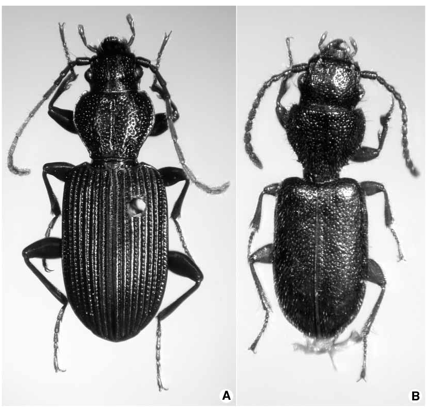
Description. Adult beetles ( Figs. 1A, 1B View FIGURE 1 ) exhibiting the recognition features of Melaenini , and following. Color of body black to testaceous, elytra concolorous or bicolored. Body form terete to depressed. Antennae and mouthparts rufotestaceous, concolorous, to bicolored, proximal antennomeres black, distal antennomeres rufous, to all antennomeres black. Legs testaceous to black.
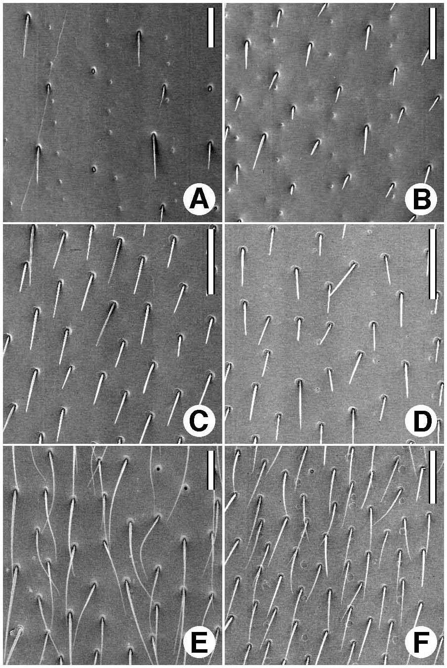
Microsculpture. Dorsal surface generally with isodiametric mesh pattern, microlines shallow, or microlines and sculpticells confined to postocular transverse impression, or scutellum; or microlines absent, surface without evident mesh pattern; or mesh pattern transverse ( Figs. 5F–G View FIGURE 5 ), microlines shallow; some appendages with fimbriate sculpticells (see generic descriptions, for details).
Luster. Body surface dull, shiny, or iridescent.
Macrosculpture. Surface generally moderately densely punctate, or smooth with only elytral striae punctate.
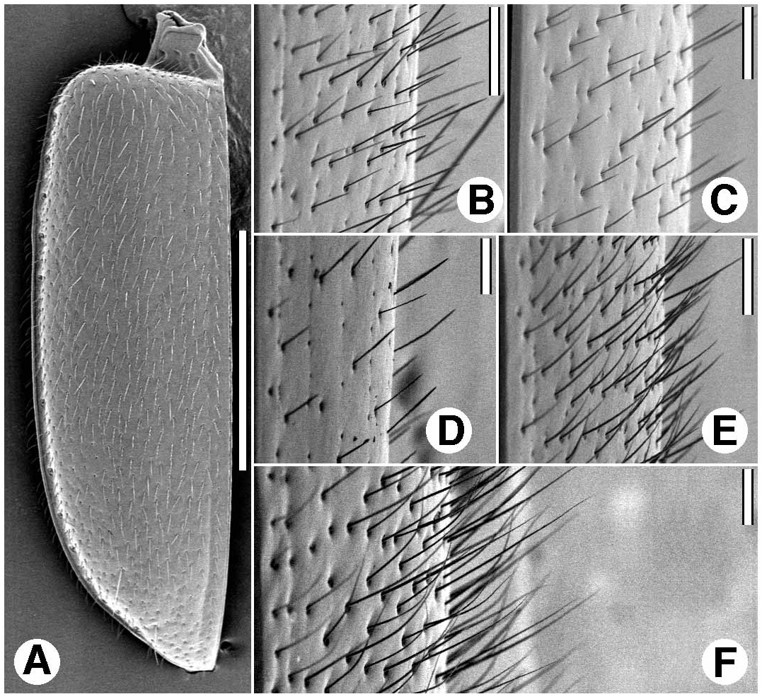
Vestiture. Body surface glabrous (except fixed setae), or with dense to very sparse covering of testaceous setae ( Figs. 2 View FIGURE 2 , 3 View FIGURE 3 ).
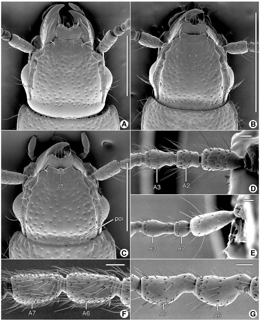
Fixed setae (body sclerites, mouthparts, elytra). Head capsule with one or two pairs of supraorbital setae; with or without row of long setae posteriad eyes, and curved forward over eye surface. Mouthparts: labrum dorsally ( Figs. 6J View FIGURE 6 , 12I View FIGURE 12 ) with row of six apical setae (as), near distal margin; mandible with single seta or with several setae in scrobe ( Figs. 6A–D View FIGURE 6 , ss, and Figs. 12A–D View FIGURE 12 , pss, sss); maxillary stipes with two setae ( Figs. 7A–B View FIGURE 7 , 12K–L View FIGURE 12 , dls, vls), palpifer with single seta ( Figs. 7A–B View FIGURE 7 , 12K–L View FIGURE 12 , pfs); labial submentum with four or more setae ( Figs. 7C View FIGURE 7 , 13A View FIGURE 13 , sms, psms, ssms); mentum with pair of paramedial setae ( Figs. 7C View FIGURE 7 , 13A View FIGURE 13 , ms); glossal sclerite distally quadrisetose or bisetose ( Figs. 7C View FIGURE 7 , 13A View FIGURE 13 , gs); labial palpomere 2 bisetose (i.e., with two long setae on anterior margin, near apex). Pronotum with single pair or two pairs of lateral setae. Elytron dorsally with or without parascutellar seta, without discal setae, laterally about 20 setae in continuous umbilicate series. Abdominal sterna IV–VI each with pair of ambulatory setae or asetose; sternum VII with ( Cymbionotum , both sexes, and Melaenus females) or without pair of setae ( Melaenus males) near posterior margin.
Head. Moderately to distinctly wide, dorsal surface moderately to slightly convex, with or without distinct transverse impression posteriad eyes; frontoclypeal suture not evident, frontal impressions hardly indicated, or broad and shallow; frontovertex each side laterad eye with distinct longitudinal sulcus extended posteriorly, to transverse groove (if present); also sharp supraorbital ridge each side extended sinuously anteriorly to mandibular fossa, and here joined to apex of frontovertical ridge; posteriolaterally, genae rather broad, broader than antennomere 1.
Eyes ( Fig. 1A–B View FIGURE 1 , 11A–C View FIGURE 11 ). Rather small, convex.
Antennae. Filiform ( Fig. 1A View FIGURE 1 ), rather long, extended posteriorly beyond basal third of elytra, or nearly moniliform ( Fig. 1B View FIGURE 1 ), shorter, extended to about basal third of elytra. Antennomeres variously proportioned, antennomeres 5–10 symmetric, slightly asymmetric, or markedly asymmetric (for details, see descriptions of genera).
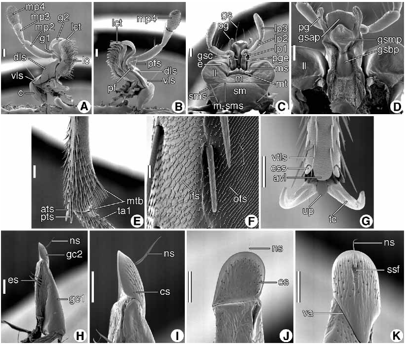
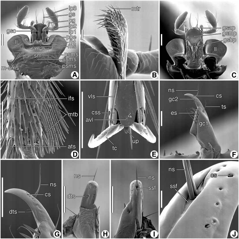
Mouthparts. ( Figs. 6A–K View FIGURE 6 , 7A–D View FIGURE 7 , 12A–L View FIGURE 12 , 13A–C View FIGURE 13 ) Labrum ( Figs. 6J–K View FIGURE 6 , 12I–J View FIGURE 12 ) transverse, distal margin very shallowly to moderately concave, or distinctly notched medially. Ventrally, epipharynx as in Figs. 6K View FIGURE 6 and 12J View FIGURE 12 : pedium (ped) triangular, either approximately equilateral or isosceles, with broad base along distal margin; parapedial projection (pp) acute or obtuse; lateral parapedial setae (lps) four to seven; extra parapedial setae (eps) either mediad or laterad parapedial ridge (pr); coeloconic sensilla (cs) relatively few to many.
Mandibles ( Figs. 6A–I View FIGURE 6 , 12A–H View FIGURE 12 ). Trigonal, narrowed distally and in dorsal aspect curved mediad, in lateral aspect curved ventrad; terebra (t) with or without series of short, diagonal sulci (dts, vts) and ventral condylar sulci (vcs); dorsolateral surface with dorsal groove (dgs) extended from scrobe (s); basal brush (bb) evident at base of occlusal margin; ventral surface with moderately long ventral groove (vg), bearing dense row of short microtrichia (mtr); also, secondary ventral groove (svg) and medial ventral depression (vd). Left mandible, dorsal surface ( Figs. 6B View FIGURE 6 , 12B View FIGURE 12 ) with sharply inflexed incisor tooth (it), terebral tooth (tt) moderately prominent, retinaculum with anterior and posterior tooth (art, prt), anterior tooth evident or not, from dorsal surface, premolar (pmt) and molar (mt) tooth evident; posterior occlusal groove (pog) distinct; ventral surface ( Figs. 6F View FIGURE 6 , 12F View FIGURE 12 ) of occlusal margin with molar ridge (mr) and its basal and occlusal extensions (bemr, omr). Right mandible ( Figs. 6C, 6G View FIGURE 6 , 12C, 12G View FIGURE 12 ) similar to left mandible in most respects, but more curved and shorter.
Maxillae ( Figs. 7A–B View FIGURE 7 , 12K–L View FIGURE 12 ). Standard for Carabidae , with basal trianguloid cardo, rectanguloid stipes with dorsal and ventral lobes (dls, vls), dimerous galea (g1, g2), and lacinia (lc) with prominent distal tooth (lct), and occlusal margin thickly setose; palpus of four articles, (mp1 4), palpomere 1 shortest and palpomere 2 longest.
Labium ( Figs. 7C–D View FIGURE 7 , 13A–C View FIGURE 13 ). Submentum (sm) continuous with gula (not shown); mentalsubmental suture (msms) distinct; mentum (m) transverse, with pair of prominent, broad lateral lobes (ll); epilobes (e) broad, mental tooth (mt) small, broad, anterior margin distinctly notched. Prementum: ligula, ventral aspect ( Figs. 7C View FIGURE 7 , 13A View FIGURE 13 ), with broad glossal sclerite (gsc) and broad longitudinal keel medially, distal margin broadly rounded, pair of paraglossae (pg), each paraglossa slender, narrow lobe, attached to glossal sclerite at base, only, surface with or without extensive covering of setalike microtrichia ( Fig. 13B View FIGURE 13 , mtr); in dorsal aspect ( Figs. 7D View FIGURE 7 , 13C View FIGURE 13 ), glossal sclerite tripartite, basal portion (gsbp) narrow, medial (gsmp) and distal (gsap) portions variously extended; palpiger (pge) moderately large; palpus with three articles (lp13), palpomere 1 short, palpomeres 2 and 3 longer, subequal, palpomere 3 broader, fusiform, apex narrowed, surface more or less setose.
Prothorax. Pronotum ( Figs. 1A, 1B View FIGURE 1 ) subcordate, markedly to slightly wider than long, broader at anterior than at posterior margin; anterior margin slightly concave, posterior margin almost straight, very shallowly sinuate; lateral margins anteriorly rounded, posteriorly sinuate; anteriolateral angles subacute, projected slightly anteriorly; posteriolateral angles acute, prominently projected or not; anterior margin not beaded, flat, posterior margin beaded or not, lateral margins narrowly beaded or not; transverse discal impressions not evident, medial longitudinal impression, distinct, evident medially, not extended to either anterior or posterior margins; lateral grooves broad to narrow; disc slightly convex, lateral declivity each side gradual or rather steep. Prosternum with intercoxal process short, broad posteriorly, ventral surface with shallow longitudinal groove.
Scutellum. Trianguloid, apex pointed.
Elytra ( Figs. 1A, 1B View FIGURE 1 ). Extended length of abdomen. Each elytron oblong, humerus broadly or narrowly rounded, preapically narrowed to narrowly rounded sutural apex, lateral margin straight, not sinuate; dorsal surface plane for most of length, apical declivity gradually to moderately steeply sloped; basal ridge narrow, not extended to edge of scutellum, anteriorly angulate ( Fig. 1A View FIGURE 1 ), or not ( Fig. 1B View FIGURE 1 ); striae not evident, or very slightly impressed, intervals flat, or striae distinct throughout length, nine in number, intervals moderately convex; parascutellar stria and diagonal portion of stria 1 absent, parascutellar setigerous puncture at base of stria 1. Plica posteriolaterally distinct.
Hind wings ( Figs. 8A View FIGURE 8 , 15A View FIGURE 15 ). Macropterous. Oblongum cell (o) large; wedge cell (w) large or absent. Veins RP 3 + 4 and AA 3 + 4 present or absent. Vein MP 4 more than, or less than, halflength of MP 3.
Legs. Articles of about average proportions for groundinhabiting carabid adults. Front tibia with antenna cleaner type B (Hlavac 1971: 57); upper spur unusually large. Middle tibia distally with sculpticells ( Figs. 7F View FIGURE 7 , 13D View FIGURE 13 , ifs, ofs) fimbriate or not. Hind coxae narrowly in contact with one another in midline. Tarsomere 5 distoventrally ( Figs. 7G View FIGURE 7 , 13E View FIGURE 13 ) with prominent lobe, rounded at apex (avl); unguitractor plate (up) narrow or broad; claws (tc) with ventral margins smooth, not serrate or pectinate. Front tarsi of males without adhesive vestiture.
Abdominal sclerites. Tergum VIII partially invaginated; with anterior projections (Roig Juñent 1998: 345, Table 2, and 357). Sterna III and IV evidently fused, with intersegmental suture fine and incomplete, not extended width of abdomen. Sterna IV–VI narrowly transversely sulcate, posteriorly. Sternum VII with posterior margin rounded, and beaded.
Male genitalia ( Figs. 8B–D View FIGURE 8 , 15B–D, 15F View FIGURE 15 , 17D–E, 17I–K, 19C–D, 19G–H, 19K, 19O, 22D–I). Phallus with base open dorsally (Roig Juñent 1998: 345, Table 2, and 357), right basal lobe absent, most of dorsal surface membranous. Endophallus with sclerite X contained wholly within phallus, large, more than half length of phallus, or much shorter than phallus; or small, basad and partially outside phallus; or absent; parameres slender, elongate, with dense row of long setae distally ( Figs. 8C–D View FIGURE 8 ), or glabrous ( Figs. 15C–D View FIGURE 15 ; cf. also van Emden 1936: 45, Fig. 11 View FIGURE 11 ).
Ovipositor ( Figs. 7H–K View FIGURE 7 , 13F–J View FIGURE 13 ). Laterotergite IX ( Fig. 15E View FIGURE 15 , lt) with or without anteriorly directed apophysis. Rami present, in Melaenus about 0.80 length of gonocoxite 1, in Cymbionotum ( Fig. 15E, r View FIGURE 15 ) 0.50–0.80 length of gonocoxite 1. Gonocoxa dimerous, gonocoxites 1 and 2 subequal ( Fig. 13F View FIGURE 13 , gc1, 2), or gonocoxite 2 much shorter than gonocoxite 1 ( Fig. 7H View FIGURE 7 , gc1, 2). Gonocoxite 1 with trichoid setae (ts) and row of ensiform setae (es). Gonocoxite 2 with subapical sensory furrow (ssf) and pair of nematiform setae (ns); without furrow pegs distad setal insertions.
Female internal genitalia ( Fig. 15E View FIGURE 15 ). Bursa copulatrix (bc) elongate, voluminous; spermathecal duct (spd) with origin near junction of bursa and common oviduct (co); spermathecal duct with epispermathecal sclerite extended from bursa copulatrix to spermathecal reservoir (Liebherr and Will 1998: 118, and 169, Appendix 2); spermathecal duct shorter or much longer than spermathecal reservoir (sp), latter reniform, or sausage shaped; spermathecal reservoir with diverticulum (di) long and convoluted. Following features absent (Liebherr and Will 1998: 169, Appendix 2): accessory gland; spermatheca 2 and its appended gland; helminthoid sclerite; villous canal; sclerotized extension of spermathecal duct; diverticula of spermathecal gland; and spermathecal digitiform process.
Way of life. Little is known about this topic. Evidently, most taxa occupy drier open habitats, but the group is represented also in the tropical rain forest vegetation zone (Table 17 and Fig. 24). Bedel (1897: 109) wrote that specimens of Cymbionotum semelederi occur on sandy clay soil on desert plains, and that they come to light on warm evenings. In contrast, specimens of C. negrei Perrault were found under bark of fallen trees.
Included taxa. This tribe includes only two genera: Melaenus Dejean (with two species), and Cymbionotum Baudi di Selve (with 20 species). The classification is outlined in Table 1.
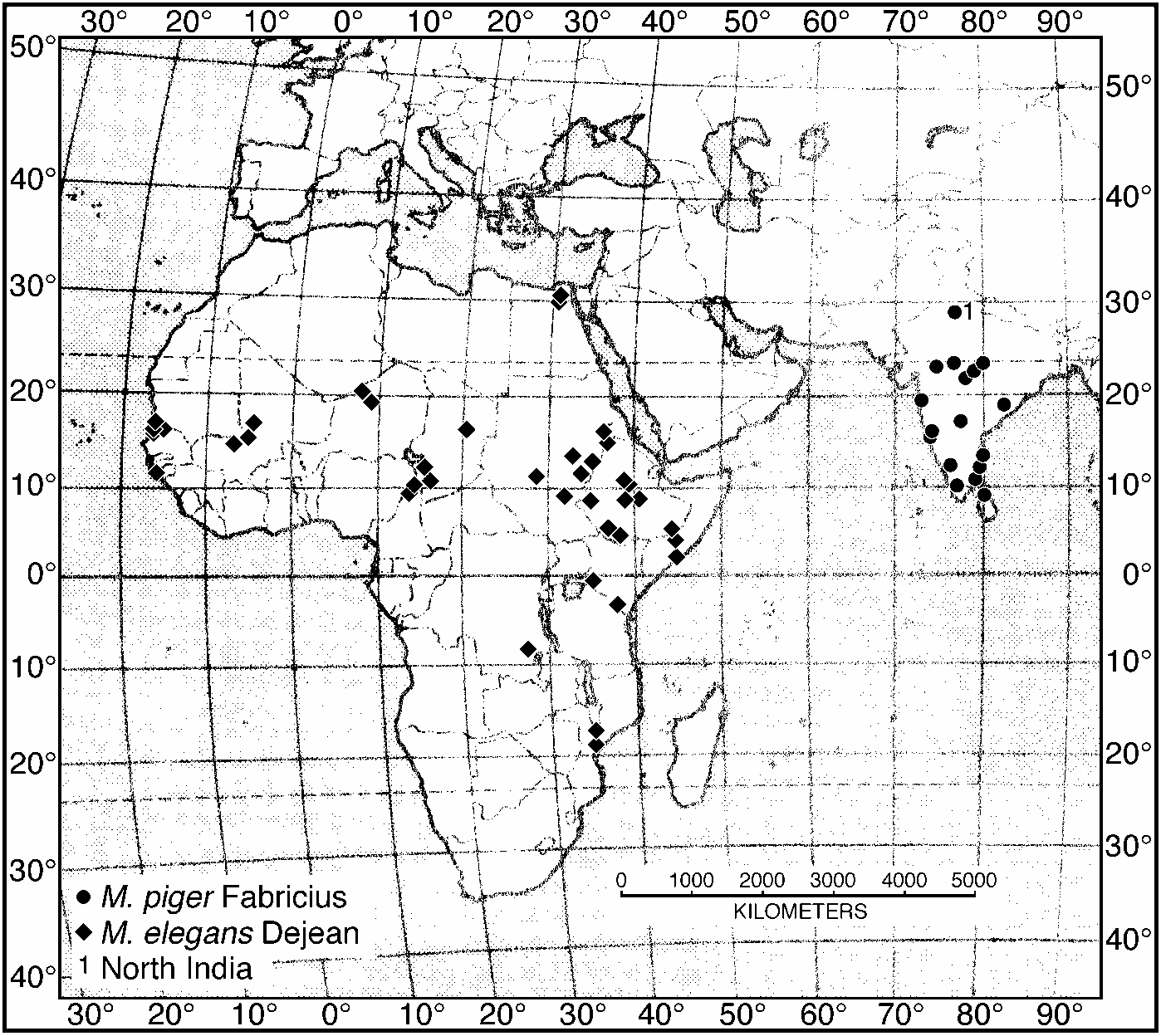
Geographical distribution ( Fig. 4 View FIGURE 4 ). The range of the Melaenini includes the Oriental, southern Palaearctic (principally south of 40º N. Lat.), and Afrotropical (principally north of the Equator) Regions of the Eastern Hemisphere ; and northernmost South America , in the Neotropical Region of the Western Hemisphere .


No known copyright restrictions apply. See Agosti, D., Egloff, W., 2009. Taxonomic information exchange and copyright: the Plazi approach. BMC Research Notes 2009, 2:53 for further explanation.
|
Kingdom |
|
|
Phylum |
|
|
Class |
|
|
Order |
|
|
Family |