
Doryphoribius citrinus ( Maucci, 1972 )
|
publication ID |
https://doi.org/ 10.5281/zenodo.277335 |
|
DOI |
https://doi.org/10.5281/zenodo.5630122 |
|
persistent identifier |
https://treatment.plazi.org/id/03D7474B-CF5E-A901-71BA-FCFED044E4C8 |
|
treatment provided by |
Plazi |
|
scientific name |
Doryphoribius citrinus ( Maucci, 1972 ) |
| status |
|
Remarks on Doryphoribius citrinus ( Maucci, 1972) View in CoL
Material examined. Holotype and five paratypes; the holotype and two paratypes deposited in the Maucci collection, “Museo Civico di Storia Naturale di Verona”, slide numbers not available; three paratypes deposited in the Binda and Pilato collection, Museum of the Department of Animal Biology “Marcello La Greca”, slide number 1200; according to Maucci, 1972, the specimens were extracted from moss on a calcareous stone wall exposed to the South, in a sunny area very close to the traffic circle “Pazin-Rjeka” of the suburbs of Gračišće, Istria.
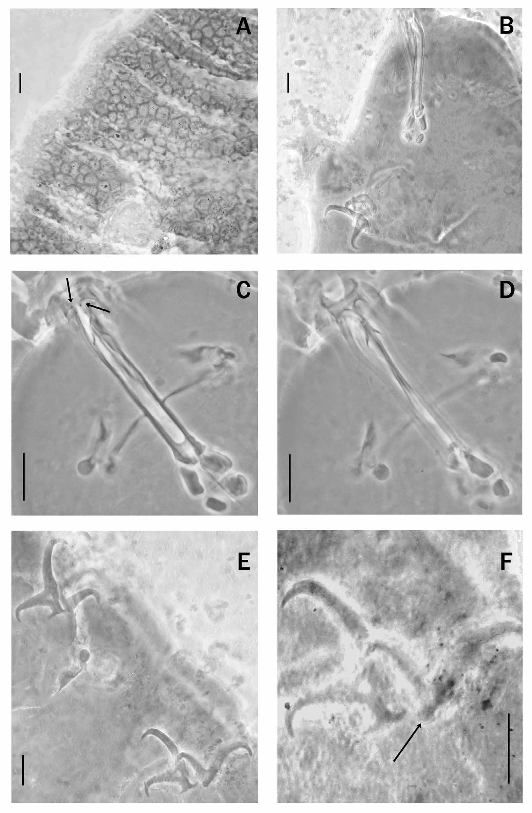
Though not very good condition, the material was better preserved than that of D. flavus so the reticulate sculpture was clearly visible ( Fig. 2 View FIGURE 2. D A). It consisted of ridges only slightly variable in width, usually narrow, delimiting a mesh that was rather variable in shape and size (up to 11.8 μm - pt = 20.3 - in a specimen 486.9 μm long). Tubercles were not formed at crossings, although the intersections may sometimes be enlarged. The mesh increased in average size from the lateral to the dorsal cuticle and from the head to the posterior portion of the body, reaching a maximum diameter between the third and the fourth pair of legs, and decreasing slightly in diameter more caudally.
The smooth swollen portions of the legs were visible, and a faint dense reticulation was visible on the external side of the first three pairs of legs, as reported by Maucci (1972).
The buccal tube appeared rather slender ( Fig. 2 View FIGURE 2. D B), dorsal teeth were present in the buccal cavity ( Fig. 2 View FIGURE 2. D C), arranged dorso-laterally, as in the paratypes of D. flavus ; ventral teeth were not visible ( Fig. 2D View FIGURE 2. D ); peribuccal papulae present, some of which were divided. Neither the buccal cavity teeth nor the peribuccal papulae were mentioned in the original description ( Maucci, 1972), and have not previously been reported. The other characters of the bucco-pharyngeal apparatus match both those observed in the type material of D. flavus and those reported in Maucci, 1972.
Claws, like those of the paratypes of D. flavus , of the Isohypsibius type ( Fig. 2 View FIGURE 2. D E), differing in shape and size on each leg; basal portions of the external claws clearly wider than those of the internal ones, accessory point and lunules present. Again, like the paratypes of D. flavus , the lunules of the inner claws ( Fig. 2 View FIGURE 2. D F) have not previously been noticed. No other sclerified structures visible on legs.
No known copyright restrictions apply. See Agosti, D., Egloff, W., 2009. Taxonomic information exchange and copyright: the Plazi approach. BMC Research Notes 2009, 2:53 for further explanation.