
Felisacus albus, Namyatova & Cassis, 2016
|
publication ID |
https://doi.org/ 10.1206/0003-0090-403.1.1 |
|
persistent identifier |
https://treatment.plazi.org/id/296A879F-563C-7563-5EA2-FB2EFED30BDE |
|
treatment provided by |
Carolina |
|
scientific name |
Felisacus albus |
| status |
sp. nov. |
Felisacus albus , sp. nov.
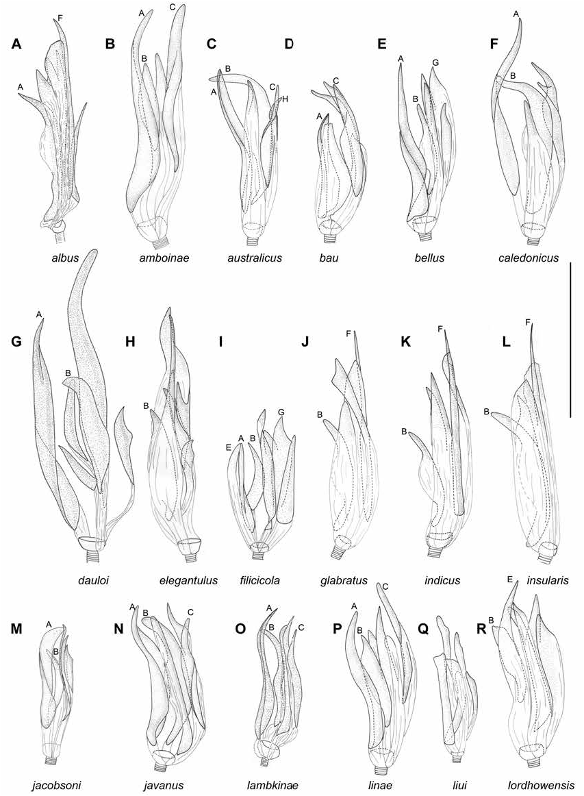
Figures 4 View FIGURE 4 , 8A View FIGURE 8 , 11A, B View FIGURE 11 , 14A View FIGURE 14 , 17 View FIGURE 17 DIAGNOSIS: Recognized by the following combination of characters: pale yellow to white coloration of dorsum with cuneus mostly red (fig. 4), antennal segment I cylindrical (as in Namyatova et al., 2016: fig. 8A), transverse depression delimiting occipital region extending laterally, vertex upraised, dorsal surface of labial segment II elongate posteriorly (as in Namyatova et al., 2016: fig. 6D); and unique combination of seven vesical spicules, having spicules B and F and lacking spicule A, and having large weekly sclerotized serrate spicule (fig. 8A).
DESCRIPTION: Male. Total length 3.7–4.3. COL- ORATION (fig. 4): Head: Mainly whitish yellow to yellow with reddish tinge and markings, dorsal surface of head darker anteriorly and along transverse depression. Eye brown with reddish tinge. Labium: Segments I–III whitish yellow to yellow, segment IV yellow. Antenna: Segments I–II yellow with reddish stripes, segments III–IV yellow to pale brown, with reddish tinge. Pronotum. Anterior part yellow with whitish yellow collar; anterior margin and punctures between anterior and posterior parts pale brown; stripe laterally reddish; posterior part whitish yellow, with brown posterior angles; scutellum and mesoscutum whitish yellow with pale brown punctures between them. Thoracic pleura. Yellow markings near forecoxa whitish yellow; scent gland evaporative area whitish yellow with yellow apex. Hemelytron: Mostly translucent, colorless; inner part sometimes with yellow tinge, translucent or opaque, with yellow to pale brown margins; corium with marking along inner margin of corium pale brown, darker anteriorly, not extending toward R+M; embolium whitish yellow, opaque, with reddish apex and pale brown outer margin; cuneus whitish yellow to yellow with reddish tinge, opaque with pale brown apex and brownish margins; membrane cell whitish yellow. Legs: Coxae whitish yellow; femora whitish yellow, yellow apically, with reddish stripe; tibiae whitish yellow to yellow with reddish stripe; tarsi pale brown. Abdomen: Whitish yellow, red dorsally. SURFACE AND VESTI- TURE: Corium smooth, with shallow and scarce punctures. Dorsum with suberect setae longer than antennal segment II diameter; antennal segment I and femora with suberect setae shorter than antennal segment II diameter; abdomen clothed with suberect mostly short simple setae. STRUCTURE AND MEASUREMENTS: Body ca. 4.2–4.3× as long as pronotum width. Head: Depression delimiting occipital region present dorsally and laterally (as in Namyatova et al., 2016: fig. 4E); distance between depression and pronotum distinctly shorter than eye diameter; longitudinal sulcus on dorsal surface longer than eye diameter; distance from eye to pronotum slightly longer than eye diameter, not swollen laterally (as in Namyatova et al., 2016: fig. 4E); vertex ca. 1.5× as wide as eye; upraised (as in Namyatova et al., 2016: fig. 6D); buccula ca. 0.2–0.25× as long as clypeus. Labium (as in Namyatova et al., 2016: figs. 6D, 9C): Reaching posterior margin of mesosternum or slightly surpassing it; segments I and II strongly reduced, together less than half as long as segment II; segment I as long as wide, segment II slightly longer than wide, its dorsal surface elongate posteriorly; segment III slightly longer than ventral side of head; segment IV ca. 1.5× as long as segment III. Antenna: Segment I cylindrical (as in Namyatova et al., 2016: fig. 8A), ca. 1.4× as long as head, ca. 1.1–1.2× as long as pronotum width; segment III slightly longer than segment II; segment IV ca. 0.3× as long as segment III. Thorax: Anterior and posterior parts subequal in length; collar distinct; posterior part slightly upraised; posterior margin concave; pronotum ca. 1.2–1.4× as wide as long and ca. 1.5–1.7× as wide as head; mesoscutum exposed. Hemelytron: Area along inner margin of clavus almost flat; inner margin of cuneus convex (as in Namyatova et al., 2016: fig. 13E), outer margin of cuneus ca. 2.5× as long as base. Abdomen: Genital capsule rotated left at right angle relative to rest of abdomen. Genitalia: Genital capsule (fig. 14A) as long as wide, its ventral wall almost twice as long as dorsal wall; posterior margin of ventral wall smooth, semioval, without outgrowth, not pointed, not curved dorsally; its apex inclined rightward; walls of genital capsule not modified; angles of paramere sockets rounded; distance between paramere sockets subequal to half width of genital capsule at base. Right paramere (fig. 11A) outer margin of apical part slightly concave; medial part more than twice as wide as basal part, bearing setae, with outer margin straight and inner margin convex; outer angle distinct, not widened; inner angle rounded, without setae; basal part ca. 0.2–0.25× as long as rest of paramere. Left paramere (fig. 11B) L-shaped; apical part not flattened, with toothlike outgrowth on posterior side medially (as in fig. 11G) and without outgrowth on dorsal surface; middle part widened, without swelling or outgrowth(s); setae present only on middle part near outer margin. Aedeagus (general view as in Namyatova et al., 2016: fig. 22I) conjunctiva weakly sclerotized; secondary gonopore placed at base of vesica in repose; sclerotization of ductus seminis around secondary gonopore shorter than wide; vesica with seven spicules, including spicules A and F (fig. 8A).
Female. Total length 3.8. COLORATION (fig. 4): Similar to male, but dorsal surface of head with reddish tinge anteriorly and along transverse depression; posterior part of pronotum whitish yellow with brown posterior margin, clavus whitish and transparent with pale brown margins. SURFACE AND VESTITURE: As in male. STRUCTURE AND MEASURE-
MENTS: Structure as in male; body ca. 3.7× as long as pronotum width; vertex in males ca. 1.8× as wide as eye; antennal segment I ca. 1.2× as long as head width, ca. 0.7× as long as pronotum width; segment II ca. 1.7× as long as head width, ca. 1.0× as long as pronotum width; pronotum ca. 1.4× as wide as long and ca. 1.8× as wide as head.
DISTRIBUTION: Known from Taiwan (fig. 17).
HOST PLANTS: Collected from Lygodium flexuosum (Schizaeaceae) .
ETYMOLOGY: The species is named for its very pale, almost white coloration, albus in Latin, meaning “white.”
DISCUSSION: Female genitalia were not dissected. Felisacus albus forms a monophyletic group with F. glabratus , F. indicus and F. insularis (figs. 1–3). All those species are similar to each other in coloration (figs. 4, 5), in the middle part of the right paramere more than twice as wide as its basal part (figs. 11A, AF, 12A, C), and in the shape and configuration of the vesical spicules (fig. 8A, J, K, L). Felisacus albus is most similar to F. insularis externally and in morphology of spicules, including spicules B and F (cf. fig. 8A with L). Felisacus insularis differs in having a yellow body (fig. 5) and the presence of five vesical spicules (fig. 8K). Felisacus glabratus and F. indicus differ from F. albus in having a mostly yellow or colorless cuneus and five vesical spicules.
MATERIAL EXAMINED: Holotype: THAI- LAND: Changwat Chiang Mai: Chomtong: Mae Klang Waterfall, 18.54944 ° N 98.56722 ° E, 28 Nov 1998, A.D. Wright, Lygodium flexuosum (Schizaeaceae) , 13 (00017861) ( AM). Paratypes: THAI- LAND: Changwat Chiang Mai: Chomtong: Mae Klang Waterfall, 18.54944 ° N 98.56722 ° E, 28 Nov 1998, A.D. Wright, Lygodium flexuosum (Schizaeaceae) , 13 (00017860), 1♀ (00017862) ( AM).
Figures 4 View FIGURE 4 , 8B View FIGURE 8 , 11C, D View FIGURE 11 , 14B View FIGURE 14 , 17 View FIGURE 17 Felisacus amboinae Woodward, 1954: 45 (original description).
DIAGNOSIS: Recognized by the following combination of characters: mostly yellow coloration of dorsum with reddish tinge on cuneus (fig. 4), cylindrical antennal segment I (as in Namyatova et al., 2016: fig. 8A), transverse depression on head extending laterally, vertex upraised, dorsal surface of labial segment II elongate posteriorly (Namyatova et al., 2016: fig. 6D); body length 3.0– 3.3 in male; cuneus ca. 2.5× as long as base; inner margin middle part of right paramere less than twice as wide as basal part and its apical part as long as medial part, in outer margin only slightly concave posteriorly (fig. 11C), vesica with four straight spicules, including spicules A, B, and C (fig. 8B).
REDESCRIPTION: Male. Total length 3.0–3.3. COLORATION (fig. 4): Head: Mostly pale brown, with reddish tinge, ventral side yellow to pale brown. Eye dark brown to black. Labium: Yellow. Antenna: Segment I yellow with reddish tinge, segment II reddish brown; segment III brown. Thorax: Pronotum and scutellum yellow; pronotum with pale brown marking close to humeral angle, anterior margin sometimes pale brown; thoracic pleura yellow; scent gland evaporative area yellow, reddish apically. Hemelytron: Mostly translucent, colorless; inner part of clavus with yellow tinge and reddish yellow margins; marking along inner margin of corium mostly yellow to pale brown, brown along apical part of clavus; embolium with pale brown or red margins; cuneus yellow with reddish tinge or margins sometimes reddish brown. Legs: Coxae yellow; femora yellow, reddish apically; tibiae mostly reddish yellow, yellow apically; tarsi yellow with segment III reddish. Abdomen: Mostly yellow. SURFACE AND VESTITURE: Body clothed with setae mostly shorter than antennal segment II diameter; dorsum with scarce setae mostly shorter than antennal segment II diameter; antennal segments I, II and femora clothed with suberect setae mostly shorter than width antennal segment II diameter; abdomen clothed with short setae. STRUCTURE AND MEA- SUREMENTS: Body ca. 4.1–4.4× as long as pronotum width. Head: Depression delimiting occipital region distinct dorsally and laterally (as in Namyatova et al., 2016: fig. 4E); distance between depression and pronotum shorter than eye diameter; longitudinal sulcus on dorsal surface subequal to eye diameter; distance from eye to pronotum subequal to or slightly longer than eye diameter, not swollen laterally (as in Namyatova et al., 2016: fig. 4E); vertex ca. 1.6–1.7× as wide as eye, upraised (Namyatova et al., 2016: fig. 6D). Labium (as in Namyatova et al., 2016: figs. 6D, 9C): Reaching middle of mesosternum; segments I and II strongly reduced; combined less than half as long as segment III; segment II slightly swollen and elongate dorsally; covering base of segment III; segment III slightly shorter than length of head ventrally; segment IV twice as long as segment III. Antenna: Segment I cylindrical (as in Namyatova et al., 2016: fig. 8A), ca. 1.6–1.7× as long as head width, ca. 1.1– 1.2× as long as pronotum width; segment II ca. 2.0× as long as head width, ca. 1.3–1.4× as long as pronotum width. Thorax: Anterior part of pronotum distinctly shorter than posterior part; collar distinct; posterior part of pronotum only slightly upraised; posterior margin of pronotum slightly concave; pronotum ca. 1.2× as wide as long and 1.4–1.6× as wide as head. Hemelytron: Area along inner margin of corium almost flat; inner margin of cuneus convex (as in Namyatova et al., 2016: fig. 13E), outer margin of cuneus ca. 2.5× as long as base. Abdomen: Genital capsule rotated left at right angle relative to rest of abdomen. Genitalia: Genital capsule (fig. 14B) ventral wall ca. 1.7× as long as dorsal wall; its posterior margin smooth, semioval, without outgrowth, rounded, not curved dorsally; apex slightly inclined leftward; sides of genital capsule not modified, angles of paramere sockets more or less rounded, not projecting; distance between paramere sockets subequal to half width of genital capsule at base; right angle of left paramere socket not projecting. Right paramere (fig. 11C) apical part distinct; apex only slightly concave posteriorly; medial part only slightly wider than basal part, bearing setae, with outer margin slightly concave and inner margin convex; outer angle distinct, widened; inner angle rounded, without setae; basal part ca. 0.15–0.2× as long as rest of paramere. Left paramere (fig. 11D) L-shaped; apical part not flattened, with toothlike outgrowth on posterior side medially and without outgrowth on dorsal surface; middle part widened, without swelling or outgrowth(s); setae placed only on middle part near outer margin of paramere. Aedeagus (general view as in Namyatova et al., 2016: fig. 2IF, G) conjunctiva weakly sclerotized; secondary gonopore placed at base of vesica in repose; sclerotization of ductus seminis around secondary gonopore shorter than wide; vesica bearing four spicules, including spicules A, B, and C (fig. 8B).
Female. Total length 3.1–3.4. COLORATION (fig. 4): Similar to male, but antennal segment IV brown; head and pronotum often with reddish tinge; tarsal segments I–II yellow and segment III pale brown to brown or entire tarsi pale brown to brown; scent gland evaporative area as in male or whitish yellow, yellow apically, sometimes uniformly yellow; abdomen yellow with red markings. SURFACE AND VESTITURE: As in male. STRUCTURE AND MEASUREMENTS: As in male, antennal segment III subequal to segment II, segment IV ca. 0.7× as long as segment III. Genitalia (general view as in Namyatova et al., 2016: fig. 23F, G): Dorsal labiate plate wider than distance between apodemes of second valvula; mostly smooth, without distinct striations, with semicircular sclerite and distinct sclerotized rings laterally; lateral oviducts placed almost medially, very close to each other, spermathecal gland placed between lateral oviducts; dorsal labiate plate with distinct tubercles, without membranous lobe medially.
DISTRIBUTION: Ambon Is., Indonesia (fig. 17).
HOST PLANTS: Unknown.
DISCUSSION: Genitalia of the male holotype and female nontype specimen sampled from the type locality were examined. Felisacus amboinae is most similar in coloration to F.albus , F. dauloi , F. filicicola , F. javanus , F. linae , F. ochraceus , and F. solomonicus (figs. 4–7), all of them being yellow with the cuneus at least partly yellow or red. Among those species, F. dauloi , F. filicicola and F. linae are similar to F. amboinae in shape of the right paramere (cf. fig. 11C with figs. 11D, AD, 12K) and distribution, however, those three species have an alternative combination of vesical spicules (cf. fig. 8B with 8G, I, P). Vesical spicules of F. amboinae are most similar to those of the Timorese species F. linae , with both species having spicules A, B, C, with spicule C distinctly posterior in position (cf. fig. 8B with 8P), although the latter species has six spicules.
MATERIAL EXAMINED: Holotype: INDONE- SIA: Maluku: Ambon Is.: Amboina, 3.66 ° S 128.166 ° E, no date provided, F. Muir, 13 (00400377) ( CAS). Paratypes: INDONESIA: Maluku: Ambon Is.: Amboina, 3.66 ° S 128.166 ° E, no date provided, F. Muir, 13 (00399741), 1♀ (00399745) ( CAS). Additional material: INDO-
NESIA: Maluku: Ambon Is.: Amboina, 3.66 ° S 128.166 ° E, no date provided, F. Muir, 4♀ (00399742–00399744, 00399746), 2 sex unknown (00399752, 00399756), 13 (00399748) ( CAS).
Figures 4 View FIGURE 4 , 17 View FIGURE 17 Felisacus auritulus Distant, 1913: 177 (original description).
DIAGNOSIS: Recognized by the following combination of characters: antennal segment I distinctly widened (as in Namyatova et al., 2016: fig. 8B), dark brown with yellow base; segment II dark brown; head and pronotum whitish yellow to yellow, dorsal surface of labial segment II elongate posteriorly (as in Namyatova et al., 2016: fig. 6D); head and pronotum mostly yellow without brown or red markings (fig. 4); body length 4; labium reaching middle of mesosternum; distance between eye and pronotum slightly shorter than eye diameter.
DESCRIPTION (based on a single, partly destroyed holotype; see also original description of Distant, 1912): Total length 4. COLOR- ATION (fig. 4): Head: Yellow to pale brown. Eye dark brown to black. Labium: Yellow to pale brown. Antenna: Segment I dark brown with yellow base, segment II dark brown. Thorax: Pronotum and scutellum yellow; thoracic pleura and scent gland evaporative area yellow. Hemelytron: Mostly translucent, colorless, inner part of clavus with yellow tinge. Legs: Coxae yellow, femora and tibiae yellow to pale brown, apices of femora and bases of tibiae somewhat darker, hind tarsus with segment I pale brown and segments II–III brown. SUR- FACE AND VESTITURE: Setae on dorsum and antennal segment I and II mostly shorter than antennal segment II diameter; setae on antennal segments I and II and femora with suberect setae mostly shorter than antennal segment II diameter. STRUCTURE AND MEASURE- MENTS: Body ca. 4× as long as pronotum width. Head: Depression, delimiting occipital region present only dorsally; distance between depression and pronotum slightly shorter than eye diameter; longitudinal sulcus on dorsal surface shorter than eye diameter; vertex ca. 3.5× as wide as eye, flat; distance from eye to pronotum slightly shorter than eye diameter, not swollen laterally. Labium (as in in Namyatova et al., 2016: figs. 6D, 9C): Reaching middle of mesosternum; segments I and II strongly reduced, combined subequal to half of segment III; segment I as long as wide, segment II slightly longer than wide, its dorsal elongate posteriorly segment III slightly shorter than length of head ventrally; segment IV twice as long as segment III. Antenna: Segment I dis- tinctly swollen (as in Namyatova et al., 2016: fig. 8B), 1.3× as long as head width, 0.6× as long as pronotum width; segment II 2× as long as head width, 0.9× as long as pronotum width. Thorax: Anterior part of pronotum distinctly shorter than posterior part; collar distinctly delimited; posterior part of pronotum slightly upraised; posterior margin of pronotum slightly concave; pronotum ca. 1.3× as wide as long and ca. 2.3× as wide as head; mesoscutum slightly exposed.
DISTRIBUTION: Seychelles (fig. 17).
HOST PLANTS: Unknown.
DISCUSSION: The description of Felisacus auritulus is based on the holotype, which is seriously damaged, with the abdomen, antennal segments III–IV, most of the hemelytra (except right clavus), and fore- and middle tibiae lost.
Felisacus auritulus is similar to F. bradi , F. ovalau , F. usingeri and F. vitilevu (figs. 4–7) in the following characters: broad antennal segment I (as in Namyatova et al., 2016: fig. 8B), dorsal surface of labial segment II elongate posteriorly and pronotum yellow (as in Namyatova et al., 2016: fig. 6D). Felisacus usingeri is most similar to F. auritulus , but the latter species differs in having the distance from eye to pronotum slightly longer than the diameter of an eye, antennal segment I is mostly yellow and is known from the Philippines. Felisacus bradi , F. ovalau and F. vitilevu differ in having a narrower antennal segment I. Additional characters that separate these species from F. auritulus include: F. bradi is known from Tahiti and its labium reaches abdominal segment VII; F. ovalau is known from Fiji and antennal segment I is mostly yellow and the labium reaches abdominal segment V; F. vitilevu is known from Fiji, and antennal segment I is mostly yellow and the labium reaches the margin of the metasternum or slightly surpasses it.
MATERIAL EXAMINED: Holotype: SEY-
CHELLES: Silhouette, Mare aux Cochons, 4.48333 ° S 55.23333 ° E, 500 m, Aug 1908, Percy Sladen Trust Expedition, 1 sex unknown (00019490) ( BMNH).


| AM |
Australian Museum |
| CAS |
California Academy of Sciences |
No known copyright restrictions apply. See Agosti, D., Egloff, W., 2009. Taxonomic information exchange and copyright: the Plazi approach. BMC Research Notes 2009, 2:53 for further explanation.