
Fergusobia cosmophyllae, Davies, Kerrie A., Giblin-Davis, Robin M., Ye, Weimin, Taylor, Gary S. & Thomas, W. Kelley, 2013
|
publication ID |
https://doi.org/ 10.11646/zootaxa.3741.1.3 |
|
publication LSID |
lsid:zoobank.org:pub:1334C8EE-C9E3-4D9A-B25A-34F62D6E76AA |
|
DOI |
https://doi.org/10.5281/zenodo.5614379 |
|
persistent identifier |
https://treatment.plazi.org/id/038EF368-FFB6-FFFA-FF03-37A0F2697F94 |
|
treatment provided by |
Plazi |
|
scientific name |
Fergusobia cosmophyllae |
| status |
sp. nov. |
Fergusobia cosmophyllae n. sp. Davies
( Figs 2 View FIGURE 2 , 9 View FIGURE 9 N, 10M)
Measurements. Table 2 View TABLE 2 .
Material examined. 12 parthenogenetic ♀, 10 pre-parasitic infective ♀, 26 ♂, and 4 parasitic ♀; roadside vegetation, Bridgewater, South Australia, Australia (35°00.30´S 138°46.00´E), a garden plant at Mylor (35º 02.33’S 138º 45.19’E), and roadside vegetation 6 km. west of Stokes Bay, Kangaroo Island, South Australia. All from multilocular shoot bud galls on Eucalyptus cosmophylla . Coll. G.S. Taylor, 16.v.2000; G.S. Taylor, 3.iii.2002; and J.T. Jennings, 8.xii.2000, respectively.
Holotype: Parthenogenetic female, together with one infective female and one male paratype on a slide deposited in the ANIC, Canberra, ACT, Australia, collected at Bridgewater, South Australia, data as above.
Paratypes: (collection data as above) deposited at WINC, The University of Adelaide, SA, Australia, 6 parthenogenetic ♀, 5 pre-parasitic infective ♀, 12 ♂, and 4 parasitic ♀; and at the USDA Nematode Collection, Beltsville, MD, USA 5 parthenogenetic ♀, 4 pre-parasitic infective ♀, and 13 ♂.
Description. Parthenogenetic female. From multilocular shoot bud galls on E. cosmophylla . Body tightly Cshaped when heat-relaxed, dorsally curved with ventral side convex and tail more curved than anterior end; similar in size to amphimictic pre-parasitic female and male; cuticle appearing smooth or faintly annulated; longitudinal striations apparent when viewed with light microscope; lateral fields not seen.
Cephalic region diameter ~70% of body diameter immediately posterior, usually offset, unstriated, ~1.5 µm long; circum-oral area flat or slightly raised. Stylet with cone 40–50% of total length, basal knobs rounded.
Orifice of dorsal pharyngeal gland ~1 or 2 µm posterior to stylet knobs. Anterior fusiform part of digestive tract occupying 62–80% (mean 70, n = 9) of body diameter, length 2.2 (1.8–2.8) times diameter; lumen of tract broadening near posterior end of gland. Pharyngeal glands enormous, occupying ~80% of body diameter, extending 42–56% (mean 48%) of total body length.
Secretory/excretory pore with non-refractile duct, about half way along length of pharyngeal gland, secretory/ excretory cell not seen. Hemizonid extending over 2 annules, 4 or 5 annules anterior to secretory/excretory pore.
Reproductive tract variable in length, usually extending part-way along dorsal pharyngeal gland (in 9 of 12 specimens examined) or to posterior end of gland; terminal cell sometimes distinctly off-set; generally outstretched, rarely flexed near tip or flexed posterior to oesophageal gland; oviduct with oocytes not in rows; uterus usually with no eggs (1 egg present in 1 specimen); vulva with wide protruding lips or flat or a depressed slit. Body narrowing gradually behind vulva. Anus flat or opening into small depression in the cuticle. Tail conoid, may be dorsally concave, length 1.5–2 times anal body diameter, tip narrow rounded.
Infective pre-parasitic female. From multilocular shoot bud gall on E. cosmophylla . Infecting mature larval stage of Fergusonina sp. or pupa. Arcuate to open C-shape when relaxed by heat, dorsally curved with ventral side convex and greatest curvature in posterior end of body; cylindroid, maximum body diameter at mid-body length or at vulva, anterior and posterior ends with similar shape; cuticle smooth, annules not seen, longitudinal striations apparent when viewed with light microscope; lateral fields not seen. Large, prominent cells in epidermis and intestine.
Cephalic region slightly offset. Circum-oral area flat or with slight central elevation, clearly separated from surrounding sectors; stylet slender, weakly sclerotised, basal knobs ellipsoid and slightly longer than wide; cone 50% of total stylet length.
Orifice of dorsal pharyngeal gland 2–3 µm posterior to stylet knobs; pharyngeal glands occupying 40–70% body diameter, extending over intestine to average 38 (31–45)% body length. Anterior fusiform part of digestive tract occupying 50–61% of body diameter, length 1.8–3 (mean 2.3, n = 5) times diameter.
Secretory/excretory pore opening near posterior of pharyngeal gland; duct not refractile, secretory/excretory cell triangular in shape, small, ~3 µm long, present just underneath epidermis. Hemizonid not seen.
Uterus packed with sperm in inseminated female; vagina at ~80 angle to longitudinal axis of body, reproductive tract extending to nerve ring; length hypertrophied in some specimens. Vulval lips slightly raised by ~2 µm. Anus obscure. Tail not hooked, length 1.1 times diameter at anus, tip almost hemispherical.
Parasitic female. Occurring in haemocoel of abdomen of fly, Fergusonina sp. Arcuate shape with broad, bluntly rounded head and tail tips. Head not off-set. No stylet; oesophagus, intestine and rectum degenerate. Reproductive tract single, greatly hypertrophied, reflexed with sections running almost full body length. Vulva a transverse slit at average 86 (77–92) % of body length.
Male. From multilocular shoot bud galls on E. cosmophylla . Body arcuate to J-shaped when relaxed by heat, tail region more or less curved ventrally, body barely narrowing posterior to cloaca. Cuticle with faint annules, longitudinal striations apparent when viewed with light microscope; lateral fields ~2 µm wide at mid-body length, flat with many broken striae, anterior end obscure.
Cephalic region offset, diameter 64–77% of body diameter immediately posterior, 1.8–2.4 µm long; circumoral area clearly separated from, and similar in length to rest of cephalic region, with lightly sclerotised framework. Stylet weakly sclerotised, with cone ~ 50% of total length, stylet knobs round, ~ 2 µm across. Anterior fusiform part of digestive tract occupying 47–74 (mean 64, n = 14) % of body diameter, length 1.9–3 (mean 2.3, n = 5) times diameter. Pharyngeal glands occupying ~70–85% of body diameter, extending over intestine to average 22–45% (mean 30%) of total body length. Lumen of intestinal tract widening posterior to pharyngeal gland.
Secretory/excretory pore opening at 80–90% of length of pharyngeal gland; duct non-refractile, secretory/ excretory cell not seen. Hemizonid extending over 2–3 annules, ~10 annules anterior to secretory/excretory pore.
Reproductive tract with single testis, variable in length, overlapping dorsal pharyngeal gland or extending part way along intestine; outstretched, rarely reflexed (1/ 26 specimens); testis, seminal vesicle and vas deferens not clearly differentiated. Bursa smooth, prominent or obscure, 10–40% of body length from tail tip, peloderan. Spicules paired, angular near middle, thick, with blunt tip, moderately sclerotised; manubrium continuous or sometimes offset, with similar width to shaft; blade notched about halfway along anterior edge, opening terminal. Inconspicuous muscles associated with cloaca. Tail short, broad, cylindroid, length 1.5 times diameter at cloaca; tip bluntly rounded to almost hemispherical.
Diagnosis and relationships. Fergusobia cosmophyllae n. sp. is morphologically characterized by the combination of a C-shaped parthenogenetic female with a short arcuate conoid tail, a broad (small a ratio) arcuate infective female with an hemispherical tail tip, and an arcuate to J-shaped male with a broad, angular spicule and short bursa.
The parthenogenetic females are similar to those of F. brittenae , F. diversifoliae n. sp., F. floribundae n. sp., F. pimpamensis n. sp., and F. ptychocarpae Davies 2008 (Taylor & Davies 2008) . The infective females are similar to those of F. brevicauda Siddiqi 1994 , F. delegatensae n. sp., F. fasciculosae Davies 2012 , F. philippinensis Siddiqi 1994 , F. ptychocarpae , F. quinquenerviae , and F. viridiflorae Davies & Giblin-Davis 2004 . Morphologically, males are similar to F. brittenae , F. fasciculosae , F. microcarpae Davies 2013 (Davies et al. 2013) , F. morrisae Davies 2012 (Davies et al., 2012b), and F. porosae Davies 2013 (Davies et al., 2013) .
The C shape of the parthenogenetic female of F. cosmophyllae n. sp. differs from that of F. curriei Fisher & Nickle 1968 , F. fisheri Davies & Lloyd 1996 , F. nervosa Davies & Giblin-Davis 2004 , F. porosae , and F. quinquenerviae (open C-shapes), from F. fasciculosae (arcuate shape), from F. r i l e y i Davies 2012 (Davies et al., 2012a) (almost straight to arcuate shapes) and from F. camaldulensae Davies 2012 (Davies et al., 2012a), F. pohutukawa Davies 2007 (Taylor et al. 2007) , and F. viridiflorae (arcuate to open C-shapes). In length, parthenogenetic females of F. cosmophyllae n. sp. (354–406 µm) are smaller than F. magna (418–780 µm) Siddiqi 1986 sensu Davies 2010 and F. indica (525–626 µm) (Jairajpuri 1962) Siddiqi 1986; and larger than F. cajuputiae Davies & Giblin-Davis 2004 (221–273 µm), F. fasciculosae (237–285 µm), F. fisheri (228–305 µm), F. jambophila Siddiqi 1986 (195–300 µm), F. leucadendrae Davies & Giblin-Davis 2004 (205–303 µm), F. morrisae (262–347 µm), F. nervosae (245–309 µm), F. philippinensis (229–310 µm), F. quinquenerviae (224–324 µm), and F. viridiflorae (259–328 µm). The stylet is larger in the parthenogenetic female of F. cosmophyllae n. sp. (8–9 µm) than in F. floribundae n. sp. (6–7 µm) and F. juliae (5–7 µm); and smaller than in F. camaldulensae (11–13 µm), F. microcarpae (9.5–11 µm), F. pohutukawa (10–11 µm), and F. rileyi (10–13 µm). The anterior fusiform part of the digestive tract of F. cosmophyllae n. sp. occupies more of the body (62–80% of body diameter) than in F. delegatensae n. sp. (45–60%). The position of the vulva (85–89%) is more posterior than in F. curriei (78–84%) and F. i n d i c a (71–84%). The shape of the posterior part of the body (conoid, arcuate, with rounded tip) of F. cosmophyllae n. sp. differs from that of F. dealbatae Davies & Giblin-Davis 2004 (conoid, straight); F. floribundae n. sp., F. morrisae , F. philippinensis and F. pimpamensis n. sp. (more slender, conoid, arcuate); F. diversifoliae n. sp. (body narrows quickly behind the vulva); and F. magna (conoid, slender, arcuate to straight). The tail tip of F. cosmophyllae n. sp. is more narrowly rounded than in parthenogenetic females of F. eugenioidae Davies 2012 (Davies et al. 2012b). Ratio c is larger (17.8–23.3) than in F. tumifaciens Currie 1937 (10–11). Ratio c’ (1.3–1.8) is smaller than in F. magna (2.5–4.8) and larger than in F. brevicauda (0.9–1.1). The parthenogenetic female of F. cosmophyllae n. sp. mostly has a shorter stylet than F. ptychocarpa Davies 2010 (Taylor & Davies 2010) (8–9 µm vs 8–13 µm), and the hemizonid is further anterior (4–5 annules vs just anterior to the secretory/excretory pore). Fergusobia cosmophyllae n. sp. lacks the large secretory/excretory cell found in F. brittenae . While morphologically similar to F. minimus n. sp., F. cosmophyllae n. sp. has a longer stylet (8–9 vs 4–8 µm in length) and the secretory/excretory pore is more posterior (158–221 µm vs 55–116 µm from the anterior end).
In being broad and having a small a ratio (4–6.8), the infective female of F. cosmophyllae n. sp. differs from all other described species except F. fisheri (6.8–12.5). Morphologically, it cannot be clearly separated from infective females of F. fisheri , having similar size and body shape; but F. cosmophyllae n. sp. tends to be broader, to have a shorter tail (15–32 vs 21–49 µm long) and a more posterior excretory pore (anterior end to pore opening 77–111 vs 66–91 µm).
The shape (arcuate to J) of the male of F. cosmophyllae n. sp. differs only from that of F. jambophila (males almost straight). Length of the male (374–502 µm) is greater than that of F. cajuputiae (286–364 µm), F. fasciculosae (274–336 µm), F. leucadendrae (254–350 µm), F. nervosae (277–312 µm), F. p o ro s a e (270–326 µm), and F. quinquenerviae (256–329 µm). Ratio a (8.4–11.4) is smaller than in F. pohutukawa (12.2–15.5). Tail shape (arcuate, with a broadly rounded tip) differs from F. philippinensis (arcuate with a truncate tip) and from F. magna , F. pohutukawa , F. ptychocarpae , F. rileyi and F. viridiflorae (more slender, straight, arcuate or C-shaped tails). The tail length (26–52 µm) in the male of F. cosmophyllae n. sp. is shorter than in F. magna (54–87 µm) and F. rileyi (58–70 µm). Ratio c’ (1.1–2.2) is larger than that of F. brittenae (0.7–1.4). The spicule (12–15 µm) is shorter than in F. brevicauda (21–27 µm), F. cajuputiae (16–20 µm), F. camaldulensae (18–22 µm), F. dealbatae (18–22 µm), F. diversifoliae n. sp. (17.5–21 µm), F. eugenioidae (23–25 µm), F. fisheri (16–20 µm), F. floribundae n. sp. (20– 23 µm), F. juliae (30–27 µm), F. minimus n. sp. (16-25 µm), F. morrisae (18–23 µm), F. nervosae (16–19 µm), F. philippinensis (19–23 µm), F. ptychocarpae (19–21 µm), F. quinquenerviae (16–20 µm), and F. viridiflorae (18–20 µm). In having an angular shape, the spicule differs from that of F. jambophila and F. pimpamensis n. sp., where it is arcuate. In F. cosmophyllae n. sp., the bursa is short (10-40% of total body length). It is longer in F. camaldulensae (50–70%), F. delegatensae n. sp. (40–68%), F. diversifoliae n. sp. (~80%), and F. pimpamensis n. sp. (61–80%). The tail tip of F. cosmophyllae n. sp. is more broadly rounded than in F. microcarpae , and the hemizonid is about 10 annules anterior to the secretory/excretory pore in F. cosmophyllae n. sp. but only 1–2 annules anterior in F. microcarpae . The male of F. cosmophyllae n. sp. has a shorter stylet (6–11 µm) than F. tumifaciens (12 µm).
Etymology. Named after Eucalyptus cosmophylla , the host plant from which the nematodes were collected.
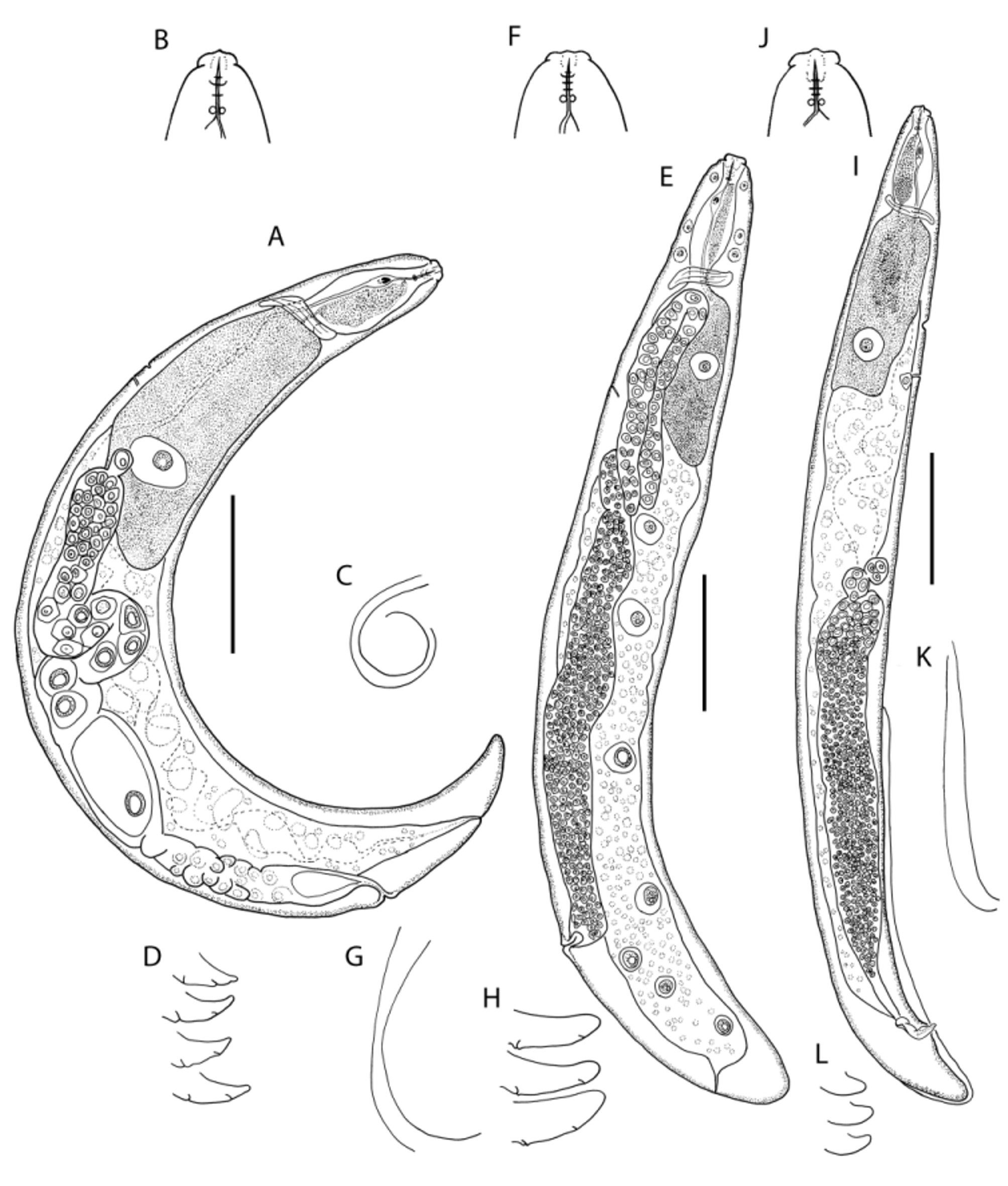

TABLE 2. Measurements (µm) of Fergusobia cosmophyllae n. sp. from large multilocular shoot bud galls on E. cosmophylla. (mean ± standard deviation (range ))
| Holotype Parthenogenetic female | Parthenogenetic females | Males | Infective females | Parasitic females | |
|---|---|---|---|---|---|
| n | 4 | 13 | 26 | 10 | 4 |
| Length | 380 | 384±14.8 (354–406) | 415±29.0 (373–502) | 412±21.3 (374–448) | 1122±114.0 (1006–1238) |
| a | 9.5 | 9.1±1.1 (7.3 – 10.5) | 11.0±1.6 (8.0 – 14.3) | 10.5±0.9 (8.4 – 11.4) | 5.9±1.3 (4.0 – 6.8) |
| b’ | 2.0 | 2.1±0.2 (1.8 – 2.4) | 3.3±0.5 (2.2 – 4.5) | 4.3±0.4 (3.5 – 4.7) | |
| c | 16.6 | 19.9±1.7 (16.6 – 23.3) | 12.9±1.9 (8.4 – 16.6) | 19.1±4.3 (13.4 – 27.6) | |
| c’ | 2.0 | 1.5±0.2 (1.3 – 2.0) | 1.5±0.3 (1.1 – 2.2) | 1.1±0.2 (0.7 – 1.2) | |
| V% | 86.0 | 86.5±1.4 (86.0 – 88.8) | 76.0±4.8 (64.0 – 81.0) | 86.0±7.7 (77.0 – 92.0) | |
| T% | 53.0±15.1 (20.0 – 78.2) | ||||
| Body diameter | 40 | 42±4.8 (36 – 50) | 38±4.3 (33 – 48) | 39±2.6 (36 – 44) | 199±35.0 (175 – 250) |
| Stylet length | 10 | 9±0.6 (8 – 10) | 8±1.1 (6 – 11) | 7±0.8 (6 – 8) | |
| Ant. End to SE pore | 97 | 99±15.6 (71 – 125) | 108±10.5 (94 – 128) | 99±10.1 (77 – 111) | |
| Tail length | 23 | 20±1.8 (16 – 23) | 32±6.3 (26 – 52) | 23±5.4 (15 – 32) | |
| Spicule length | 13.5±0.7 (12 – 15) |
No known copyright restrictions apply. See Agosti, D., Egloff, W., 2009. Taxonomic information exchange and copyright: the Plazi approach. BMC Research Notes 2009, 2:53 for further explanation.