
Fergusobia diversifoliae, Davies, Kerrie A., Giblin-Davis, Robin M., Ye, Weimin, Taylor, Gary S. & Thomas, W. Kelley, 2013
|
publication ID |
https://doi.org/ 10.11646/zootaxa.3741.1.3 |
|
publication LSID |
lsid:zoobank.org:pub:1334C8EE-C9E3-4D9A-B25A-34F62D6E76AA |
|
DOI |
https://doi.org/10.5281/zenodo.5614383 |
|
persistent identifier |
https://treatment.plazi.org/id/038EF368-FFBE-FFE2-FF03-312FF2677D8B |
|
treatment provided by |
Plazi |
|
scientific name |
Fergusobia diversifoliae |
| status |
sp. nov. |
Fergusobia diversifoliae n. sp. Davies
( Figs 5 View FIGURE 5 , 9 View FIGURE 9 I, 10I)
Measurements. Table 4 View TABLE 4 .
Material examined. 14 parthenogenetic ♀, 6 pre-parasitic infective ♀, and 15 ♂; roadside vegetation, Meningie, South Australia, Australia (35°40´S 139°20´E). From multilocular shoot bud galls on Eucalyptus diversifolia . Collected K.A. Davies, 29.viii.1996 and 4.iii.2000.
Holotype: A parthenogenetic female, on a slide with an infective female and a male paratypes, deposited in the ANIC, Canberra, ACT, Australia, collection data as above.
Paratypes: (collection data as above) deposited at the WINC, The University of Adelaide, SA, Australia, 10 parthenogenetic ♀, 4 pre-parasitic infective ♀, 10 ♂; and the USDA Nematode Collection, Beltsville, MD, USA 3 parthenogenetic ♀, 1 pre-parasitic infective ♀, and 3 ♂.
Description. Parthenogenetic female. From multilocular shoot bud galls on E. diversifolia . Shape variable, dorsally curved with ventral side convex to form arcuate, open C or C-shape; similar in size to amphimictic preparasitic female and to male; body tapering posterior to vulva to form a short conoid tail. Cuticle with obscure annules, ~1 µm wide, longitudinal striations apparent when viewed with light microscope; lateral fields not seen.
Cephalic region diameter ~70–80% of body diameter immediately posterior, offset, 1.2–1.5 µm long, unstriated; rounded outline and flat or slightly raised circum-oral area in lateral view. Stylet 8–9 µm long, with cone 40–50% of total length, basal knobs 2–3 µm across at base, round.
Orifice of dorsal pharyngeal gland ~1 µm posterior to stylet knobs. Anterior fusiform part of digestive tract diameter 59–68% of body diameter, length 2.3 (2.2–2.5) times diameter; lumen of tract broadening posterior to dorsal pharyngeal gland. Pharyngeal glands enormous, occupying ~75–85% of body diameter, extending 48 (33– 67) % of total body length.
Secretory/excretory pore 87–123 µm from anterior end with duct leading to large, ellipsoid secretory/excretory cell ~10–15 µm long that may push pharyngeal gland aside. Duct opening at about 30% of length of pharyngeal glands. Hemizonid extending over one or two annules, 8 annules anterior to secretory/excretory pore.
Reproductive tract variable in length, extending part-way along dorsal pharyngeal gland or to nerve ring; outstretched or with one or two flexures in region of nerve ring or gland; oviduct usually with two oocytes per row; uterus containing no eggs or one egg (in 3 of 13 specimens examined); vulva with barely protruding lips or a depressed slit. Tail short, conoid, length 1–2 times anal body diameter, tip rounded.
Infective pre-parasitic female. From multilocular shoot bud gall on E. diversifolia . Infecting mature larval stage of Fergusonina sp. or pupa. Arcuate shape when heat-relaxed, dorsally curved with ventral side convex; maximum body diameter at mid-body length, body tapering only slightly posterior to vulva. Cuticle appears smooth with obscure annules ~1 µm wide, longitudinal striations apparent when viewed with light microscope; lateral fields not seen.
Cephalic region diameter ~65–85% of diameter immediately posterior, offset, ~1 µm long; circum-oral area flat or slightly raised; stylet slender, 9–11 µm long, weakly sclerotised with ellipsoid basal knobs longer than wide, 1–2 µm across; cone 40% of total length.
Orifice of dorsal pharyngeal gland 2 µm posterior to stylet knobs. Anterior fusiform part of digestive tract diameter 40–50% of body diameter, length 2.8 times (2.5–3.3) diameter. Pharyngeal glands occupying ~50% body diameter, extending over intestine to average 29 (25–34) % body length.
Secretory/excretory pore opening 80–130 µm posterior to anterior end of body, about 50% along length of pharyngeal glands; secretory/excretory cell not seen. Hemizonid extending over 3 or 4 annules, 6–8 annules anterior to secretory/excretory pore.
Uterus packed with sperm in inseminated female; vagina at right angle to longitudinal axis of body; reproductive tract extending to nerve ring, sometimes hypertrophied in length. Vulval lips small, slightly raised, with large muscles associated with vagina. Tail short, broad, length 1–2 times diameter at anus, tip almost hemispherical.
Male. From multilocular shoot bud galls on E. diversifolia . Body arcuate to J-shaped when relaxed by heat, tail region more or less curved ventrally or ventrally concave. Cuticle weakly annulated, annules ~1µm wide, with strong longitudinal striae apparent when viewed with light microscope; lateral fields not seen.
Cephalic region diameter 75–85% of diameter immediately posterior, offset, ~2 µm long; circum-oral area flat, with lightly sclerotised framework; stylet 8–9 µm long, with cone 50% of total length, round stylet knobs 1–2 µm across.
Anterior fusiform part of digestive tract occupying 50–73% of body diameter, length 2.2 (1.7–3.1) times diameter. Pharyngeal glands occupying ~35–80% of body diameter, extending over intestine to 30 (26–34) % of total body length.
Secretory/excretory pore opening 98–129 µm posterior to anterior end of body, at ~50 % length of pharyngeal gland; obvious but not highly refractile duct, secretory/excretory cell not seen. Hemizonid extending over 2–3 annules, about 8 annules anterior to secretory/excretory pore.
Reproductive tract with single testis, usually extending to nerve ring; outstretched or reflexed at posterior of pharyngeal gland or in region of nerve ring; testis, seminal vesicle and vas deferens not clearly differentiated.
Bursa smooth or crenate, peloderan; prominent or obscure; arising anterior to secretory/excretory pore. Spicules paired, angular near their middle, moderately sclerotised; manubrium not offset, wider than shaft, blade tapering gradually to truncate tip; opening terminal or just sub-terminal. Inconspicuous muscles associated with cloaca. Tail ventrally curved, length 1.6–2.4 times diameter at cloaca, tip rounded.
Diagnosis and relationships. Fergusobia diversifoliae n. sp. is morphologically characterized by the combination of a C-shaped parthenogenetic female with a conoid tail, an arcuate infective female with a hemispherical tail tip, and arcuate, C- or J-shaped male with angular spicules and long peloderan bursa.
The parthenogenetic female is most similar to F. brittenae , F. cosmophyllae n. sp., F. delegatensae n. sp., F. floribundae n. sp., F. pimpamensis n. sp. and F. ptychocarpae . The infective female is most similar to F. brittenae , F. camaldulensae , F. cosmophyllae n. sp., and F. delegatensae n. sp. The male is most similar to those of F. delegatensae n. sp., F. eugenioidae , F. floribundae n. sp., F. pimpamensis n. sp., and F. ptychocarpae .
From phylogenetic analyses based on sequences of D2/D3 and COI, F. diversifoliae n. sp. is genetically close to Fergusobia species isolated from multilocular shoot bud galls on E. amygdalina , E. delegatensis ( F. delegatensae n. sp.) and E. racemosa , and from flower bud galls on E. obliqua , all from the subgenus Eucalyptus (Ye et al. 2007; Davies et al. 2010a).
In shape (arcuate to open C-shaped to C-shaped), the parthenogenetic female of F. diversifoliae n. sp. differs from F. r i l e y i (almost straight to arcuate). In length (302–475 µm), the female is smaller than F. indica (525–626 µm); but larger than F. cajuputiae (221–273 µm), F. fasciculosae (237–285 µm), and F. jambophila (195–300 µm). In having a flat or slightly raised circum-oral area, F. diversifoliae n. sp. differs from F. camaldulensae , F. eugenioidae , F. jambophila , F. juliae , F. magna , and F. pohutukawa , in which it is peaked. The stylet (8–11 µm) is longer than in F. floribundae n. sp. (6–7 µm) and F. juliae (5–7 µm). The vulva is more posterior (84–91%) than in F. nervosae (81–83%) and F. tumifaciens (81%). The shape of the body posterior to the vulva (almost straight, conoid, narrowing sharply) in F. diversifoliae n. sp. differs from that of F. brevicauda , F. cajuputiae , F. camaldulensae , F. fasciculosae , F. leucadendrae , and F. nervosae (arcuate or curved, with broadly rounded tips), F. dealbatae , F. floribundae , F. magna , F. morrisae , F. philippinensis , F. pimpamensis n. sp., and F. ptychocarpae (more slender), and F. pohutukawa (straight, conoid, may have mucron). The parthenogenetic female of F. diversifoliae n. sp. is separated from those of F. brittenae by the large secretory/excretory cell in the latter, which also tends to have a more slender tail. In having the hemizonid more distant (8 annules) from the secretory/ excretory pore, the parthenogenetic female of F. diversifoliae n. sp. differs from that of F. cosmophyllae n. sp. (4–5 annules), F. dealbatae (4–6), F. delegatensae sp. nov (5), F. microcarpae (2–3), F. porosae (2–3), F. quinquenerviae (2–6), and F. viridiflorae (4). The parthenogenetic female of F. diversifoliae n. sp. is mostly longer (302–475 µm) than that of F. fisheri (228–305 µm); and to have a more posterior vulva (84–91%) than in F. curriei (78–84%). It is close to F. minimus n. sp., but lacks the swollen duct of the secretory/excretory system, and the secretory/excretory pore is more posterior (87–123 µm vs 55–116 µm from the anterior end of the body).
The shape of the infective female (arcuate) of F. diversifoliae n. sp. differs from that of F. magna , F. brittenae and F. curriei (open C); from F. fisheri (barely J); from F. eugenioidae , F. juliae , F. morrisae , and F ptychocarpae (strongly curved in posterior region); and from F. r i l e y i (almost straight). It is shorter (357–473 µm) than F. magna (537–633 µm); and longer than F. cajuputiae (239–309 µm), F. dealbatae (307–347 µm), F. fasciculosae (268–332 µm), F. leucadendrae (227–291 µm), F. porosae (277–300 µm), and F. quinquenerviae (259–325 µm). The stylet (9–11 µm) is longer than in F. brevicauda (8–8.5 µm), F. cajuputiae (7–8 µm), F. cosmophyllae n. sp. (6–8 µm), F. floribundae n. sp. (5–8 µm), F. microcarpae (6–7 µm), and F. minimus n. sp. (4–5 µm). Ratio a (8.2–12) of the infective female is larger than F. cosmophyllae n. sp. (4–6.8). The shape of the body posterior to the vulva (straight with a hemispherical tip) differs from that F. pimpamensis n. sp. (arcuate or curved with a hemispherical tip); and from F. philippinensis (with truncate tail tip). Morphologically, the infective female of F. diversifoliae n. sp. is close to that of F. delegatensae n. sp., but can be separated because it has a continuous, compared to an offset, cephalic region. The position of the hemizonid (6–8 annules anterior to the secretory/excretory pore) separates it from F. camaldulensae (immediately in front), F. nervosae (2 annules), and F. viridiflorae (1 annule).
The shape of male F. diversifoliae n. sp. (arcuate, C- or barely J-shaped) differs from that of F. brittenae , F. curriei , F. juliae , and F. ptychocarpae (J-shape); and from F. jambophila and F. porosae (almost straight). The body length (413–459 µm) is greater than in F. cajuputiae (286–364 µm), F. fasciculosae (274–336 µm), F. jambophila (200–390 µm), F. leucadendrae (254–350 µm), F. microcarpae (311–398 µm), F. nervosae (277–312 µm), F. philippinensis (280–390 µm), F. p o ro s a e (270–326 µm), and F. quinquenerviae (256–329 µm). The stylet is shorter (7–9 µm) than in F. magna (9.5–13 µm), F. pohutukawa (10–11 µm) and F. r i l ey i (11–13 µm). Ratio a of male F. diversifoliae n. sp. (10.8–14.6) is larger than that of F. morrisae (8.5–10.7) and F. tumifaciens (8–9). The tail shape (arcuate) differs from that of F. viridiflorae (more slender, straight or arcuate); from F. cosmophyllae n. sp. (broader, arcuate), and from F. philippinensis (arcuate, with truncate tip). The tail is shorter in the male of F. diversifoliae n. sp. (33–46 µm) than in F. magna (54–87 µm), and F. pohutukawa (50–61 µm). The spicule length (17.5–21 µm) is shorter than in F. eugenioidae (23–25 µm); and longer than in F. cosmophyllae n. sp. (12–15 µm). In having an angular shape, the spicule differs from that of F. jambophila , F. pimpamensis n. sp. and F. rileyi n. sp., where it is arcuate; and the angle is larger than in spicules of F. delegatensae n. sp. The anterior of the bursa is near the excretory pore of F. diversifoliae n. sp., separating it from F. delegatensae n. sp. (near the head); F. brevicauda (33% of the body length from the head), F. camaldulensae (50–70%), F. dealbatae (30–50%), F. fisheri (20–55%), F. floribundae n. sp. (23–55%), and F. minimus n. sp. (12–28 %).
Etymology. Named after Eucalyptus diversifolia , the host plant from which the nematodes were collected.
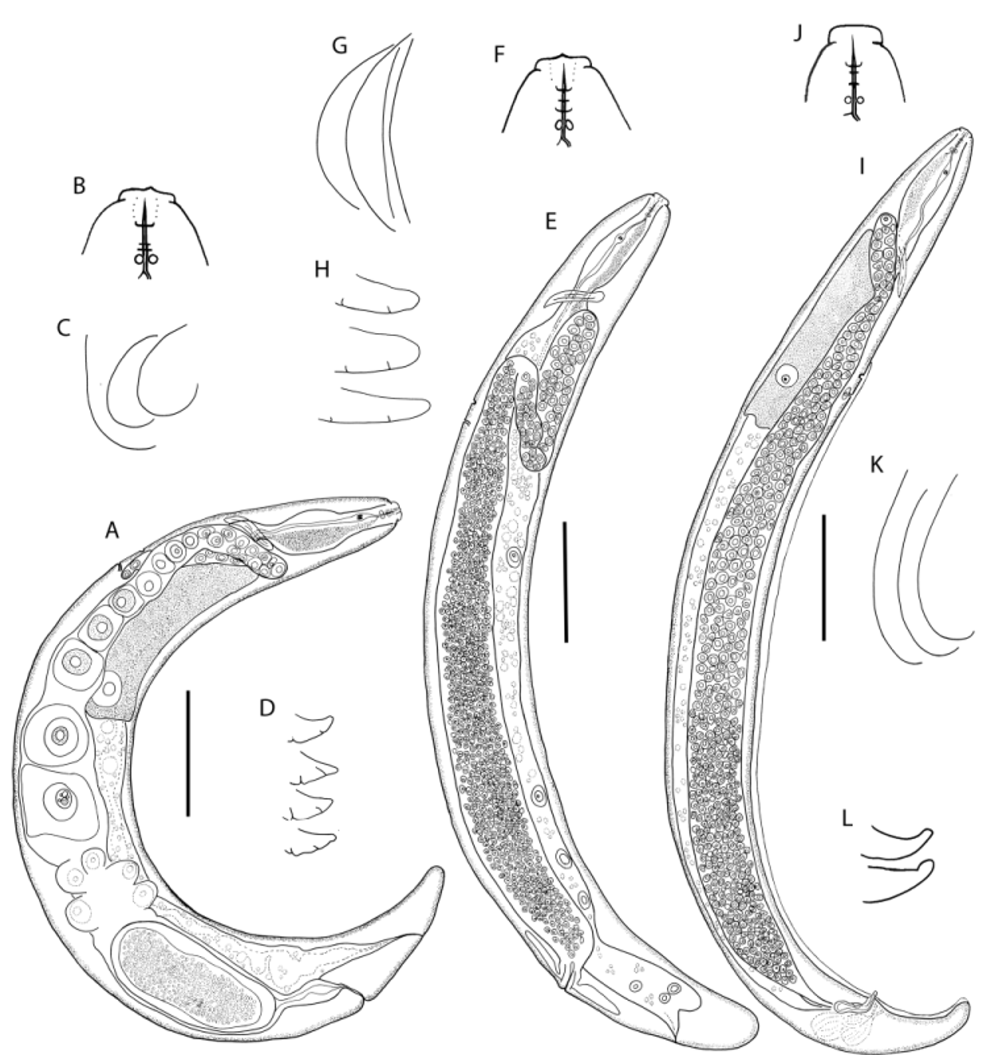

TABLE 4. Measurements (µm) of Fergusobia diversifoliae n. sp. from large multilocular shoot bud galls on E. diversifolia. (mean ± standard deviation (range )).
| Holotype Parthenogenetic female | Parthenogenetic females | Males | Infective females | |
|---|---|---|---|---|
| n | 14 | 15 | 6 | |
| Length | 447 | 424±47 (302–475) | 434±13 (413–459) | 420±41 (357–473) |
| a | 8.6 | 8.6±1.0 (6.8–9.8) | 11.7±1.0 (10.8–14.6) | 10.1±1.4 (8.2–12.0) |
| b’ | 2.1 | 2.1±0.3 (1.5–3.0) | 3.2±0.3 (2.9–3.8) | 3.5±0.5 (2.9–4.0) |
| c | 19.1 | 19.1±5.5 (10.7–27.9) | 10.6±0.8 (9.2–12.4) | 15.0±1.8 (12.0–17.5) |
| c’ | 1.4 | 1.4±0.4 (0.9–2.2) | 2.0±0.2 (1.6–2.4) | 1.1±0.3 (0.7–1.1) |
| V% | 87.1 | 87.1±2.2 (83.7–90.6) | 81.8±2.2 (79.6–85.7) | |
| T% | 77.4±5.5 (64.5–81.9) | |||
| Body diameter | 50 | 50±7.0 (37–67) | 37±2.7 (32–41) | 42±5.8 (32–48) |
| Stylet length | 9 | 9.2±0.9 (8–11) | 8±0.6 (7–9) | 10±0.8 (9–11) |
| Ant. End to SE pore | 105 | 105±12.5 (87–123) | 109±7.2 (98–128) | 106±18.9 (80–130) |
| Tail length | 26 | 24±6.1 (15–33) | 41±3.0 (33–46) | 29±5.6 (23–39) |
| Spicule length | 20±1.1 (17–21) |
No known copyright restrictions apply. See Agosti, D., Egloff, W., 2009. Taxonomic information exchange and copyright: the Plazi approach. BMC Research Notes 2009, 2:53 for further explanation.