
Fergusobia pruinosae Davies, 2018
|
publication ID |
https://doi.org/ 10.11646/zootaxa.4399.1.1 |
|
publication LSID |
lsid:zoobank.org:pub:3F04719B-9132-4C5A-9305-89A102BEF6C9 |
|
DOI |
https://doi.org/10.5281/zenodo.5966565 |
|
persistent identifier |
https://treatment.plazi.org/id/8D1C2802-C231-C820-FF30-FA606C6461C6 |
|
treatment provided by |
Plazi |
|
scientific name |
Fergusobia pruinosae Davies |
| status |
sp. nov. |
Fergusobia pruinosae Davies n. sp. apud MSp 62 ( Davies et al. 2012a)
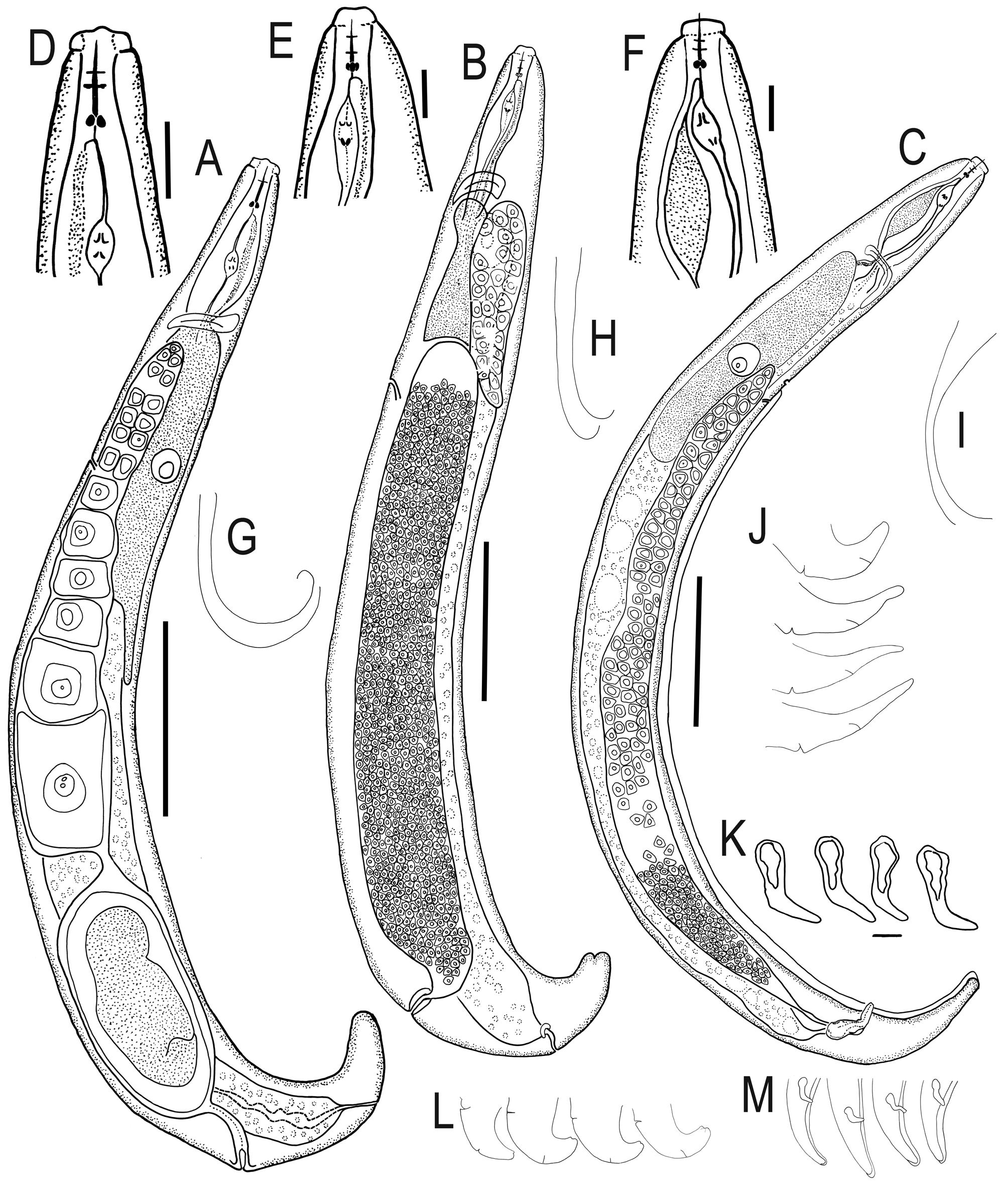
( Fig. 3 View FIGURE 3 )
Measurements. Table 4.
Material examined. Holotype: Parthenogenetic female, Tarara Bar, Carlton Hill Station, near Kununurra, WA, Australia (15°33.29´S 128°40.27´E). From roadside vegetation; flower bud galls on Eucalyptus pruinosa Schauer , collected K.A. Davies, 31.v.2001. On a slide with a paratype infective female and a male, deposited in the ANIC, Canberra, ACT, Australia GoogleMaps .
Paratypes: Vouchers (collection data as above) deposited at the WINC, The University of Adelaide, SA, Australia, 5 parthenogenetic ♀ s, 4 infective females and 5 Ƌs on slides numbered WINC 004288-89 View Materials ( WNC 2209 View Materials ) ; at the Western Australian Museum , Perth, WA, Australia, 9 parthenogenetic ♀ s, 4 infective females and 10 Ƌ s on slides; and at the USDA Nematode Collection, Beltsville, MD, USA 1 parthenogenetic ♀ and 1 Ƌ on a slide. Twenty parthenogenetic ♀ s, 10 pre-parasitic infective ♀ s and 16 ♂ s examined.
Description. Parthenogenetic female. Body an open C-shape, with most curvature posterior to vulva; relatively broad (a 6.8–13.6, mean 10.9); of similar size to males and slightly larger than infective females; body narrows behind anus to form a conoid tail ( Fig. 3A View FIGURE 3 ). Cuticle not swollen when fixed, sub-cuticle with strong longitudinal striae. Lateral fields faint, apparently with broken striae.
Cephalic region ~ 80% diameter of body at anterior end, off-set, 1.5–3 µm high, unstriated; rounded outline in lateral view, circum-oral area barely raised ( Fig. 3D View FIGURE 3 ). Amphids not seen. Stylet strongly sclerotised, with cone 30– 40% of length, basal knobs just higher than wide, 2–3 µm wide at base, rounded.
Orifice of dorsal pharyngeal gland ~ 1–2 µm posterior to stylet knobs. Anterior fusiform part of digestive tract occupying 69 (52–80)% body diameter, length 3.2 (2.8–3.6) times diameter; lumen of tract broadens gradually in distal half of dorsal pharyngeal gland and remains distinct for ca 70% of body length. Pharyngeal glands enormous, extending over intestine, occupying 79 (74–87)% of body diameter, distance from head to posterior end of glands being 40 (30–49)% of total body length. Gland nucleus large, with large nucleolus.
Secretory/excretory pore opens opposite nucleus of pharyngeal gland; duct obscure; secretory/excretory cell not seen. Hemizonid over ~2 annules, 5 or 6 annules anterior to pore ( Fig. 3A View FIGURE 3 ).
Reproductive tract extending to nerve ring; flexed in 8/ 20 specimens examined (number of flexures varying from 1 to 5); oviduct usually with two oocytes per row at proximal end; quadricolumella smooth; uterus with no or one egg; vulva a simple transverse slit with flat lips; no vulval plate. Anus pore-like. Tail conoid, curved, shape varying from slender to broader; concave on ventral side; length 1–2 times anal body diameter; tip bluntly or broadly rounded ( Fig. 3J View FIGURE 3 ).
Infective pre-parasitic female. Almost straight to J-shaped when relaxed by heat; relatively broad; maximum body diameter in posterior half of body length; body tapers sharply posterior to anus ( Fig. 3B View FIGURE 3 ). Cuticle obscurely annulated, <1 µm wide; longitudinal striae apparent with light microscope; lateral fields not seen.
Cephalic region offset, sub-rectangular in shape; circum-oral area flat; stylet slender, weakly sclerotised with small basal knobs higher than wide; <2µm wide; cone ~ 30% of length.
Orifice of dorsal pharyngeal gland often obscure, ~1 µm posterior to stylet knobs. Anterior fusiform part of digestive tract little expanded, occupying 33–73% of body diameter, length 3.8 (2.9–5.0) times diameter. Pharyngeal glands obscure, extending over intestine, occupying ~60% body diameter, distance from head to posterior end of glands being 32 (28–35)% of total body length.
Secretory/excretory pore opens posterior to pharyngeal glands; duct obscure. Hemizonid obscure, either immediately or 2 annules anterior to pore.
Uterus packed with sperm in inseminated females, occupying ~60% of body length; vagina perpendicular to body axis, appears to be surrounded by strong muscles; reproductive tract hypertrophied in some specimens. Vulva a transverse slit, vulval lips flat, no vulval plate present. Anus an obscure pore. Tail short; length 1–1.5 times diameter at anus, tip distinctly notched ( Fig. 3L View FIGURE 3 ).
Male. Body arcuate to C-shaped when relaxed by heat, tail region slightly curved ventrally ( Fig. 3C View FIGURE 3 ). Cuticle clearly annulated, annules ~1µm wide; strong longitudinal striae apparent with light microscope; lateral fields faint, lines not discernible.
Cephalic region occupying ~75–80% anterior body diameter, offset, ~ 2–3 µm high; circum-oral area flat, with lightly sclerotised framework; stylet with cone ~40% of length, round knobs <2 µm wide.
Orifice of dorsal pharyngeal gland ~ 1 µm behind knobs. Anterior fusiform part of digestive tract occupying 69 (56–80)% of body diameter, length 2.8 (2.5–3.1) times diameter. Pharyngeal glands extending over intestine, occupying 64 (51–81)% of body diameter, distance from head to posterior end of glands being 33 (25–41)% of total body length.
Secretory/excretory pore opens opposite nucleus of pharyngeal gland; duct obscure; secretory/excretory cell not seen. Hemizonid lens-like, extending over two annules, 3–5 annules in front of secretory/excretory pore.
Reproductive tract with single testis, variable in length, usually extends part way along gland; testis, seminal vesicle and vas deferens not clearly differentiated. Bursa apparently leptoderan, smooth; may be prominent or obscure; arises 70–84% along length of body, in front of secretory/excretory pore but posterior to hemizonid. Spicules paired, angular (>90°) at ~ 40–50% of length, with manubrium and shaft longer than blade; moderately sclerotised; manubrium similar to or wider than shaft, may or may not be offset dorsally; blade narrows evenly to bluntly rounded tip; opening terminal ( Fig. 3K View FIGURE 3 ). Inconspicuous muscles associated with cloaca. Tail slender, arcuate, ventrally concave, conoid; length 9–11 times diameter at cloaca; bluntly to broadly rounded tip ( Fig. 3M View FIGURE 3 ).
Diagnosis and relationships. Fergusobia pruinosae n. sp. is morphologically characterized by the combination of a medium sized, open C-shaped parthenogenetic female in which the cuticle does not swell upon fixation, having a strongly sclerotised stylet, with a more or less narrowly conoid tail with a bluntly or broadly rounded tip; an infective female that is arcuate to J-shaped with a notched tail tip; and an open C-shaped male with a stout, angular spicule and bursa arising near the secretory/excretory pore. Morphologically, F. pruinosae n. sp. is similar to F. leptospermum , F. pauciflorae n. sp. and F. pimpamensis. While DNA sequences are not available for F. pruinosae n. sp., it was collected from FBGs on E. pruinosa ( Symphyomyrtus , section Adnataria). Thus, its host plant is not close to those of the nematodes mentioned above (respectively, collected from Leptospermum, Eucalyptus section Cineraceae, and Angophora ), suggesting that it is a true species. In addition, the infective female of F. pruinosae n. sp. has a distinctly notched tail that is not present in other described species. The male of F. pruinosae n. sp. differs from that of F. leptospermum in having a shorter bursa (arising 70–84 vs 95% along body length). The hemizonid is more anterior in the parthenogenetic female of F. pruinosae n. sp. than in that of F. pauciflorae n. sp. The male of F. pruinosae n. sp. has a body that is more curved than that of F. pauciflorae n. sp., and the structure of the spicule differs (being more arcuate in F. robustae n. sp.). The morphometrics of the parthenogenetic female of F. pruinosae n. sp. and F. pimpamensis overlap, but they are separated on the form of the cephalic region (wider in F. pruinosae n. sp. relative to body diameter), and in F. pruinosae n. sp. the stylet is more strongly sclerotised than in F. pimpamensis . The male of F. pruinosae n. sp. has a narrower tail tip and a less arcuate spicule than that of F. pimpamensis .
The parthenogenetic female of F. pruinosae n. sp. (body an open C shape) differs from that of F. armillarisae , F. dealbatae , F. decorae , F. janetae n. sp., F. linariifoliae , and F. rileyi (straight to arcuate); and from that of F. brevicauda , F. brittenae , F. cosmophyllae , F. diversifoliae , F. fasciculosae , F. floribundae , F. gomphocephalae , F. indica , F. leucoxylonae , F. magna , F. microcarpae , F. minimus , F. morrisae , F. schmidti , F. planchonianae , F. porosae , F. ptychocarpae , F. viminalisae , and F. viridiflorae (more curved, C-shaped). The parthenogenetic female of F. pruinosae n. sp. has a relatively long tail (37–50 µm long), separating it from all described species except F. floribundae , F. indica , F. janetae n. sp., F. pimpamensis , F. magna , and F. rileyi . In having a non-extensile uterus, the female differs from that of F. armillarisae , F. brevicauda , F. camaldulensae , F. jambophila , F. linariifolia , F. magna , F. rileyi , F. tolgaensis , in which it is extensile. In having cuticle which does not swell upon fixation; the female differs from F. armillarisae , F. decorae , F. pohutukawa , and F. viridiflorae in which it does. The parthenogenetic female of F. pruinosae n. sp. is smaller than that of F. indica (respectively, 363–427 vs 525–626 µm long), and has a smaller stylet (7–11 vs 12–15 µm long).
The infective female of F. pruinosae n. sp. differs from all described Fergusobia spp. in having a strongly notched tail tip. While the infective female of some F. gomphocephalae also have notched tails, they have smaller bodies than those of F. pruinosae n. sp. (L 222–298 vs 314–398 µm).
In shape (open C with a narrow, conoid tail), the male of F. pruinosae n. sp. differs from those of F. leucadendrae (open C shape with tail tip that is broadly rounded); F. armillarisae , F. camaldulensae , F. dealbatae , F. decorae , F. jambophila , F. janetae n. sp., F. linariifolia , F. obliquae n. sp., F. pauciflorae n. sp., F. pohutukawa , F. rileyi , and F. tolgaensis (straight to arcuate); from F. juliae and F. schmidti (J-shaped); and from F. brevicauda , F. cajuputiae , F. colbrani , F. curriei , F, leucoxylonae , F. microcarpae , F. minimus , F. nervosae , F. porosae , F. ptychocarpae , F. viminalisae , and F. viridiflorae , in which males have a C-shaped body with a broader tail. In having a flat circum-oral area, the male is separated from F. camaldulensae , F. jambophila , F. sporangae , and F. tolgaensis in which the oral area is raised. The length of the tail in the male of F. pruinosae n. sp. is longer than in that of F. colbrani and F. gomphocephalae (respectively, 37–50 vs 28–36 and 20–33 µm). The shape of the tail (arcuate with a broadly rounded tip) differs from that of F. philippinensis (truncate tip). In the male of F. pruinosae n. sp., the bursa arises at 69–84% of body length, near the excretory pore, separating them from species in which it arises at less than 50% (including F. brevicauda , F. brittenae , F. cosmophyllae , F. curriei , F. decorae , F. eugenioidae , F. fisheri , F. floribundae , F. leucoxylonae , F. microcarpa , F. minimus , F. nervosae , F. quinquenerviae , F. porosae , F. robustae n. sp., F. rosettae , F. schmidti , F. sporangae , and F. tumifaciens ). The bursa is longer in F. leptospermum , arising at ~95% of body length. The spicule is more heavily sclerotised in the male of F. pruinosae n. sp. than in that of F. diversifoliae and F. morrisae , in which it is more slender in form. The male of F. pruinosae n. sp. is larger than that of F. morrisae (respectively, 387–487 vs 291–373 µm long), and usually smaller than that of F. magna (respectively, 387–487 vs 446–588 µm long).
Etymology. Named after Eucalyptus pruinosa , the plant species from which the nematodes were collected.
No known copyright restrictions apply. See Agosti, D., Egloff, W., 2009. Taxonomic information exchange and copyright: the Plazi approach. BMC Research Notes 2009, 2:53 for further explanation.
|
Kingdom |
|
|
Phylum |
|
|
Class |
|
|
Order |
|
|
Family |
|
|
Genus |