
Gigantohierax suarezi Arredondo & Arredondo, 2002a
|
publication ID |
https://doi.org/ 10.11646/zootaxa.4780.1.1 |
|
publication LSID |
lsid:zoobank.org:pub:D6CC1683-8BF0-4ABF-ABFE-3EC63E66AE5C |
|
DOI |
https://doi.org/10.5281/zenodo.3856797 |
|
persistent identifier |
https://treatment.plazi.org/id/039EF96A-FFEE-224F-ED83-FB18FC6DFC57 |
|
treatment provided by |
Plazi |
|
scientific name |
Gigantohierax suarezi Arredondo & Arredondo, 2002a |
| status |
|
† Gigantohierax suarezi Arredondo & Arredondo, 2002a
Suárez’s Giant Eagle; Águila Gigante de Suárez
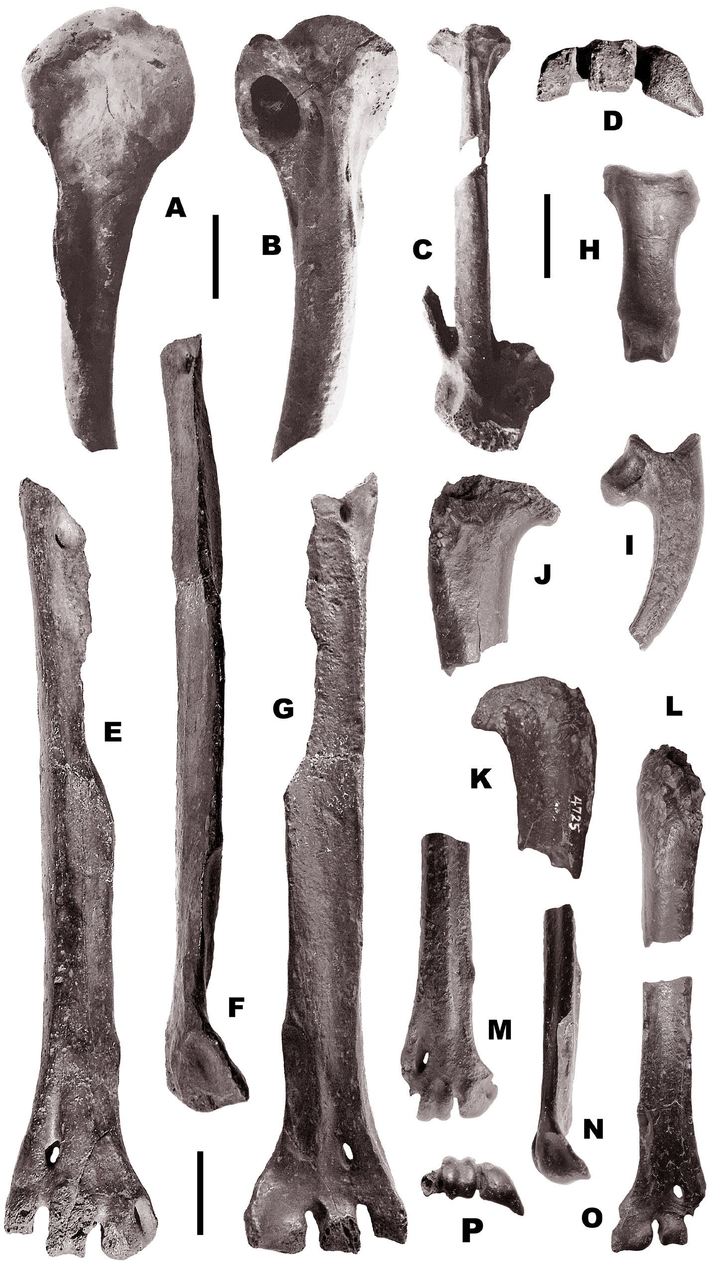
( Figures 11 View FIGURE 11 : A–I; Table 6)
Gigantohierax suarezi Arredondo & Arredondo, 2002a , Poeyana no. 470–475 [for 1999], p. 10.
Referred material. San Felipe I: Proximal halves of right, MNHNCu 75.4721, and left, MNHNCu 75.4722, humeri; fragmentary right carpometacarpus, MNHNCu 75.4724; left carpometacarpus without metacarpal III, MNHNCu 75.4723; right tarsometatarsus without proximal end and part of the medial border, MNHNCu 75.4728; distal half of left tarsometatarsus, MNHNCu 75.4729; distal end of left tarsometatarsus without trochlea metatarsi IV, MNHNCu 75.4730; ungual phalanges of left digit I, MNHNCu 75.4731, MNHNCu 75.4777; right, MNHNCu 75.4733, and left, MNHNCu 75.4732, metatarsal I.
Emended diagnosis. The largest species in the genus Gigantohierax .
Description. The specimens of Gigantohierax suarezi described here do not differ in size or qualitative characters from those recovered in other Cuban deposits. This material differs from all fossil and living accipitrids known in America by its huge size ( Arredondo & Arredondo 2002a; Table 6; Suárez et al. in prep.), being similar in this respect to the Haast’s Eagle from New Zealand (see Holdaway 1990). It differs from other American taxa of Accipitridae examined (see Appendix 1) by having humerus (Fig. A–B) small in relation with the remaining elements of the skeleton, with shaft well arched. In palmar view the bicipital furrow is very shallow and expanded, but a small deep area exists distad to the head. Ligamental furrow inexistent, being indicated by a very small and round scar, located well proximad. Bicipital surface rounded (or convex), reduced, and bicipital crest angulated distally. Deltopectoral
crest projected nearly perpendicular to the surface of the shaft. In anconal view, the proximal end is expanded. Head not flattened, very large and wide (or expanded mediolaterad). Capital groove wide but very shallow. The surface below (distad) to the head is very wide and flat. Attachment of supraspinatus being a very large, deep groove that gouges the shaft. As a result of this, the median crest is very prominent, and a well-developed ridge is characteristic in this portion of the shaft. In the proximal third, the medial side of the shaft is completely flat, and is more triangular in cross section at this point than in other eagles or hawks examined (see Appendix 1). Line of latissimus dorsi anterioris well developed, protruding posteriorly from shaft (in two specimens available to evaluate this character) at the level of the distal portion of the deltopectoral crest. Pneumatic foramen very large, its diameter much larger than the midshaft diameter. The external border of the shaft is acute distad (or below) to the bicipital crest, as a result of a compression (anteroposterior) that produce the posterolateral flat surface of the shaft at this point. Carpometacarpus ( Fig.11C View FIGURE 11 ) short and heavy. This element is even larger and more robust than in Titanohierax gloveralleni Wetmore, 1937 (paratype, proximal end of carpometacarpus MCZ 2258) or “ Amplibuteo ” woodwardi and “ A ”. hibbardi (slightly more than the half of the length of the latter taxa). In lateral view, the metacarpal I is very wide at its base. The metacarpal III is thinner and located more medially, being much less extended laterad. The proximal symphysis is longer in internal view. In external (or lateral) view, there is a deep depression with a bony wall in the internal side, not present in T. gloveralleni . The internal face of the metacarpal I is markedly excavated. The space between metacarpals I and III is large. Metacarpals II and III well separated. Metacarpal II compressed anteroposteriorly. The trochleae are long. In internal view, the proximal symphysis is short and projected in an open angle. Tarsometatarsus ( Fig.11 View FIGURE 11 D–G) elongated, larger than the femur (see measurements of this element in Arredondo & Arredondo 2002a: table 1). The proximal articulation and the medial proximal half of this element is unknown in the material from the tar seeps in study (but known from other Cuban localities). In anterior view, the shaft is long and columnar where both proximal and distal ends are proportionately small. Both anterior metatarsal borders are rounded, or convex, pronounced at both sides of the well excavated anterior metatarsal groove. Outer proximal foramen very large, close in size to the distal foramen (the inner is unknown in this material), perforating the bone from anterior to posterior face. Tubercle for tibialis anticus located central and high, slightly diagonal papilla separated from the anterolateral metatarsal border by a well grooved space. Distally the shaft is flat (by an anteroposterior compression), expanded above the trochleae. Outer extensor groove short and deep above (or proximad) to the distal foramen. Trochleae proportionately small in relation with the remaining bone, flaring well distad and abruptly from shaft (more evident in trochlea metatarsi II), and almost uniform in size. Notch between trochleae metatarsorum II and III slightly thinner. Trochlea metatarsi III short (depicting a nearly squared figure), slightly bended laterally, and with a well-defined groove. The outer ring of the latter is the distalmost point of the bone, and the trochlea metatarsi IV the shortest (less distad extended). In lateral view, the shaft is slightly arched and facies subcutanea lateralis ungrooved, being variable from a nearly flat to strongly convex surface.At distal end, the trochlea metatarsi IV is short and ovoid in shape, with posterior wing rounded, poorly projected posterodistad. At the base of the former trochlea, in the posterior half of the border of the shaft, a well-marked groove is present (at this level the bone is extremely rounded anteriorly). In posterior view, the outer proximal foramen is again about the same size of the distal foramen. Posterior metatarsal groove wide, moderately excavated proximad, very shallow distad. Crista plantaris medialis much less prominent distad than crista plantaris lateralis, although the former is better defined reaching distad the proximal end of the metatarsal facet. Metatarsal facet high (proximad), greatly excavated and expanded through its length, being deeper proximad. It is oriented mediad with prominent edges, being the border (medial border of the bone) very thin and acute. At the same level, but in the outer side, a wide and well-excavated (deeper distad) groove turns laterally and reaches the base of the trochlea metatarsi IV. The trochlea metatarsi III possess a moderate sized posterior articular surface, widely grooved, and located in an excavated, neck-like base. Trochlea metatarsi IV also well grooved, having an articular surface much more proximad than in the latter, as a result of its shortness. Trochlea metatarsi II wider at its base, which is relatively flat (or poorly excavated). The wing of this trochlea is located higher (proximal) than it’s distal contour. The distal end is expanded, produced by the flat and wide base of the trochlea metatarsi II (more noticeable than in anterior view). The internal and external intertrochlear notches are wider in posterior view than in anterior view, and both excavate (well proximad) the bone at both sides of the base (or neck) of the trochlea metatarsi III. In medial view, the metatarsal facet is facing medially, and as a result of this medial bending of the metatarsal facet onto the medial border, the crista plantaris medialis is quite visible in its distal portion (because it is less posteriorly located). The medial border along the metatarsal facet is just a thin, acute, and sharp edge, not having the flat surface found in most of the members of Accipitridae . In distal view, the trochleae are massive, aligned near the horizontal plane describing a very smooth arch. Trochleae metatarsorum II and IV with reduction of their posterolateral wings. Trochlea metatarsi III widely grooved through its articular surface. Trochlea metatarsi IV less grooved, only in the posterior half. Metatarsal I with wide digital condyle. The proximal phalanx ( Fig. 11H View FIGURE 11 ) is relatively short, massive, and also wide. Ungual phalanx ( Fig. 11I View FIGURE 11 ) with arc less pronounced (versus Arredondo & Arredondo 2002a:10) than in Aquila or Harpia , and close in this condition to the species of “ Amplibuteo ”.
Measurements. Humerus.—proximal width: 36.7; depth of head: 11.2; least width and depth of shaft at midpoint:10.8–10.3. Carpometacarpus.—total length: 89.1; proximal width of metacarpal II: 8.0. For measurements of the tarsometatarsus, see Table 6.
Comments. This is the largest known species of Accipitridae in America, either extinct or living ( Arredondo & Arredondo 2002a). It represents the diurnal size equivalent of Ornimegalonyx oteroi . In Cuba, its remains are found frequently in Quaternary cave deposits (Suárez pers. obs.). Gigantohierax is a valid genus, distinct from Titanohierax and other Buteonine hawks (Suárez et al. in prep.). Recently, Steadman et al. (2019) recorded a large eagle from Hispaniola ( Haiti and the Dominican Republic), based on two fragmentary tibiotarsi and several phalanges recovered in cave deposits of the island. Examination of the photographs, measurements, and descriptions published there (op. cit., figs. 1–4), reveals that the supratendinal bridge in the largest of the two tibiotarsi (Museo Nacional de Historia Natural, Santo Domingo, MNHNSD FOS 24.001), is located more horizontally than in the smaller specimen, character that agrees, along with the huge size of the bone, with G. suarezi . The phalanges also coincide in their size with G. suarezi . The distal half of the left tibiotarsus mentioned above, are referred to the Cuban genus and identified herein as Gigantohierax sp., extending the ancient range of distribution of this genus to Hispaniola. The smaller tibiotarsus represents another species, one with a more vertical supratendinal bridge than in the former, and probably known from the West Indies. Therefore, in the material recorded for Hispaniola by Steadman et al. (2019), two large-sized accipitrids are represented, instead of one, as the authors concluded. Another species in the genus Gigantohierax from Cuba, also known from Las Breas de San Felipe, is represented by elements comparable to those known in other taxa of the Antillean Subregion and the continent.
No known copyright restrictions apply. See Agosti, D., Egloff, W., 2009. Taxonomic information exchange and copyright: the Plazi approach. BMC Research Notes 2009, 2:53 for further explanation.
|
Kingdom |
|
|
Phylum |
|
|
Class |
|
|
Order |
|
|
Family |
|
|
Genus |
Gigantohierax suarezi Arredondo & Arredondo, 2002a
| Suárez, William 2020 |
Gigantohierax suarezi
| Arredondo & Arredondo 2002 |