
Ischnopelta guarani Rosso & Campos
|
publication ID |
https://doi.org/ 10.11646/megataxa.6.2.3 |
|
DOI |
https://doi.org/10.5281/zenodo.5753400 |
|
persistent identifier |
https://treatment.plazi.org/id/03828787-2C10-FFAA-FF77-F996FEBF003D |
|
treatment provided by |
Plazi |
|
scientific name |
Ischnopelta guarani Rosso & Campos |
| status |
sp. nov. |
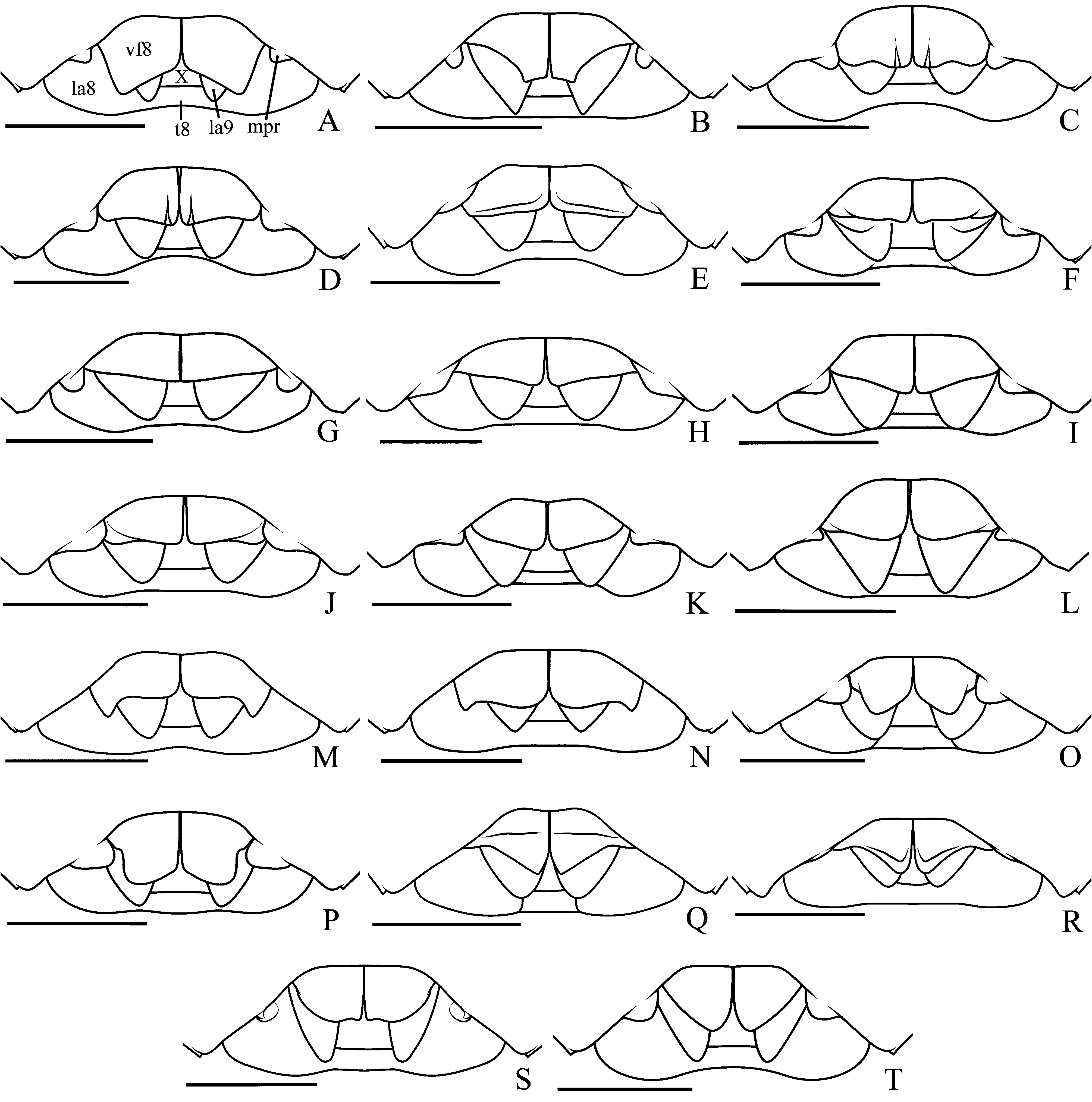
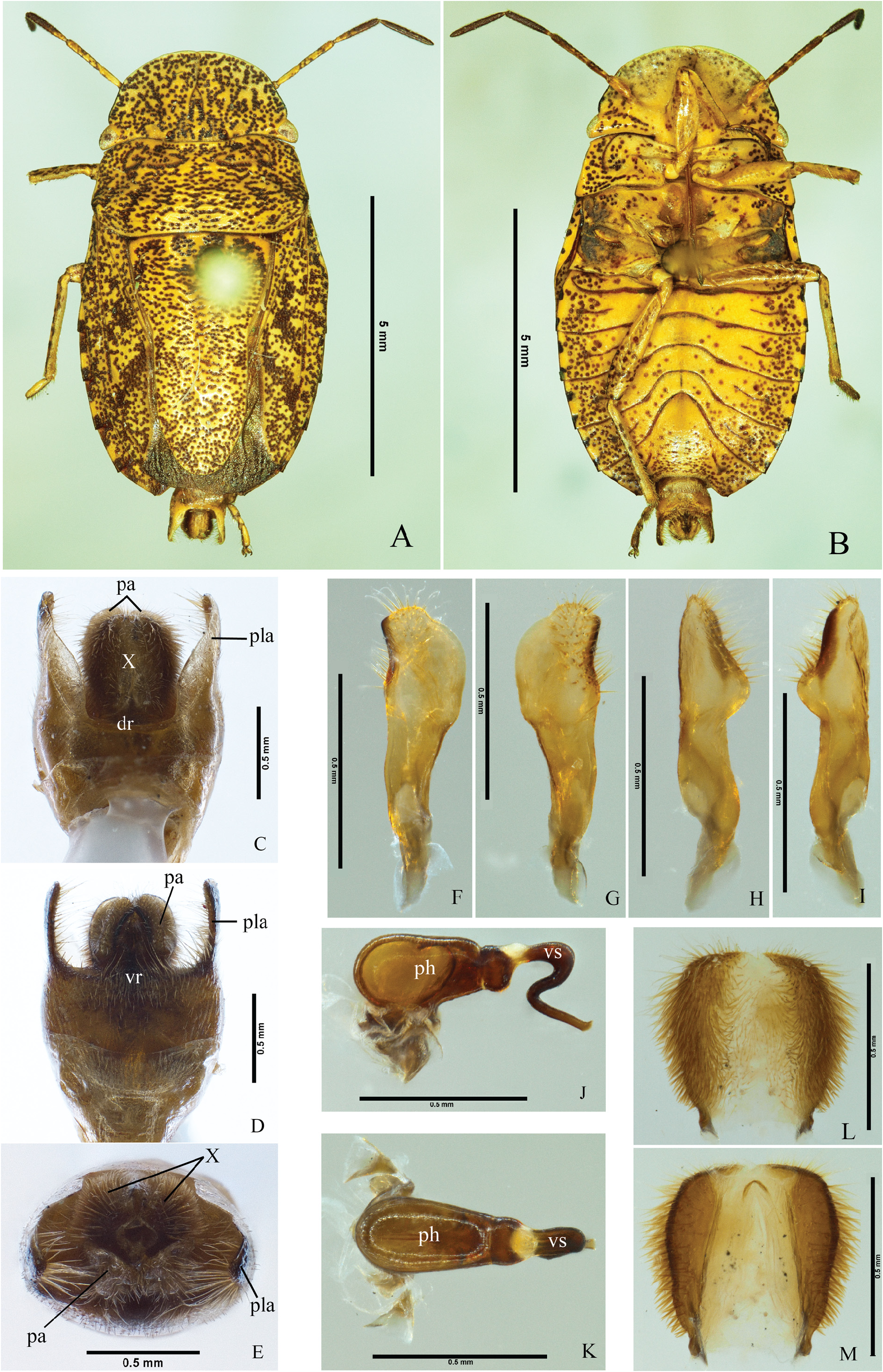
Ischnopelta guarani Rosso & Campos , sp. n. ( Figs. 5S View FIGURE 5 , 27–28 View FIGURE 27 View FIGURE 28 )
Etymology. The epithet is a tribute to the Guarani, a native people of South America, in whose original lands occurs the species.
Type locality. ARGENTINA, Corrientes, Laguna Brava [-27.4957, -58.6441] GoogleMaps .
Holotype. Male. ARGENTINA, Corrientes, Laguna Brava (7 km E Corrientes, Route 5), 18.I.1989, C.W. & L.B. O’Brien & G. Wibmer. Deposited at Museo de La Plata , Universidade Nacional de La Plata ( MLPA), La Plata, Argentina.
Paratypes. 5 males and 2 females. BOLIVIA, Santa Cruz, Warnes (5 km SSE Warnes, Rio Selva Hotel ), 2 males, 20–21.X.2000, Morris & Wappers, [- 17.561111, -63.1994444], (J.E. Eger, Private collection) GoogleMaps ; PARAGUAY, Alto Paraguai ( Grand Chaco, 250 km West Paraguay River), 1 male, 28. VI.1936, Alberto Schulze, [-23.366667, -59.6667], ( USNM); GoogleMaps Central, Capiatá , 1 female, 15.I.1991, G. Arriagada, [-25.3500, -57.4167], (J.E. Eger, Private collection); GoogleMaps Vila Elisa , 1 female, 2.XII.1939, Dernier Coll., [-25.3667, -57.6167], ( MLPA) GoogleMaps ; ARGENTINA, Formosa, Laishi ( Riacho Tohué ), 2 males, 11.I.1939, Dernier Coll., [-26.408333, -58.258333], ( MLPA) GoogleMaps .
Description. The overall somatic morphology is as described for I.scutellata ,except for the following features. Head. Antennae: segment I dark yellowish with brown blotches; segments II and II dark yellowish with brown punctures, ventral surface brown in some specimens; segments IV and V dark brown, proximal portion of segment IV dark yellowish with brown punctures in some specimens; segments ratio: I=II<III<IV<V.
Thorax. Scutellum: post-frenal lobe margins subparallel to the distal half. Hemelytra: conspicuous spot at apex of radial vein.
Abdomen pale-yellow; dark spots at the lateral of urosternites elongated and subtriangular, both wide and subequal in length.
Male. Apical margin of membrane of hemelytra convex; median portion of the posterior margin of urosternite VII concave; urosternite VII reaching anteriorly the imaginary line connecting the spiracles of urosternite V. Genitalia. Pygophore with dorsal and ventral rim slightly concave ( Figs. 27C View FIGURE 27 , dr; 27D, vr). Posterolateral angles 1.4 times longer than the rest of the pygophore, perpendicular to the frontal plane and subparallel, apices slightly convergent, basal portion of the dorsal margin less sclerotized and folded to the interior of the pygophore ( Fig. 27C–E View FIGURE 27 , pla). Setae in a band on the ventral rim, part of the lateral surface of the pygophore, and the lareral of the posterolateral angles; setae long on ventral rim and ventral margin of the posterolateral angles. Segment X longer than wide, not reaching the apex of the posterolateral angles and parameres; oval and weakly emarginated apically; lateral margins sclerotized and densely covered with long setae; mid-longitudinal portion membranous with short and sparse setae ( Figs. 27C–E, X; 27L–M View FIGURE 27 ). Parameres claviform, oblique to the frontal plane, outer margin sinuous, convex on apical portion; inner margin sinuous, with a shallow cavity more sclerotized on the distal portion; apical margin convex forming a convergent process with the inner margin; dorsal and ventral surfaces sinuous, setae covering a narrow band on the sclerotized area of the inner margin ( Figs. 27D View FIGURE 27 , pa; 27F–I). Cup-like sclerites externally visible, apices rounded and subparallel. Phallus: vesica sharply sinuous, proximal portion directed posteriorly, sharply curved ventroanteriorlly on median portion, and curved ventroposteriorlly on distal portion; basal portion laterally widened, short, dorsally flat, ventrally expanded, and gradually narrowing posteriorly; secondary gonopore beveled ( Fig. 27J–K View FIGURE 27 ).
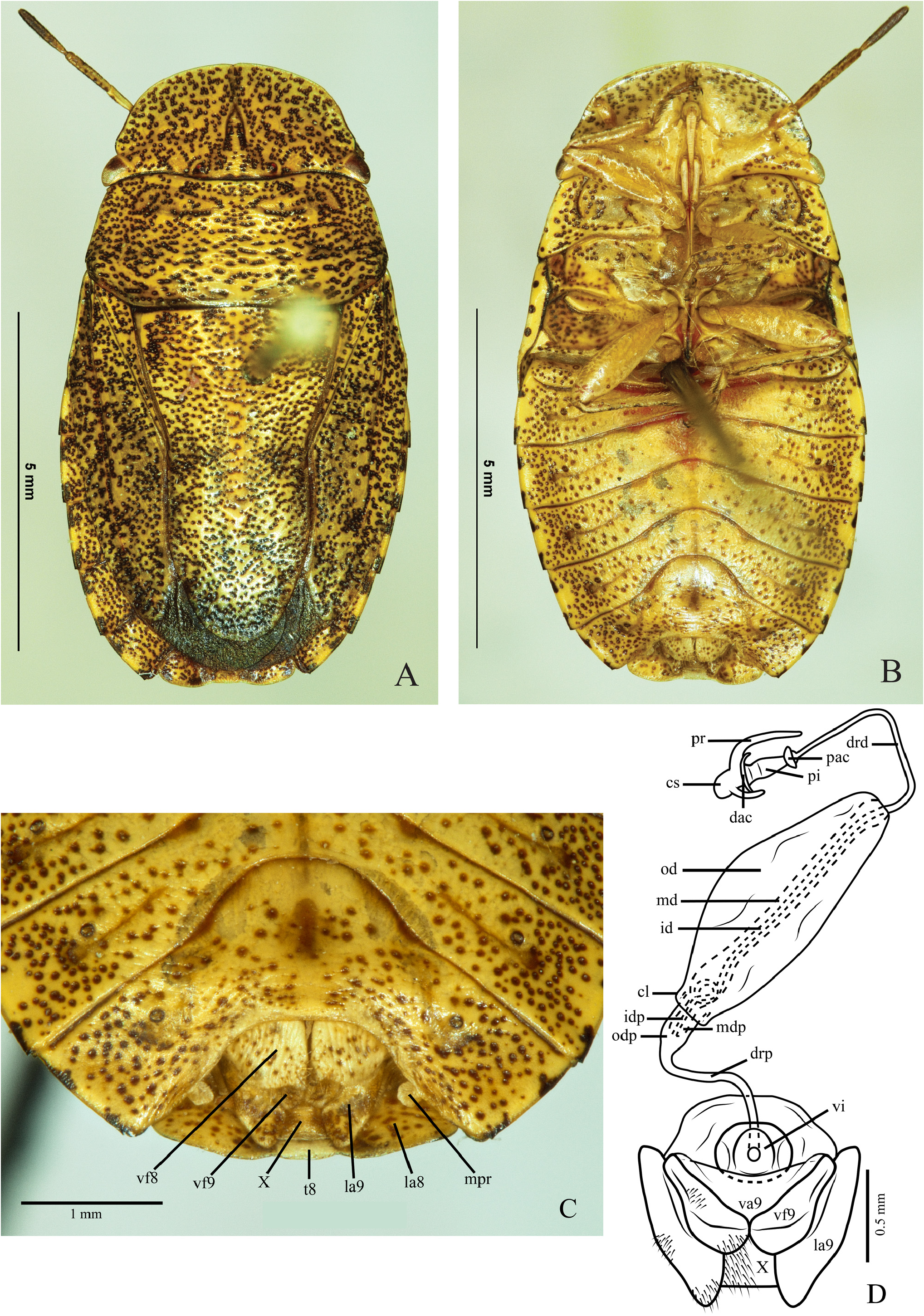
Female. Membrane of hemelytra not reaching the posterior margin of mediotergite VIII, posterior margin convex; posterior margin of mediotergite VIII and of & Campos, sp. n. specimens evaluated (n).
urosternite VII, and projections of urosternite VII as described for I. scutellata , but the later perpendicular to the surface of urosternite VII ( Fig. 28C View FIGURE 28 , mpr). Genitalia. Valvifers VIII wider than long; posterior margin sinuous; sutural margins subrectilinear and dorsally folded; surface convex longitudinally, dark yellowish with brown punctures, setae on distal half of sutural margins and on posterior margin; longitudinal grooves narrow and shallow ( Figs. 5S View FIGURE 5 ; 28C View FIGURE 28 , vf8). Valvifers IX partially covered by valvifers VIII; lateral margin sinuous, midbasal portion of ventral surface without setae ( Fig. 28D View FIGURE 28 , vf9). Laterotergites IX not reaching the posterior margin of mediotergite VIII; lateral margin convex; setae on median portion of lateral margin ( Fig. 28C–D View FIGURE 28 , la9). Thickening of vaginal intima subcircular, median area broadly oval and membranous ( Fig. 28D View FIGURE 28 , vi). Vesicular area anterior to the collar 1/8 of the posterior portion; median duct anterior to the collar with slight proximal widening ( Fig. 28D View FIGURE 28 , mdp), median duct posterior to the collar with proximal and distal widening ( Fig. 28D View FIGURE 28 , md), inner duct coiled in the proximal widening ( Fig. 28D View FIGURE 28 , id). Distal ductus receptaculi narrower than proximal one, and 0.9 times the length of the vesicular area posterior to the collar ( Fig. 28D View FIGURE 28 , drd, drp). Pars intermedialis barrel-shaped ( Fig. 28D View FIGURE 28 , pi); annular crests directed to the ductus receptaculi, the proximal slightly larger than half the diameter of the distal one ( Fig. 28D View FIGURE 28 , dac, pac). Capsula seminalis oval, with two filiform projections, one laterobasal long and sinuous, and the other shorter, midlateral, both directed to the pars intermedialis ( Fig. 28D View FIGURE 28 , cs, pr).
Measurements: Table 11.
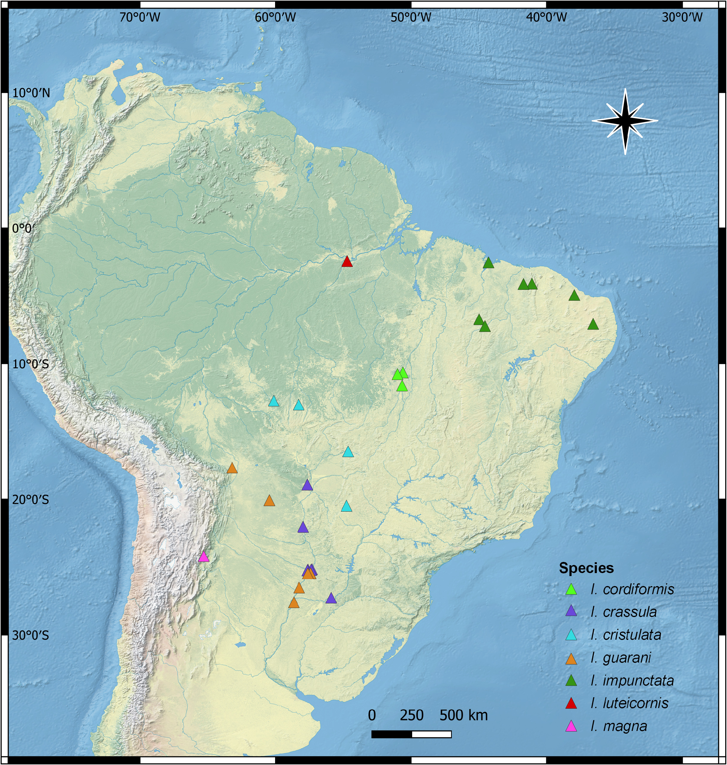
Distribution. Bolivia (Santa Cruz), Paraguay (Alto Paraguay, Central), Argentina (Formosa, Corrientes) ( Fig. 7 View FIGURE 7 ).
Comments. Ischnopelta guarani sp. n. ( Fig. 27A– B View FIGURE 27 ; 28A–B View FIGURE 28 ) is similar to I. paiagua sp. n. ( Fig. 38A–B View FIGURE 38 ), differing by the labrum inserted slightly posteriorly to half the distance between the anterior margin of the eyes and the apex of mandibular plates, the wider abdominal lateral blotches, and the shape of the parameres ( Figs. 27F–I View FIGURE 27 ; 38F–I View FIGURE 38 ).

| MLPA |
Argentina, La Plata, Universidad Nacional de La Plata, Museo de la Plata |
| VI |
Mykotektet, National Veterinary Institute |
| USNM |
Smithsonian Institution, National Museum of Natural History |
No known copyright restrictions apply. See Agosti, D., Egloff, W., 2009. Taxonomic information exchange and copyright: the Plazi approach. BMC Research Notes 2009, 2:53 for further explanation.
|
Kingdom |
|
|
Phylum |
|
|
Class |
|
|
Order |
|
|
Family |
|
|
Genus |