
Jasmineira princei ( McIntosh, 1916 ) Tovar-Hernández, 2007
|
publication ID |
https://doi.org/ 10.1080/00222930701250912 |
|
persistent identifier |
https://treatment.plazi.org/id/03B1879F-3011-5C7A-FE84-E4BBC715F9AE |
|
treatment provided by |
Felipe |
|
scientific name |
Jasmineira princei ( McIntosh, 1916 ) |
| status |
comb. nov. |
Jasmineira princei ( McIntosh, 1916) View in CoL new combination
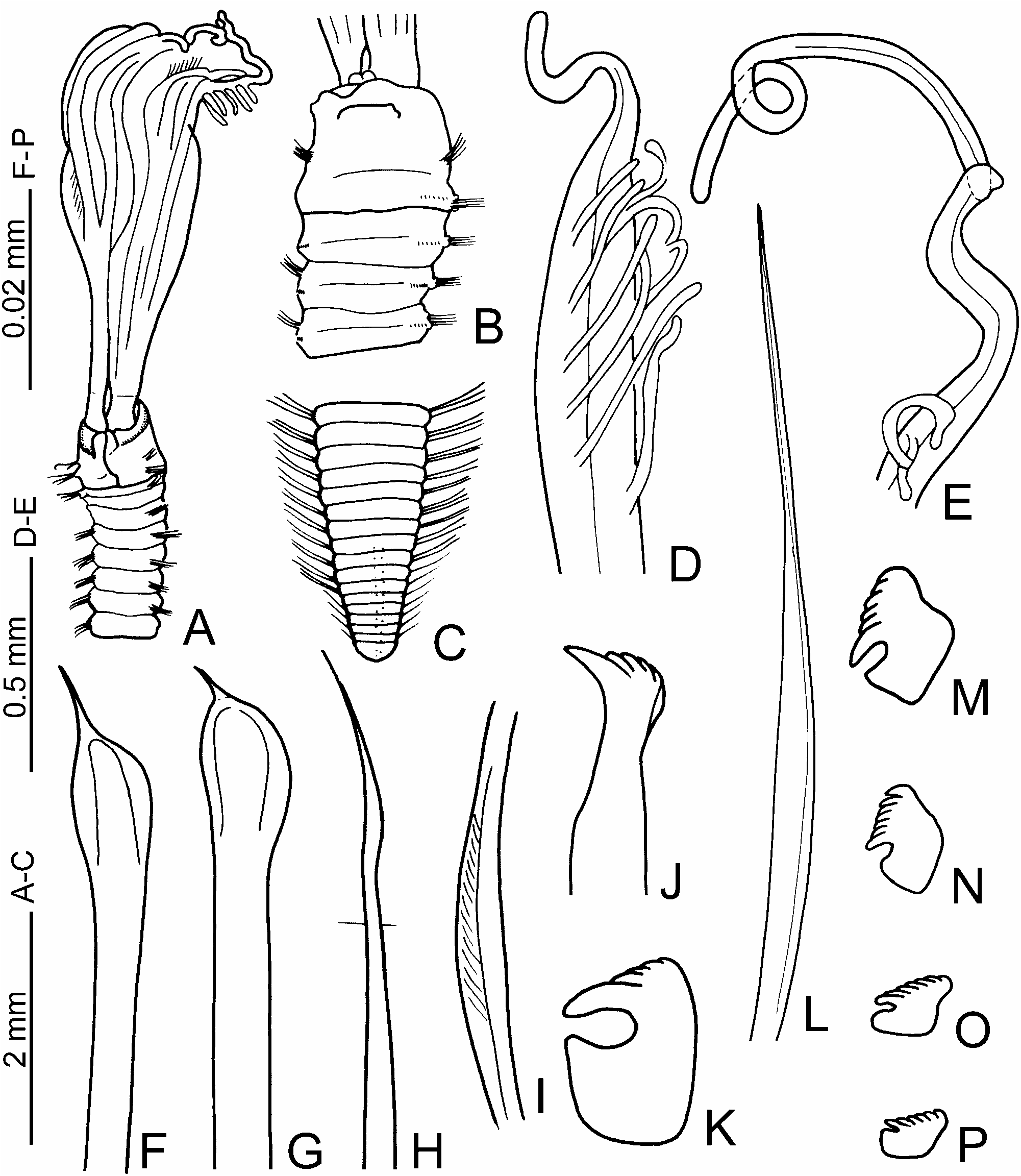
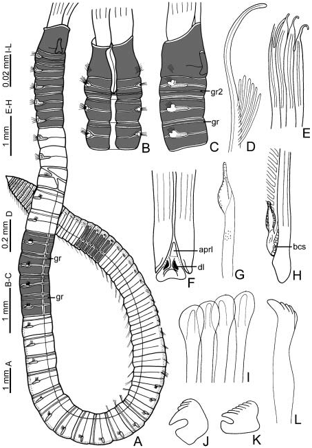
( Figure 18A–G View Figure 18 )
Chone princei McIntosh 1916, p 60 View in CoL –63, Plate 3, Figures 3–8 View Figure 3 View Figure 4 View Figure 5 View Figure 6 View Figure 7 View Figure 8 .
Material examined
Type material. Quebec [ BMNH 1921.5 .1.4373 (lectotype, two paralectotypes), Gulf of Saint Lawrence , coll. Dr Whiteaves, 1872] .
Additional material. Euchone analis KrØyer, 1856 [ USNM 333 View Materials , probable type] .
Redescription (in parentheses variation observed in paralectotypes)
Colour, body shape, and size. Body cream coloured. Trunk cylindrical. Body length 23 mm (18–25), width 2 mm (2–2.2). Tube is a smoothly rounded, firm structure of mud which coats the internal chitinous lining.
Branchial lobes and branchial crown. Insertion of the branchial lobes exposed beyond collar. Branchial crown length 15 mm (14–18). Base of the branchial lobes prominent. Radioles: 13 pairs (12). Pinnules long, of similar size along radioles. Radiolar tips short ( Figure 18C View Figure 18 ). Palmate membrane absent. Narrow radiolar flanges. Dorsal lips elongate, triangular, midrib present, lateral lamellae broad dorsally. Dorsal pinnular appendages absent. Ventral lips small, rounded. Ventral radiolar appendages: five pairs (three), the inner one about three-quarters the length of branchial crown, the remainder about one-half the length of branchial crown.
Peristomium . Anterior peristomial ring lobe not exposed beyond collar, distally entire. Posterior peristomial ring collar: antero-dorsal margin deeply incised, forming two welldeveloped dorsal pockets; two dorsal vascular loops; entire length of mid-dorsal collar margins forms a broad gap; lateral margin entire ( Figure 18B View Figure 18 ); ventral margin incised, forming two trapezoidal-shaped lappets ( Figure 18A View Figure 18 ); ventral margin higher than dorsal ( Figure 18B View Figure 18 ). Ventral shield bean-shaped, two times longer than wide, divided transversely ( Figure 18A View Figure 18 ). Ratio of posterior peristomial ring collar length versus chaetiger 2 length, in lateral view: 2:1 ( Figure 18B View Figure 18 ).
Thorax. Chaetiger 1: elongate, narrowly hooded chaetae. Chaetigers 2–8: notopodia— elongate, narrowly hooded chaetae; one anterior row with bayonet chaetae ( Figure 18D View Figure 18 ); two posterior rows with paleate chaetae, palea long with medium-sized mucro ( Figure 18E View Figure 18 ); neuropodia—one row of acicular uncini with the main fang surmounted by three rows of teeth equal in size, occupying half the length of main fang; hood present ( Figure 18F View Figure 18 ). Glandular ridge on chaetiger 2: narrow ( Figure 18A, B View Figure 18 ).
Abdomen. Abdominal segments: 52 (48). Anterior segments: two transverse rows of elongate, narrowly hooded chaetae; uncini with the main fang surmounted by teeth equal in size, breast reduced to narrow swelling, handles long ( Figure 18G View Figure 18 ). Posterior segments: very elongate, narrowly hooded chaetae, 25% longer than in anterior segments; uncini similar to those in anterior abdomen. Pygidium with a triangular, posterior margin.
Gametes. Lectotype female with oocytes in anterior abdomen, visible through the body wall. Paralectotype female, oocytes from the second thoracic segment to the last thoracic segment.
Methyl green staining. The anterior margin of collar is not coloured dorsally, ventrally, or laterally. The ventral shield of collar is dark coloured. The posterior and basallateral margins of collar are dark. The ventral shield of the first thoracic chaetiger is dark with anterior longitudinal grooves ( Figure 18A View Figure 18 ). Each thoracic segment is uniformly coloured ventrally ( Figure 18A View Figure 18 ); laterally, each segment has a dark rectangle in the upper and lower side of the torus ( Figure 18B View Figure 18 ); dorsally, colourless.
Remarks
In this study, a lectotype and two paralectotypes are designated from syntypes according to Article 74.4, International Commission on Zoological Nomenclature (2000). Chone princei is redescribed and transferred to the genus Jasmineira due to the fact that the original description lacked some critical details diagnostic for Jasmineira . The redescription provided here shows that in J. princei : (1) insertion of the branchial lobes is exposed beyond collar; (2) pinnules are similar-sized along radioles; (3) palmate membrane is absent; (4) radiolar flanges are narrow; (5) dorsal lips are elongate, triangular with radiolar appendages; (6) dorsal pinnular appendages are absent; (7) anterior peristomial ring lobe is not exposed beyond collar; (8) dorsal pockets are well developed with vascular loops in the peristomium; (9) abdominal uncini have the main fang covered by teeth of equal size, breast reduced to narrow swelling, and handles long; and (10) oocytes are distributed in thorax and anterior abdomen.
Jasmineira View in CoL is often confused with Fabrisabella Hartman, 1969 View in CoL , because both have abdominal uncini with a reduced narrow breast and long handle; however, they differ in that Fabrisabella View in CoL lacks dorsal pinnular appendages and bayonet chaetae; both are present in Jasmineira ( Fitzhugh 1989) View in CoL .
McIntosh (1916), in his description of Chone princei View in CoL , referred to a ‘‘glandular tubular organ’’ located at the dorsal peristomium; this structure is called ‘‘vascular loops’’ in the redescription given here. The vascular loops are circular cameras situated dorsally in each side of the peristomium; inside each camera there is an S- or C-shaped vessel; their structure and function has not been studied yet, but it is very probable that they are vascularized by the central blood vessel. The vascular loops have also been recorded in the genus Fabrisabella ( Fitzhugh 1989) View in CoL , and in some specimens of Euchone analis View in CoL ( Figure 18O View Figure 18 ).


No known copyright restrictions apply. See Agosti, D., Egloff, W., 2009. Taxonomic information exchange and copyright: the Plazi approach. BMC Research Notes 2009, 2:53 for further explanation.
|
Kingdom |
|
|
Phylum |
|
|
Class |
|
|
Order |
|
|
Family |
|
|
Genus |
Jasmineira princei ( McIntosh, 1916 )
| Tovar-Hernández, María Ana 2007 |
Chone princei
| McIntosh WC 1916: 60 |