
Lepidocyrtinus barnardi Womersley, 1934
|
publication ID |
https://doi.org/ 10.11646/zootaxa.4898.1.1 |
|
publication LSID |
lsid:zoobank.org:pub:3F66CE1D-1036-4226-BC28-029F9961E069 |
|
DOI |
https://doi.org/10.5281/zenodo.4392561 |
|
persistent identifier |
https://treatment.plazi.org/id/7B6687A7-FFFB-FF99-FF12-D569FB91F991 |
|
treatment provided by |
Plazi |
|
scientific name |
Lepidocyrtinus barnardi Womersley, 1934 |
| status |
|
Lepidocyrtinus barnardi Womersley, 1934 View in CoL
Figs 2 View FIGURE 2 , 13–17 View FIGURE 13 View FIGURE 14 View FIGURE 15 View FIGURE 16 View FIGURE 17 , Tables 1 View TABLE 1 and 4 View TABLE 4
Lepidocyrtinus cooperi var. barnardi Womersley, 1934: 461 View in CoL , fig. 10, South Africa, Cape Town, Kirstenbosch, not examined. Seira (Lepidocyrtinus) barnardi ; Yosii 1959: 12, figs 6A–I, South Africa, Cape Town, Kirstenbosch.
Seira barnardi ; Coates 1968: 454; Janion-Scheepers et al. 2015: 68, Africa South (catalog).
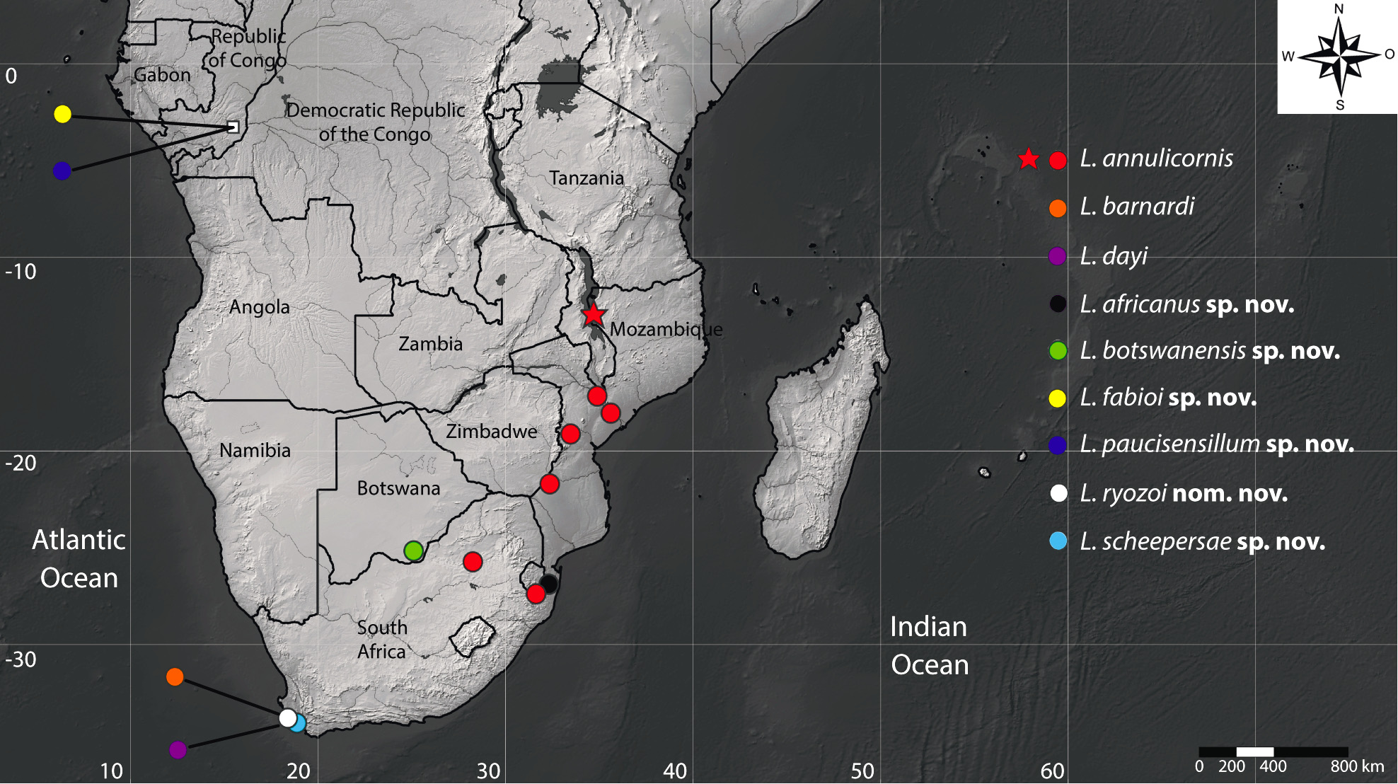
Typological note. One cotype of four specimens used in the original description of Lepidocyrtinus cooperi var. barnardi by Womersley (1934) is deposited at SAM/IZIKO (Scheepers, personal communication). We could not analyse this material, but our specimens fit the descriptions of Womersley (1934) and Yosii (1959) and they were collected from the type locality ( Fig. 2 View FIGURE 2 ). For this reason, the redescription is justified.
Examined material. 1 male and 4 females in slides and 1 specimen in alcohol ( INPA): South Africa, Cape Town, Table Mountain National Park, Newlands , 33°57’51.9”S, 18°26’25.1”E ( Fig. 2 View FIGURE 2 ), in Pine Plantation, 200 m, 12.ix.2008, Leaf Litter, C Uys coll. GoogleMaps 3 females in slides and 170 specimens in alcohol ( CC / UFRN): 33°57’58.3”S, 18°26’31.6”E, in Afrotemperate Forest , 230 m, 15.i.2009, Sugar-baited Ant Trap, C Uys coll. GoogleMaps 23 specimens in alcohol ( INPA): Spilhaus, Cecilia Ravine , 33°59’53.7”S, 18°24’51.6”E, in Fynbos, 520 m, 23.i.2009, Sugar-baited Ant Trap, C Uys coll. GoogleMaps
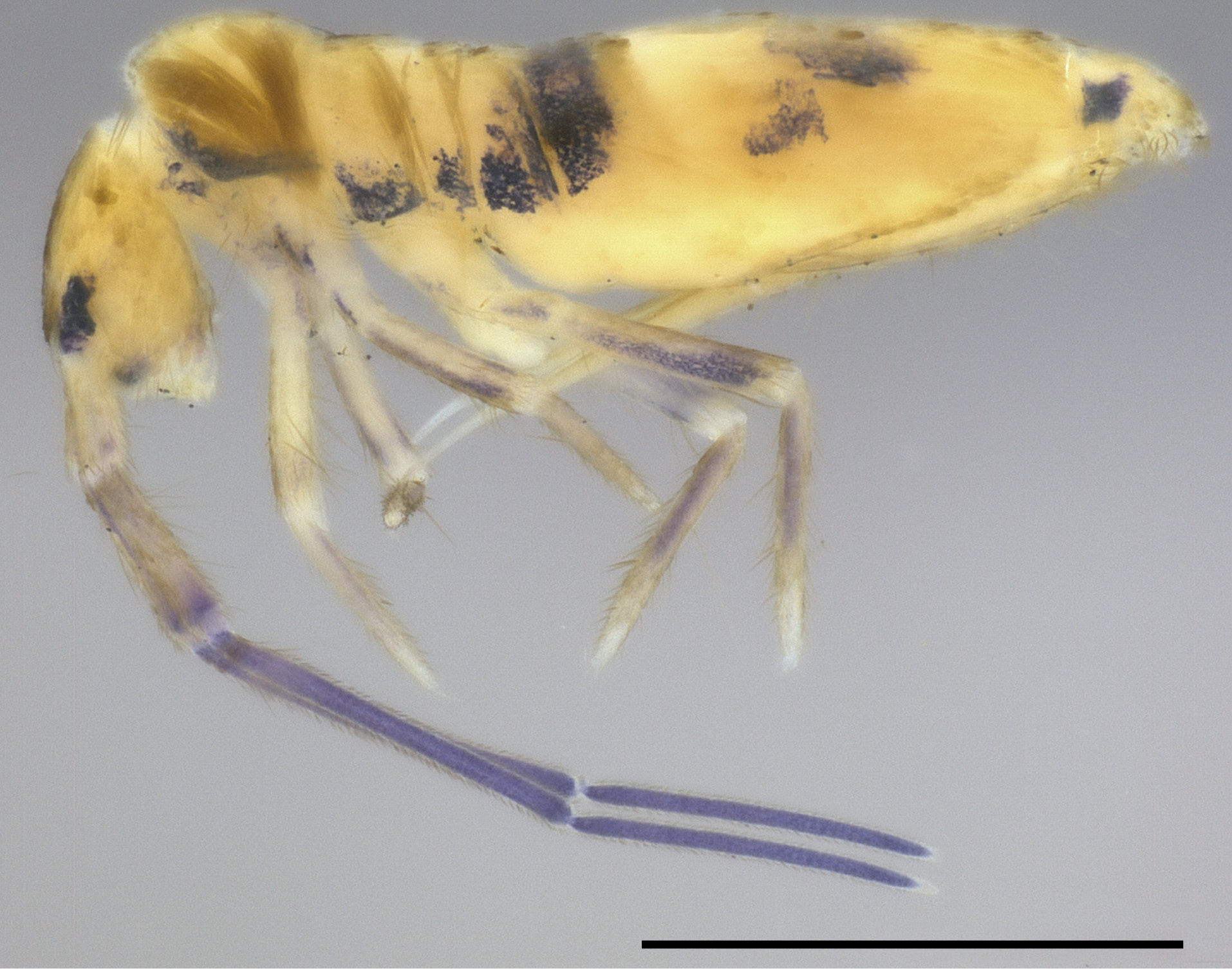
Diagnosis. Body with dark blue pigments on Ant III–IV, Th II laterally up to one transverse band on Abd III, and spots on Abd IV–V and legs ( Fig. 13 View FIGURE 13 ); head mac A0, A2–3, A5, M1–2, M4i, S0–6 and Pp5 present; prelabral chaetae smooth; labial papilla E with l.p. apically pointed and not reaching the base of a.a. ( Figs 14 View FIGURE 14 D–F); basolateral and basomedian labial fields with chaetae R (smaller than others) ciliated, e and l2 smooth, L1 ciliated or smooth; Th II not projected anteriorly and with 17 anterior, 9 median (m1ip, m4i–4p present) and 15–16 posterior mac (p2ep, p2ep2, p3 as mac); Th III–Abd II with 10, 5–6, 4 central mac respectively; Abd IV with 10–11 central mac and 11 lateral mac ( Figs 15 View FIGURE 15 , 16B View FIGURE 16 ); unguis a.t. present or absent; unguiculus with pe lamella serrated and with small proximal tooth; collophore anteriorly with 4–5 spine-like chaetae and 4 ciliated chaetae apically acuminate, posteriorly without spines, smooth chaeta present ( Fig. 17E View FIGURE 17 ); manubrium ventrally with 2/2 subapical chaetae; manubrial plate without blunt mac; dens with with 13–19 blunt mac apically nipple-like ( Figs 17 View FIGURE 17 F–G).
Description. Total length (head + trunk) of specimens 2.21–3.22 (n=4). Specimens pale white with dark blue pigment on distal Ant II to Ant IV, anterior head, lateral Th II to Abd II which extends to most of the Abd III, central spots on Abd IV and V, coxa II–III, and trochanter to proximal half of tibiotarsus; eyepatches black ( Fig. 13 View FIGURE 13 ). Scales present on Ant I to basis of Ant III, dorsal and ventral head, dorsal thorax and abdomen, legs (except empodia), anterior collophore, ventral and dorsal manubrium and dens ventrally.
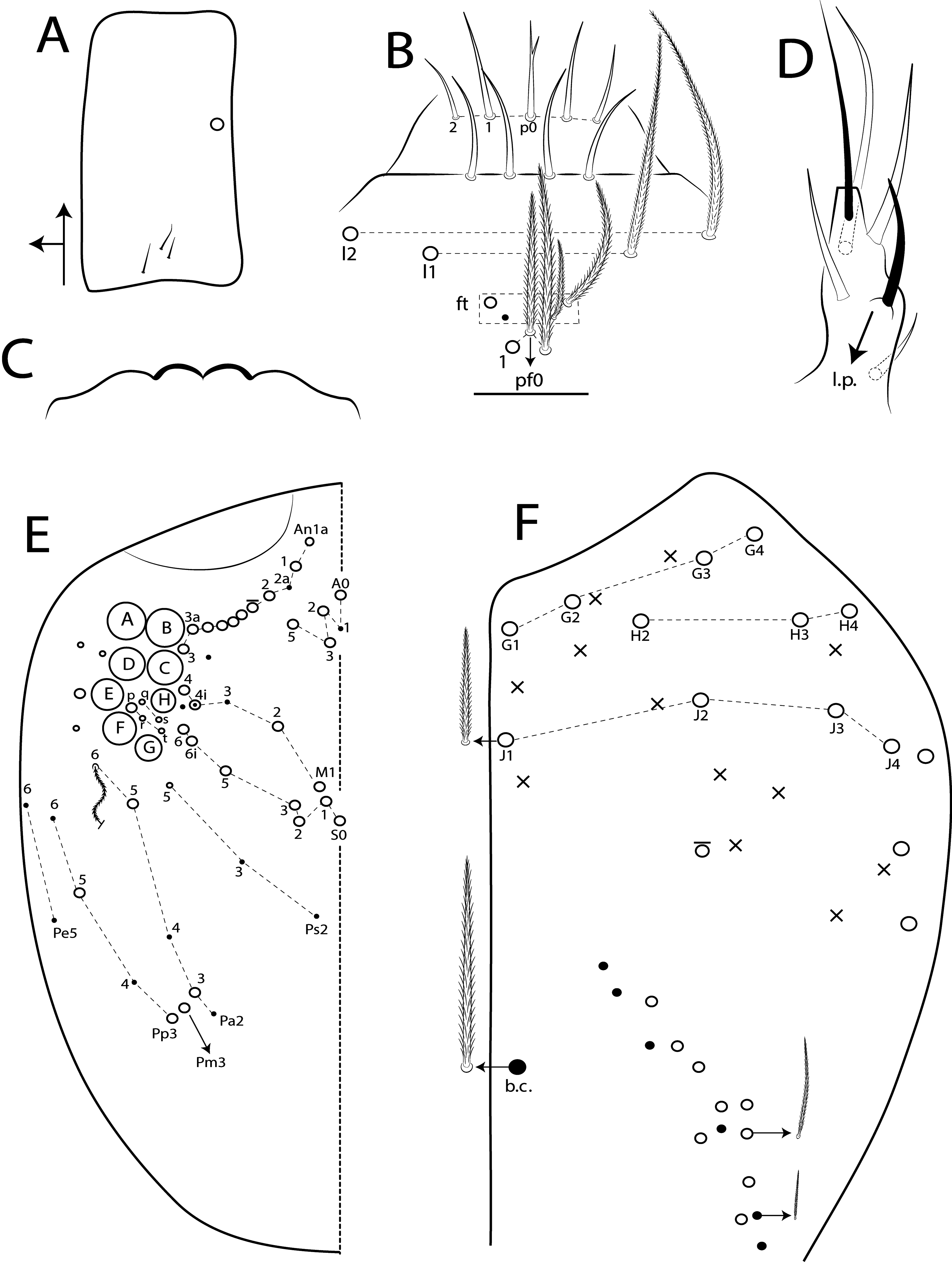
Head. Antennae subequal to trunk length ( Fig. 13 View FIGURE 13 ); ratio antennae: trunk = 1: 1.04; antennal segments ratio as I: II: III: IV = 1: 1.57–1.82: 2.92–3.24: 2.97 (Ant IV present in one specimens). Ant IV annulated on distal one seventh, with apical bulb apically bilobed. Ant III annulated on distal two thirds, apical organ with 2 rods elongated, 3 spiny guard sens, s-blunt sens of different sizes and ciliated chaetae ( Fig. 14A View FIGURE 14 ). Ant I dorsally with 3–4 median mac and 3 smooth mic at the base, without spines ( Fig. 14B View FIGURE 14 ). Eyes A and B larger, G and H smaller, others subequal, and with 5 ‘IO’ chaetae; head dorsal macrochaetotaxy with 9–10 An, 4 A, 4 M, 8 S and 1 Pp mac ( Fig. 14F View FIGURE 14 ). Clypeal formula with 4 (l1–2), 8 (ft), 5 (pf1–2) ciliated chaetae, l1–2 and pf0–1 larger, l1 apically acuminate, others subequal ( Fig. 14C View FIGURE 14 ). Prelabral chaetae smooth, inner chaetae eventually with two small median filaments ( Fig. 14D View FIGURE 14 ). Labral p0 chaeta with median filament unequal in length. Labral inner and outer papillae rounded, not projected (as in Fig. 19C View FIGURE 19 ). Maxillary palp with smooth t.a. and b.c. weakly ciliated, thicker and 1.19 longer than t.a. Labial papilla E with l.p. apically pointed and not reaching the base of a.a. ( Fig. 14E View FIGURE 14 ). Basolateral and basomedian labial fields with chaetae M1–2, R (smaller than others) ciliated, e and l2 smooth, L1 ciliated or smooth. Ventral head with about 25 ciliated chaetae, postlabial formula with 4 (G1–4), 2 (H2, H4), 3 (J1–3) chaetae, 5 thin posterior chaetae with different lengths, and 3 larger chaetae, 1 b.c. surrounding the cephalic groove ( Fig. 14G View FIGURE 14 ).
Thorax chaetotaxy ( Figs 15 View FIGURE 15 A–B). Th II not projected anteriorly, a, m and p series with 17, 9 and 15–16 mac, respectively. Th III a, m and p series with 4, 1 and 8 mac, respectively. Ratio Th II: III = 1.69–153: 1 (n=4).
Abdomen chaetotaxy ( Figs 15 View FIGURE 15 C–D, 16A–C). Abd I a, m and p series with 0–1, 5 and 0 mac, respectively. Abd II a, m and p series with 1, 4 and 0 mac, respectively. Abd III a, m and p series with 0–1, 3 and 1 mac, respectively. Abd IV with 10–11 central mac on A–T series and 11 lateral mac on E– Fe series; at least 12 posterior sens (ps type I, others type II) and 11 posterior mes. Abd V a, m and p series with 1, 4 and 6 mac, respectively. Ratio Abd III : IV = 1: 3.87–4.23 (n=4).
Legs. Subcoxa I with 5 chaetae and 3 psp; subcoxa II with an anterior row of 12 chaetae, posterior row of 7 chaetae, 1 posterior chaeta and 8 psp; subcoxa III with one row of 12 chaetae, 2 anterior chaetae and 2 posterior psp ( Figs 17 View FIGURE 17 A–C). Trochanteral organ with about 94 spine-like chaetae ( Fig. 17D View FIGURE 17 ). Tibiotarsus normal or subdivided on distal two thirds, outer side with 3 large chaetae apically acuminate, inner side with 8 mac weakly ciliated. Unguis with 3–4 inner teeth, b.t. smaller than m.t. in length, and a minute a.t., present or absent. Unguiculus with all lamellae smooth and acuminate, except pe serrated and with a small proximal tooth; ratio unguis: unguiculus = 1: 0.52. Tibiotarsal smooth chaeta 1.32 larger than unguiculus and tenent hair 0.88 smaller than unguis outer edge.
Collophore. Anterior side with 19–22 chaetae, 4–5 proximal spine-like chaetae, 1 thin and 7–9 normal ciliated chaetae, 4 ciliated chaetae apically acuminate and 3 mac distally; posterior side with 12 chaetae, 8 thin ciliated chaetae widely distributed and 3 ciliated and 1 smooth chaetae distally; lateral flap with about 42–44 chaetae, 4–5 smooth and 38–39 ciliated ( Fig. 17E View FIGURE 17 ).
Furcula. Manubrium ventral formula with 0, 0, 0, 2/2 (subapical), 24–28 (apical) ciliated chaetae plus approximately 18 elongated apical scales per side ( Fig. 17F View FIGURE 17 ); manubrial plate with 3 ciliated mac apically acuminate (no blunt), 11 ciliated chaetae of different sizes and 3 psp. Dens dorsally with one proximal row of 13–19 blunt mac finely ciliated and apically nipple-like ( Fig. 17G View FIGURE 17 ).
Remarks. Lepidocyrtinus barnardi resembles L. dayi in Th II normal (not projected anteriorly) and with anterior mac group sparce, prelabral chaetae mostly smooth, labral papillae rounded and discrete, dorsal chaetotaxy of Th II–III, and manubrial plate without blunt mac ( Tables 3–4). However, L. barnardi differs from this species by color pattern with one transversal band on Abd III and one central spot on Abd IV, while in L. dayi most of the body is pigmented ( Figs 13 View FIGURE 13 , 18 View FIGURE 18 ). They also differ in head by basomedian labial field with chaeta e smooth (ciliated in L. dayi ), labial papilla E with l.p. not reaching the base of a.a. (surpassing the base in L. dayi ), and dorsal chaetotaxy with S4 mac and Pa3, Pm3 and Pp3 absent (opposite in L. dayi ) ( Figs 14 View FIGURE 14 E–F, 19D–E). In dorsal chaetotaxy they differ by Th III with 10 central and 3 lateral mac (11 and 2 respectively in L. dayi ), Abd I generally with 6 central mac (a2 present or absent, m4 present) and Abd II with 4 central mac, while in L. dayi there are 5 central mac (m4 absent) on Abd I and 3 central mac (m3e absent) on Abd II ( Figs 15 View FIGURE 15 B–D, 20B–D). Other features that differ are: unguis a.t. often present (absent in L. dayi ), manubrium ventral formula with 0, 0, 0, 2/2 chaetae (1, 2, 2, 0/ 2 in L. dayi ), and dens with 13–19 blunt mac (only 1 in L. dayi ) ( Figs 17 View FIGURE 17 F–G, 22F–G).


| INPA |
Instituto Nacional de Pesquisas da Amazonia |
| CC |
CSIRO Canberra Rhizobium Collection |
| UFRN |
Universidade Federal do Rio Grande do Norte |
| V |
Royal British Columbia Museum - Herbarium |
No known copyright restrictions apply. See Agosti, D., Egloff, W., 2009. Taxonomic information exchange and copyright: the Plazi approach. BMC Research Notes 2009, 2:53 for further explanation.
|
Kingdom |
|
|
Phylum |
|
|
Class |
|
|
Order |
|
|
Family |
|
|
Genus |
Lepidocyrtinus barnardi Womersley, 1934
| Cipola, Nikolas Gioia, Morais, José Wellington De & Bellini, Bruno Cavalcante 2020 |
Seira barnardi
| Janion-Scheepers, C. & Deharveng, L. & Bedos, A. & Chown, S. L. 2015: 68 |
| Coates, T. J. 1968: 454 |
Lepidocyrtinus cooperi var. barnardi
| Yosii, R. 1959: 12 |
| Womersley, H. 1934: 461 |