
Leptostylochus elongatus Bock, 1925
|
publication ID |
https://doi.org/ 10.5281/zenodo.178319 |
|
DOI |
https://doi.org/10.5281/zenodo.6251694 |
|
persistent identifier |
https://treatment.plazi.org/id/1D4987D2-8F1B-FFB8-32BA-ABFAB35AE33F |
|
treatment provided by |
Plazi |
|
scientific name |
Leptostylochus elongatus Bock, 1925 |
| status |
|
Leptostylochus elongatus Bock, 1925 View in CoL
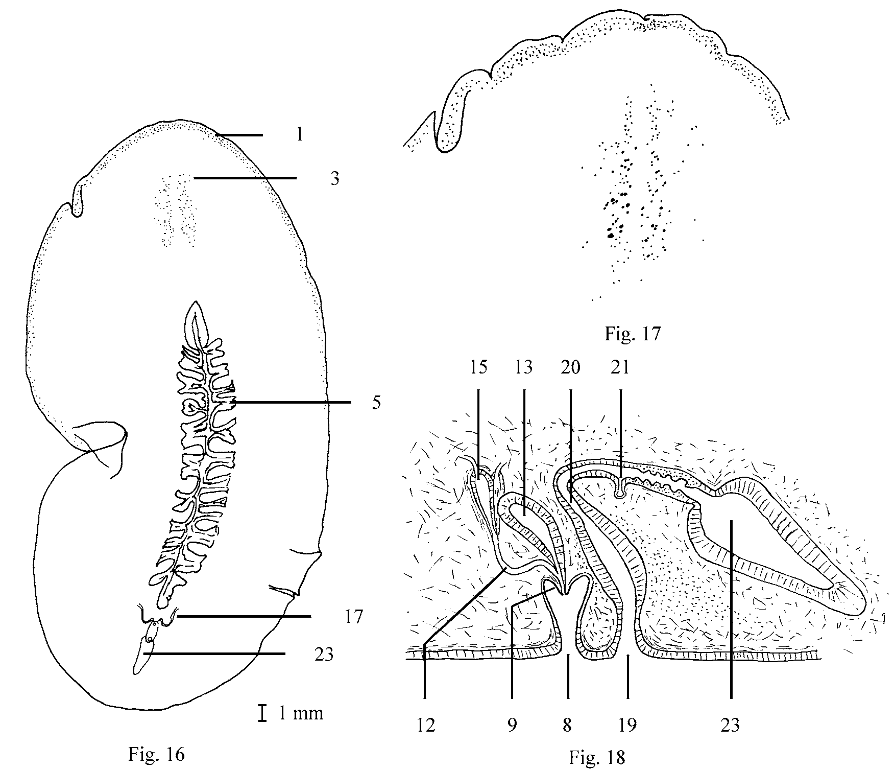
( Figures 16–18 View FIGURES 16 – 18 )
Material
Fifteen specimens were collected from the north island, three from Whangateau Harbor, six from Point Chevalier, two from intertidal rocks in front of the Island Bay Marine Laboratory, two at Karaka Bay, Port Nicholson, one at Castle Point and one at Te Raekaihau Point. Three specimens collected under rocks at Lower Portobello south island.
Morphology
External features: The largest specimen measured 40mm long by 10mm wide. Most specimens ranged from 20 to 33mm long and 5 to 8mm wide. Tentacles are lacking. The dorsal surface is a gray brown lighter at the margins and gray ventrally. Small marginal eyes form a narrow band that extends posteriorly about one third the length of the body. Cerebral eyes extend from behind the cerebral organ as two elongate groups the may merge forming a narrow v-shaped group. The long, heavy ruffled pharynx occupies one-half the body length. The mouth is located in the posterior one-third of the pharyngeal pocket. The intestinal branches do not anastomose.
Reproductive anatomy: The gonopores are located very close together. The male gonopore leads into a short antrum into which a short conical shaped penis projects. The penis is unarmed. The testes occupy a dorsal position and are interspersed with the ovaries. The sperm ducts are ventral and lateral to the pharyngeal pocket twisting and curving as they progress posteriorly. Posterior to the pharyngeal pocket the sperms duct curve medially then dorsally where the ducts become inflated with a thin muscular layer forming spermiducal bulbs. Now as ejaculatory ducts they unite to form a common ejaculatory duct that curves ventrally then posteriorly to receive the prostatic duct prior to entering the base of the penis papilla. The small prostatic vesicle which extends dorsally and anteriorly has a smooth interior with a muscular wall which is transversed by extracapsular gland ducts. The female gonopore leads into the vagina which receives the secretions of the shell glands and continues dorsally and anteriorly to a position above the prostatic vesicle turning dorsal and posterior to receive the common oviduct. The duct of Lang’s vesicle, with a ridged lining, continues to enter into the large Lang’s vesicle.
Two specimens as wholemounts, one stained and one unstained, and a specimen as a set of sagittal sections have been deposited with the California Academy of Science. CAS Nos. 173084, 173085 and 173086 respectively.
No known copyright restrictions apply. See Agosti, D., Egloff, W., 2009. Taxonomic information exchange and copyright: the Plazi approach. BMC Research Notes 2009, 2:53 for further explanation.