
Leydigia (Neoleydigia) macrodonta Sars, 1916
|
publication ID |
https://doi.org/ 10.11646/zootaxa.2082.1.1 |
|
persistent identifier |
https://treatment.plazi.org/id/03BE87A4-4C79-5257-CE97-E1167A55FCAD |
|
treatment provided by |
Felipe |
|
scientific name |
Leydigia (Neoleydigia) macrodonta Sars, 1916 |
| status |
|
VII. Leydigia (Neoleydigia) macrodonta Sars, 1916 View in CoL
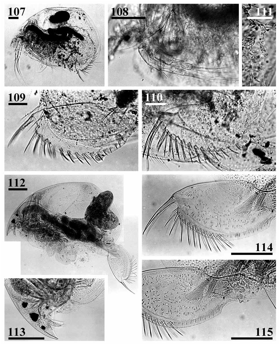
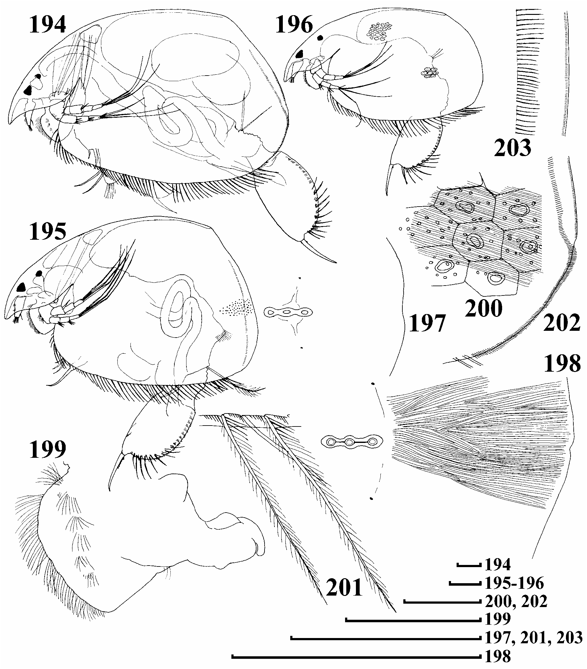
( Figs 112–115 View FIGURES 107–115. 107–111 , 194 View FIGURES 194–203 –223)
Leydigia macrodonta Sars, 1916, p. 328 View in CoL –329, Pl. 37: figs 2, 2a, 2b;
Leydigia macrodonta macrodonta Sars View in CoL in Smirnov 1971, p. 454, fig. 560.
Not Leydigia macrodonta var. louisi Jenkin, 1934, p. 283 View in CoL –285, fig. 14.
Not L. macrodonta macrodonta Sars View in CoL in Fernando and Kanduru 1984, Tab. 1; Jeje, 1988, p. 113 –115, figs 1–8.
Not L. macrodonta longiseta Chen View in CoL Shou-zhong, 1992, p. 165–167 (in Chinese), p. 167–168 (in English), figs 1–10; Chen Shou-zhong, 1993, p. 14.
Type locality. "A small pool in the Cape Flats, alongside the railway line between Retreat and Lakeside" ( Sars 1916), Republic of South Africa.
Lectotype (selected here). The adult parthenogenetic ♀, on slide GOS F9222 labelled "S.A.Mus. I".
Paralectotypes (selected here).
(1) Labelled "S.A.Mus. I": 1 parth. ♀, on the same slide GOS F9222 with the lectotype.
(2) Labelled " Cape of Good Hope": 3 parth. ♀♀, slide GOS F9218 (bad condition); 4 parth. ♀♀, slide GOS F9217 (bad condition); 9 parth. ♀♀, slide GOS F9216 (bad condition); 18 parth. ♀♀, slide GOS F9215 (bad condition) .
Possible type material. Unknown locality: 1 parth. ♀, slide GOS F9221 (bad condition); 3 parth. ♀♀, slide GOS F9220 (bad condition); 1 parth. ♀, slide GOS F9219 (bad condition).
Note on typification. Sars (1916) referred only to a single locality (see above), but he was mistaken: in reality he also had a material from Cape of Good Hope, present in his collection now, and these slides were marked as "type locality". By contrast, I did not find any samples or slides marked as it is reported above, while there were slides marked as "S. A. Mus I", also marked as "type locality". Keeping in mind, that only Dr. Purcell from the South African Museum sent samples to Sars, I selected lectotype on a slide marked as "S. A. Mus I".
Other material examined. Republic of South Africa. KwaZulu-Natal: Summerhill farm Dam, Frere, coll. 21.10.1994 by K. Martens, tube NNS 2002-095 (28º52'57''S, 29º44'47''E) GoogleMaps . Eastern Cape: Watercatchment Dam, McClear, 28.03.1993 by K. Martens, tube NNS 2002-124 (31º06'29''S, 28º20'09''E); Jamieson major Dam, Grahamstown, col. 07.12.1989 by K. Martens, tube NNS 2002-187 (33º19'15''S, 26º26'30''E) GoogleMaps . Western Cape: Verlorenvlei station 3 (first bridge), Elandsbaai, coll. 15.04.1993 by K. Martens and Davies, tube NNS 2002-154 (32º19'12''S, 18º21'38''E); Soetendalsvlei wetland, coll. 25.08.2000 by G. Jones, tube NNS 2000-226 (34º43'59''S, 19º58'15''E). Unknown state: C. S.I. R GoogleMaps . Stream Survey, Ben 12, coll. 07.03.1955 by A. D. Harrison and B. R . Allanson, tube NHM 1959.7.9.126–134.
Diagnosis. Parthenogenetic female. Body subovoid to triangular-ovoid, maximum height in posterior half, dorsal margin almost straight in posterior part, postero-dorsal angle distinct, posterior margin without 'step' in ventral portion ( Figs 112 View FIGURES 107–115. 107–111 , 194–196 View FIGURES 194–203 ). No coarse striation on valves, fine striation delicate ( Figs 198, 200 View FIGURES 194–203 ). Head small, compound eye small, ocellus always bigger than eye ( Fig. 113 View FIGURES 107–115. 107–111 ). PP = 2.5–4 IP, lateral head pores about 1.2 IP distance from midline ( Figs 197–198 View FIGURES 194–203 ). Labral keel subquadrangular, posterior margin with a single tuft of setules basally, anterior margin with long fringe of setules from base to apex, also five lateral groups of setules on sides ( Fig. 199 View FIGURES 194–203 ). Setae at middle of ventral margin of valve with long setules, short setules between their bases ( Fig. 201 View FIGURES 194–203 ). In posterior portion of the margin, setae asymmetrically setulated, posteriormost seta long. Submarginal setules of ventral portion of posterior margin short, fine, in continuous row; in middle of margin, robust, long, and sparse ( Fig. 202 View FIGURES 194–203 ). Marginal membrane with minute 'setules' ( Fig. 203 View FIGURES 194–203 ).
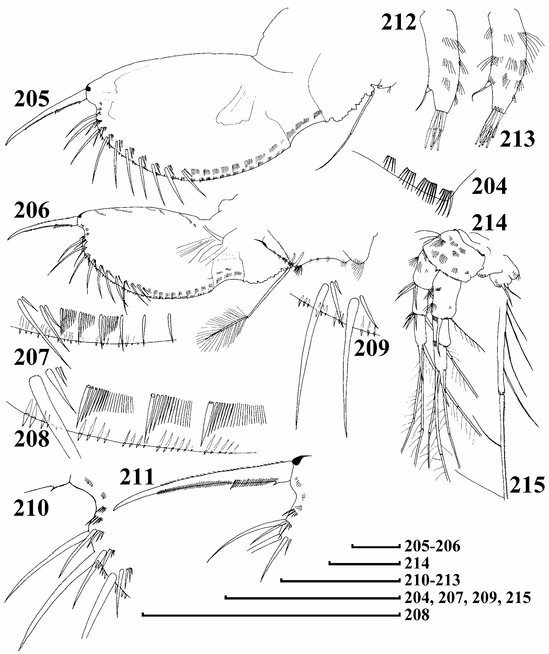
Abdomen with dorsal projection basally and a row of stout setules on distal margin of second segment ( Figs 204, 206 View FIGURES 204–215 ). Postabdomen subovoid, elongate ( Figs 114 View FIGURES 107–115. 107–111 , 205–206 View FIGURES 204–215 ), with regularly convex ventral margin, preanal margin markedly longer than anus, in general convex, with numerous small hillocks, preanal and postanal angles well defined, small distal margin present ( Fig. 115 View FIGURES 107–115. 107–111 ). Postanal marginal denticles in about 19–22 clusters, no marginal denticles on anal margin. About 10 fascicles of stout lateral setae, 2–5 setae in each fascicle, marginalmost setae of each fascicle significantly larger than the rest, 3–4 times as long as second seta, next 1–3 setae, if present, fine ( Figs 207–209 View FIGURES 204–215 ). About 12–14 fascicles of lateral setules on basal half of postanal and anal margin. Postabdominal claw long, slightly curved in distal half or straight, no setules at base, basal spine rudimentary, but seems to be always present ( Figs 210–211 View FIGURES 204–215 ).
Antenna I stout, not reaching tip of rostrum, with 4 transverse rows of long setules on anterior face, and analogous series on lateral and posterior face, no setules at end ( Figs 212–213 View FIGURES 204–215 ). Sensory seta arising about 1/4 of way from distal end. Largest aesthetasc half as long as appendage, reaching tip of rostrum. Antenna II with 3 long and stout spine-like setules on first, and 3–4 of these setules on second endopod segment ( Fig. 214 View FIGURES 204–215 ). Apical swimming setae with basal and distal segments unilaterally armed with stout, long setules, minute setules along other side of distal segment, no chitinous insertions within distal segments ( Fig. 215 View FIGURES 204–215 ). Basal and lateral seta subequal in size, both shorter than apical setae.
Trunk limb I ( Fig. 216 View FIGURES 216–222 ) with ODL large, bearing three series of fine setules and a long seta with unilaterally setulated distal segment. IDL with 4 medial clusters of fine setules, and 3 marginal clusters of short, robust setules. First IDL seta small, with numerous minute setules distally; second and third setae unequal in length and setulation ( Fig. 217 View FIGURES 216–222 ). Endite 3 with long seta 1 and small receptor near base, endite 2 with two longer soft setae (e–f) subequal in length, seta 2 naked, with long sensullum near base. Two ejector hooks of subequal size. Trunk limb II with exopodite ovoid, small, with tuft of short setules, eight scrapers (in one atypical female 9 scrapers were found, see Fig. 218 View FIGURES 216–222 ), distalmost scraper with naked basal segment, on distal lobe with basal tuft of long setules, distalmost seta in filter plate with inflated basal segment. Trunk limb III with trapezium-shaped exopodite, with four setae and small rudiment of seta 3, seta 2 with distal segment setulated bilaterally with short setules, filter plate with distalmost and basalmost setae smaller than others. Trunk limb IV with subovoid exopodite, with six setae, setae 2 long, seta 1 short, both armed with long, fine setules ( Fig. 221 View FIGURES 216–222 ); filter plate with distalmost seta with greatly inflated basal portion, all setae with inflated tips. Trunk limb V with semi-circular exopodite, on inner face of limb, a rudimentary, setulated seta and slender, unilaterally setulated seta, distal armature of gnathobase as lobe ( Figs 222 View FIGURES 216–222 –223).
Ephippial female, male. Unknown.
Size. Juvenile and adult parthenogenetic females from Soetendalsvlei wetland 710–920 µm (n = 6); from Watercatchment Dam 685–1280 µm (n = 30), up to 1.2 mm according to Sars (1916).
Differential diagnosis. L. macrodonta is the only species with the preanal margin of postabdomen longer than the anal margin. Also, only in 2 species of L. (Neoleydigia), L. macrodonta and L. propinqua , are the lateral head pores far from the major pores, but L. macrodonta has four large and one rudimentary setae on exopodite III.
Taxonomical comments. This species has rarely been reported, and usually has been treated ( Jenkin 1934; Smirnov 1971) as a relative of. L. leydigi . Investigators suggested it has a significant basal spine on the postabdominal claw, though Sars (1916) commented only on "a minute denticle at the base [of the claw]". Due to this incorrect idea, two members of the subgenus L. ( Leydigia ), namely (1) L. macrodonta var. louisi Jenkin, 1934 and (2) L. macrodonta longiseta Chen Shou-zhong, 1992, were established as its variety and subspecies. In reality, 1 is a good species, and 2 may be a valid species, far from L. (N.) macrodonta (see above). Jeje (1988) described L. ciliata in his "redescription of L. macrodonta macrodonta "; Jeje's ideas on the morphology of this species were generated without study of the types.
Distribution. Southermost portion of African continent, where it is relatively common. An Asian record ( Fernando & Kanduru 1984) is dubious.

| R |
Departamento de Geologia, Universidad de Chile |
| NHM |
University of Nottingham |
No known copyright restrictions apply. See Agosti, D., Egloff, W., 2009. Taxonomic information exchange and copyright: the Plazi approach. BMC Research Notes 2009, 2:53 for further explanation.
|
Kingdom |
|
|
Phylum |
|
|
Class |
|
|
Order |
|
|
Genus |
Leydigia (Neoleydigia) macrodonta Sars, 1916
| Kotov, Alexey A. 2009 |
L. macrodonta macrodonta Sars
| Jeje, C. Y. 1988: 113 |
Leydigia macrodonta macrodonta Sars
| Smirnov, N. N. 1971: 454 |
Leydigia macrodonta var. louisi
| Jenkin, P. M. 1934: 283 |
Leydigia macrodonta
| Sars, G. O. 1916: 328 |