
Leydigia (Neoleydigia) microps Sars, 1916
|
publication ID |
https://doi.org/ 10.11646/zootaxa.2082.1.1 |
|
persistent identifier |
https://treatment.plazi.org/id/03BE87A4-4C76-5251-CE97-E31F7BCEFA4A |
|
treatment provided by |
Felipe |
|
scientific name |
Leydigia (Neoleydigia) microps Sars, 1916 |
| status |
|
V. Leydigia (Neoleydigia) microps Sars, 1916 View in CoL
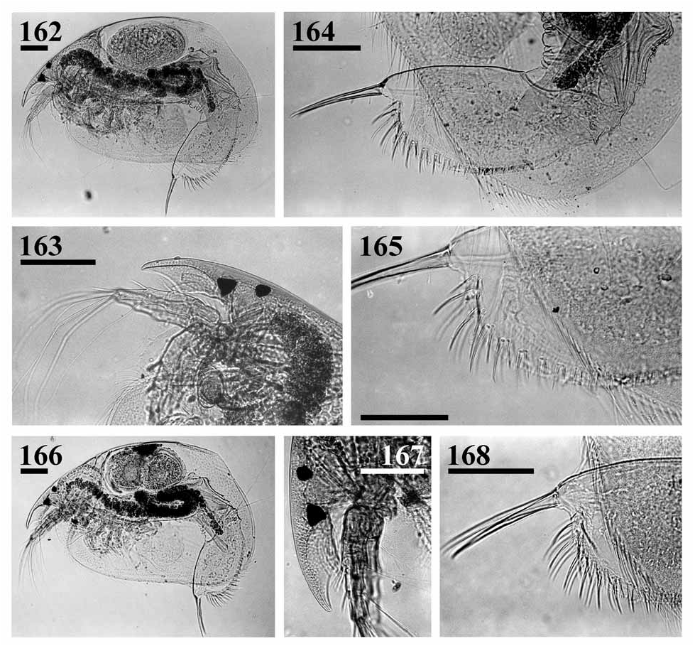
( Figs 162–193 View FIGURES 162–168 View FIGURES 169–182 View FIGURES 183–193 )
Leydigia microps Sars, 1916, p. 330 View in CoL –331, Pl. 38: figs 2, 2a–b; Smirnov 1971, p. 460, figs 571–572.
? Leydigia microps Sars View in CoL in Harding 1961, p. 45
Type locality (according to newly selected lectotype). Green Point Common, Republic of South Africa. Sars (1916) did not specify a type locality. He hatched L. microps from "mud taken at three different localities, viz. Green Point Common, Klipdam, and neighbourhood of Bergvliet".
Lectotype (selected here). Green Point Common, the adult parth. ♀, 900 µm, on slide GOS F 9223.
Paralectotypes (selected here).
(1) Green Point Common, 4 parth. ♀♀, slide GOS F 9223 (well-preserved, on same slide as lectotype); 6 parth. ♀♀, slide GOS F 9229.
(2) Cape of Good Hope , 1 parth. ♀, slide GOS F 9224 (bad condition); 5 parth. ♀♀, slide GOS F 9226 (bad condition); 1 parth. ♀, slide GOS F 9227 (bad condition); 1 parth. ♀, slide GOS F 9228 (bad condition) .
Possible type material. Unknown locality, 2 ♂♂, slide GOS F 9225 (relatively bad condition).
Other material examined. Republic of South Africa: unknown locality, C. S.I. R ., Stream Survey , Val 150A, coll. 12.06.1956 by A. D. Harrison and B. R . Allanson , slide NHM no number, and tube NHM 1959.7 .9.124.
Diagnosis. Parthenogenetic female. Body triangular-ovoid; maximum height in the middle or in posterior half ( Figs 162, 166 View FIGURES 162–168 , 169–170 View FIGURES 169–182 ), dorsal margin slightly convex in posterior part, postero-dorsal angle well-defined, although rounded, posterior margin without a 'step' in ventral portion. Valves coarsely striated, especially in postero-dorsal portion. A distinct fine striation covers head shield and valve surface ( Figs 171, 174 View FIGURES 169–182 ). Head small, compound eye particularly small, ocellus 1.5–3 times larger eye in diameter ( Figs 163, 167 View FIGURES 162–168 ). Major and lateral pores on plate without striation, lateral head pores about 0.5 IP distance from midline, somewhat anterior to level of central pore ( Fig. 171 View FIGURES 169–182 ). Labral keel with well-defined apex, subtriangular, posterior margin with 3 groups of long setules, anterior margin with long setules, almost reaching apex, and groups of long setules located submarginally ( Figs 172–173 View FIGURES 169–182 ). On valves, setae at middle of ventral margin with long setulation, no setules between their bases ( Fig. 174 View FIGURES 169–182 ). Posterior marginal setae setulated distinctly asymmetrically, posteriormost seta short ( Figs 175–176 View FIGURES 169–182 ). Submarginal setules of posterior margin fine, of approximately similar size throughout, not organized into groups. No marginal membrane.
Postabdomen subquadrangular, robust ( Figs 164–165, 168 View FIGURES 162–168 , 177–180 View FIGURES 169–182 ), preanal margin very short, somewhat undulated, preanal and postanal angle well-defined. Postanal margin from slightly convex to slightly concave, distal margin from well- to ill-defined. Postanal marginal denticles in about 15 clusters, 2–3 series of marginal denticles on anal margin ( Fig. 181 View FIGURES 169–182 ). About 9–13 fascicles of stout lateral setae, up to 4 setae in each fascicle distally, 2 setae in each fascicle in middle of margin, marginalmost setae significantly larger than second seta. About 6–11 fascicles of lateral setules on basal half of postanal and anal margin. Postabdominal claw long, slightly curved, 0–3 setules at base, no basal spine ( Figs 165 View FIGURES 162–168 , 182 View FIGURES 169–182 ).
Antenna I thick, not reaching tip of rostrum, with 4 transverse rows of long setules on anterior face, and two fascicles of fine, long setules on posterior face, no setules at tip ( Fig. 183 View FIGURES 183–193 ). Sensory seta arising close to distal end. Largest aesthetasc markedly longer than half of length of appendage, reaching tip of rostrum in some specimens. Antenna II with 3 stout spine-like setules on first, and 2–3 of these setules on second endopod segment ( Fig. 184 View FIGURES 183–193 ). Apical swimming setae without chitinous insertions within distal segments. Basal and distal lateral setae subequal in size, similar in size to apical setae.
Trunk limb I ( Figs 185–186 View FIGURES 183–193 ) with ODL large, bearing series of fine setules and a long seta with unilaterally setulated distal segment. IDL with 3 marginal clusters of robust setules, first seta small, naked; second and third setae unequal in length, with similar setulation. Endite 3 with three soft setae (a–c) and long, bent seta 1, small receptor near its base; endite 2 with one short (d) and two long (e–f) soft setae unequal in length, and small seta 2, no sensillum near base; endite 3 with three soft setae (g–i) and short seta 3. Two ejector hooks of similar size. Trunk limb II with exopodite ovoid, small, with a group of long, fine setules. Distalmost scraper (1) with naked basal segment, on distal lobe with basal group of long setules ( Fig. 187 View FIGURES 183–193 ). Trunk limb III exopodite sub-rectangular, with seven setae, setae 1 and 2 with distal segment asymmetrically setulated, setae 3–4 rudimentary ( Figs 188–189 View FIGURES 183–193 ). Trunk limb IV with subovoid exopodite, with six setae, setae 1 and 2 relatively long ( Fig. 190 View FIGURES 183–193 ). All setae of filter plate with inflated tips. On inner face of limb V, two setulated setae of subequal length, basalmost slender, with a hillock near base ( Fig. 191 View FIGURES 183–193 ). Distal armature of gnathobase as a lobe, two setae in 'filter plate'.
Ephippial female. Unknown.
Adult male. Only 2 males on a slide, in relatively bad condition and somewhat deformed, were studied, but not in detail ( Figs 192–193 View FIGURES 183–193 ). According to Sars' (1916) pictures and description, body subtriangular, dorsal margin straight in posterior 2/3 of length, head large, rostrum as in female, eye smaller than ocellus. Postabdomen elongate, significantly narrowing distally, with inflated base for penis and postabdominal claws, ventral margin almost straight. Preanal margin short, pre- and post-anal angles ill-defined, postanal margin with distinct concavity, a dorso-distal angle present, but rounded, a short distal margin defined. Marginal denticles reduced, lateral setae occupy distal 2/3 of postanal portion, these long, robust, organised in groups of 2 in distalmost portion, singular in middle, and small, in clusters more basally. Postabdominal claws slender, almost straight. Penis slender, somewhat shorter than claw, almost straight. Antenna I, trunk limb I not studied.
Size. Lectotype 900 µm, juvenile and adult parthenogenetic females from Green Point Common 560–900 µm (n = 5), adult male, 542 µm (n = 1); juvenile and adult parthenogenetic females from unknown locality ("Stream Survey") 470–640 µm (n = 5), Sars (1916) reported length up to 0.86 mm.
Differential diagnosis. L. microps is an animal with a series of unique traits among all well-studied species: (1) on labral keel, lateral fascicles of setae located on anterior margin; (2) no marginal membrane on posterior margin of valve; (3) antennular sensory seta located very near distal end; (4) on limb I, seta 1 on endite 3 long; (5) on limb III, setae 1 and 2 with distal segments setulated specially asymmetrically; (6) on limb IV, setae 1 and 2 longer than in relatives; (7) male postabdomen strongly widening distally and with singular lateral setae instead of fascicles. Also, there are 7 setae on exopodite III, a state otherwise found only in L. propinqua , which is very different from L. microps . Also, only L. macrodonta has a similar distal portion of the postabdomen, though its postabdomen has a more convex ventral margin and a long preanal margin.
Taxonomical comments. L. microps has never been redescribed from original material since its description by Sars (1916). In his revision Smirnov (1971) reproduced illustrations from Sars, and an extract from his description. This species has been reported several times from other South African localities (e.g. Harding 1961), but I am not sure that authors determined this species according to Sars's understanding.
Distribution. Relatively rare: known only from southernmost Africa.


| R |
Departamento de Geologia, Universidad de Chile |
| NHM |
University of Nottingham |
No known copyright restrictions apply. See Agosti, D., Egloff, W., 2009. Taxonomic information exchange and copyright: the Plazi approach. BMC Research Notes 2009, 2:53 for further explanation.
|
Kingdom |
|
|
Phylum |
|
|
Class |
|
|
Order |
|
|
Genus |
Leydigia (Neoleydigia) microps Sars, 1916
| Kotov, Alexey A. 2009 |
Leydigia microps
| Harding, J. P. 1961: 45 |
Leydigia microps
| Smirnov, N. N. 1971: 460 |
| Sars, G. O. 1916: 330 |