
Libanasa brachyura Karny, 1928
|
publication ID |
https://doi.org/ 10.11646/zootaxa.3946.1.5 |
|
publication LSID |
lsid:zoobank.org:pub:5E11419C-46F9-49DF-8ED9-08340F8740E5 |
|
DOI |
https://doi.org/10.5281/zenodo.6098567 |
|
persistent identifier |
https://treatment.plazi.org/id/03ED9175-FFA5-FF95-18C5-E0A1EA00FD68 |
|
treatment provided by |
Plazi |
|
scientific name |
Libanasa brachyura Karny, 1928 |
| status |
|
Libanasa brachyura Karny, 1928 reinstated species
Libanasa brachyura Karny, 1928
Libanasa signatus: sensu Johns, 1997
Material examined. Holotype ♀ nymph. labelled (1) Libanasa brachyura det Karny Type (in Karny's hand) (2) Coll. Karny (handwritten) (3) Coll. Karny (printed) ( NHMW) Dar es Salaam, Tanzania. There is also a so called "allotype" which is a very small female nymph ( NHMW).
2 ♂♂, 1 ♀, 1 ♂ nymph, 1 ♀ nymph, Kazimzumbwi Forest Reserve, Kisarawe District, Tanzania 39o 3’ E, 6o 57’ S. Coll. FRONTIER Tanzania, Jan–Feb. 1991 ( ZMUC). 1 ♂ nymph, 3 ♀♀ nymphs, Kambai Forest Reserve, Muheza District, (Tanga Region), Tanzania. 38o 41’ E, 4o 59’ S. coll. FRONTIER Tanzania, Jan–Feb 1991 ( ZMUC). 1 ♂, 1 ♀, 1 ♂ nymph, 1 ♀ nymph, Tanzania, East Usambara Mountains, Zigi Trail, 4.97 S, 38.67 E, 450 m, wet forest, March 1999, August 2001, October 2006, December 2011 (collection C Hemp). 8 ♂♂, 10 ♀♀, 2 ♂ nymphs, Tanzania, West Usambara Mountains, Lutindi forest, 4.67 S 38.33 E, 1250 m, submontane forest, March 2014, October 2014, December 2014 (collection C Hemp).
Redescription male. Colour. —reddish-brown to paler golden brown on venter, darker over central vertex and central third of pronotum, posterior margins of all tergites slightly darker, more so in centre than at sides, these margins with faint indications of paler spots.
Body smooth, shining ( Fig. 1 View FIGURE 1 ). Setae sparse, except on the tarsi. Head and antennae.—Scape swollen on inner side, 2nd and 3rd units fused, the 3rd elongate, 21–23 basal antennomeres almost bare, others pilose. Mandibles: no ventral process extending ventrad beside labial base in front of lacinia base. Maxillary palps: MP4 swollen in distal 66–70% and for the most part the swelling is also pilose; MP5 fully pilose. Thorax and legs. —All nota with narrow marginal rim; ventral edge of mesepipleuron forming a weakly projecting rounded flap. Stridulatory organ: Male: T1 with ca 40 short points (or sharp pegs) scattered over lateral surface, smaller closer to pleura; T2 with ca 36 short points over posterior half of lateral surface; T3 with ca 40 short points over posterior half of lateral surface those close to posterior margin broader and aligned dorso-ventrally; T4 with ca 8 very small points, T5 with 1–4 points; T6–9 lacking points and all tergites without fine matt area associated with points as seen in other species; pleural surfaces with very few points; adjacent surface of hind femur smooth, lacking spinules or ridges (25x magn.).
Coxal spines narrow, sharp. Front leg: inner coxal margin with a single pale, thick swelling surmounted by a single long seta; femur lacking spines below; no apical spines; tibia with prominent, ovoid tympanum on both sides, and slightly swollen nearby; long, moveable, dorsal spine at ca 0.4x from base; 4 ventral pairs the proximal pair close to the distal apex of tympanum; 2 dorsal and 2 ventral apical spurs. Middle leg: inner coxal margin with a single pale, thick swelling surmounted by a single long seta; femur lacking spines below and at apex; tibia with a dorsal proximal pair of articulated spines, placed almost symmetrically, a single retrolateral spine before midpoint and another pair distad at ca 0.8; 2 dorso-apicals and 2 ventro-apicals; 4 ventral pairs placed almost symmetrically. Hind leg: inner coxal margin with two pale, thick swellings, the inner about twice the size of the outer, each surmounted by a single long seta; femur broad at base narrowing to a long almost cylindrical neck, the ventral ridges being reduced; 12 or 13 lateral "muscle" or "chevron" ridges, ca 21 ventro-lateral visible muscle block insertion patches; ventral ridges poorly developed, lack spines and have few setae; single small retrolateral apical genual spine; tibia with weak dorsal ridges, the 10–11 non-articulated, spines small, sharp and entirely lacking insertion rings, set along distal 2/3rds of tibia, arranged irregularly in pairs; apical spurs fully moveable within insertion rings; dorsal subapical pair relatively short, prolateral apical slightly shorter than metatarsus, retrolateral apical reaching midpoint of 2nd tarsomere; short ventroapical pair and even shorter subapical ventral pair ( Fig. 4 View FIGURE 4 ); usually 1 ventral spine just beyond midlength. Prosternum: presternite indicated by a field of setae, basisternite weakly bilobed; furcosternite broad, triangular, the pit at each lateral corner; postfurcasternite bipartite, weakly sclerotised. Mesosternum; spinasternite small, free; presternite weakly sclerotised, setose; basisternite with pair elongate blunt cones reaching almost the length of adjacent coxa, each with a single, long seta; furca and spinasternite fused, forming a sclerotised cavity, presumably fusing with the postfurcasternite (if present).
Metasternum: spinasternite fused with mesosternite (see above); presternite distinct, inset into centre of basisternite; triangular, with median longitudinal depression; basisternite thus almost divided, with pair of narrower elongate blunt cones set slightly closer than mesosternal pair, each with a single, long seta; furcosternite deep with cavity, overhung by a slightly swollen median lobe on first abdominal sternite. Abdomen. - Cerci: ♂ and ♀ similar, long, thin and narrowing to a sharp, setose apex (i.e. no bare sclerotised tip).
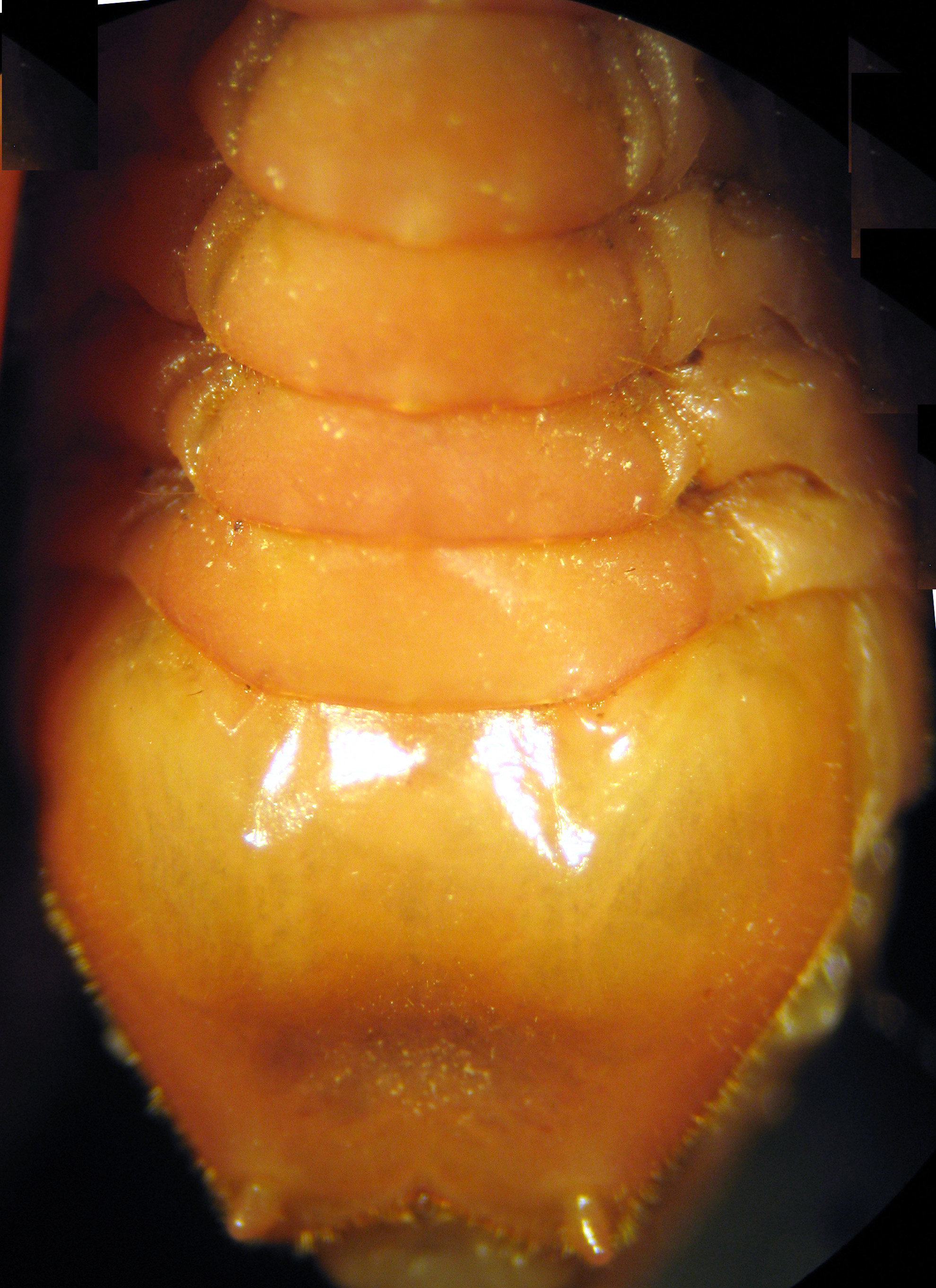
Male abdominal sternites: broadening distad ( Fig. 3 View FIGURE 3 ), smooth, each almost bare over 2/3rds of anterior surface, otherwise with fine, short setae. Terminalia: T9 with 2 slightly swollen and dark, sclerotised, submedian lobes, the falci of T10 widely separated and hooked over these lobes ( Fig. 2 View FIGURE 2 ); supra-anal plate simple, semi-lunar, its margin slightly thickened; paranal plates relatively short, each apex turned dorsad and with two very short spinous hooks: subgenital plate massive ( Fig. 3 View FIGURE 3 ), 2.5x length of adjacent sternite, broad at base the corners close to tergite, extending as a rounded bulbous plate that is weakly emarginate in centre, styles very short, not extending beyond margin of plate; fine short setae over entire lateral and posterior margins and extending onto adjacent surfaces, the central surface almost bare; inner walls of endophallus can expand and expose numerous short, narrow sclerotised plates which have 3–6 very fine points on their surface ( Fig. 2 View FIGURE 2 ), its dorsal surface with many irregular transverse rows of pointed spicules.
Female. Stridulatory organ: far fewer and sparser pegs. Tracheal spiracles: pronotal subcircular, closed by three subequal lobes, anterior, posterior and ventral; mesonotal ovoid, with two lobes. 1st abdominal enlarged closed by two apparently weakly sclerotised lobes which have scattered sclerotised points these also over the adjacent pleural surfaces; other abdominal spiracles nearly equal in size circular or slightly ovoid, the posterior two (7th & 8th segments) within a notch formed by a small extension of adjacent tergite behind spiracle.
Female subgenital plate massive, basal corners close to T8, narrowing to a broad, weakly bilobed plate, the median emargination short, inner ridge weak lacking a median extension that would fit in the triangular space at the base of the ovipositor; dorsal and ventral valves of equal length, smooth-edged, gently curved; paired inner valves almost as long; dorsal valve with setae over dorsal half of internal surface. The adult female has a median sclerotised strip separate from the margin of the sixth sternite and adjacent pocket, yet appears to lack a pair of glands. Two presumed juvenile females (penultimate instar ovipositor ca 17 mm) have the ventral valves distinctly shorter than the dorsal valves but both pairs appear fully sclerotised. Also under the margin of the 6th sternite there are pair of sclerotised rings and blackened internal structure suggestive of a gland. No other features indicate that these females could be of another species. Egg: heavily sclerotised, dark red-brown, elongate oval, finely scuptured hexagonal surface.
Measurements. Lengths (large adults preserved, abdomen flexible or broken): body 35–40 mm, antennae 80– 85 mm, pronotum 10.5 mm, hind leg femur: 28 mm, tibia: 26 mm; tarsus: 9 mm; metatarsus: 3.7 mm; male cerci 9 mm, female cerci 10 mm, ovipositor 20–23 mm; egg: 5.0 x 1.8 mm. Three slightly smaller specimens are treated here as penultimate instars, but if not, they represent size variation within populations.
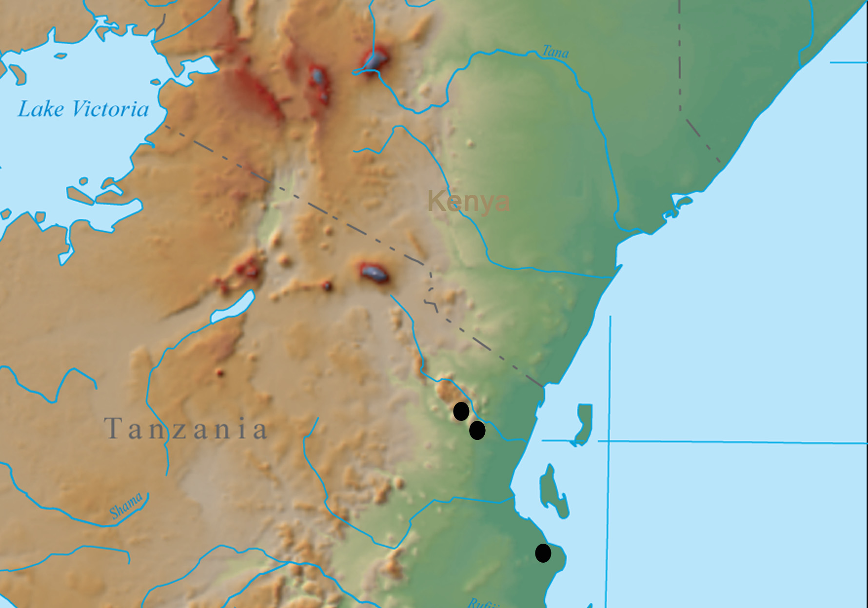
Distribution. Tanzania (West Usambara Mountains, East Usambara Mountains, coastal Tanzania between Tanga and Dar es Salaam) ( Fig. 7 View FIGURE 7 ).
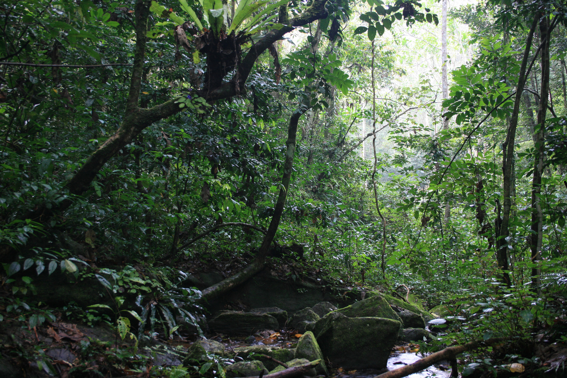
Habitat. Coastal, wet evergreen lowland to submontane forest ( Figs. 9 View FIGURE 9 , 10 View FIGURE 10 ).
Biology. Carnivorous night-active species. One proventriculus, of usual anostostomatid construction ( Maskell 1928; Judd 1948), contained a mass of fine irregularly broken, hyaline tubes, probably guard hairs from plant leaves, and numerous femora, tibiae, setae and macerated portions of bodies of lepidopteran or coleopteran larvae; three small (<1 mm), chewed heads with prominent hemispherical eyes were reminiscent of small lathridiids. Very little material which could be from soft plant tissue was present. Individuals are found among forest litter and on branches of understory vegetation up to 1–1.5 m. Mating. Males produce large spermatophores ( Fig. 8 View FIGURE 8 ) and stay together with females in mating position for a period of approximately 0.5 to several hours. Many adult specimens and several mating pairs were seen on the Zigi Trail in the East Usambara Mountains in December 2011. One female dissected held only five, deeply sclerotised eggs within the abdomen perhaps indicating that eggs are laid between then and February during the wetter season of the year.


No known copyright restrictions apply. See Agosti, D., Egloff, W., 2009. Taxonomic information exchange and copyright: the Plazi approach. BMC Research Notes 2009, 2:53 for further explanation.
|
Kingdom |
|
|
Phylum |
|
|
Class |
|
|
Order |
|
|
Family |
|
|
Genus |
Libanasa brachyura Karny, 1928
| Johns, Peter M. & Hemp, Claudia 2015 |
Libanasa signatus: sensu
| Johns 1997 |
Libanasa brachyura
| Karny 1928 |