
Marilia cunhaporanga, Camargos & Pes & Hamada, 2020
|
publication ID |
https://doi.org/ 10.11646/zootaxa.4853.1.1 |
|
publication LSID |
lsid:zoobank.org:pub:F4107225-9653-4407-BC4A-F6D9C26A9F93 |
|
DOI |
https://doi.org/10.5281/zenodo.4410246 |
|
persistent identifier |
https://treatment.plazi.org/id/DA608C21-6921-9F30-FF76-FF4CFCDCFB3E |
|
treatment provided by |
Plazi |
|
scientific name |
Marilia cunhaporanga |
| status |
sp. nov. |
Marilia cunhaporanga sp. nov.
( Figs 10–13 View FIGURE 10 View FIGURE 11 View FIGURE 12 View FIGURE 13 , 29 View FIGURE 29 )
Diagnosis: Marilia cunhaporanga sp. nov. is similar to M. crea Mosely 1949 in their contiguous eyes and the lateral sutures of segment IX forming a large median plate in lateral view. However, the 2-4-2 tibial spur formula of M. cunhaporanga sp. nov. differentiates it from M. crea , which has a 2-4-4 formula.
The larvae are similar to those of M. manicorei sp. nov. by the dark line pattern on the frontoclypeal sutures. However, the presence of much pigmentation on the thoracic sclerites and the dark spots on the abdomen differentiate M. cunhaporanga sp. nov. from M. manicorei sp. nov., which has no distinct thoracic or abdominal pigmentation or spots.
Adult: Male forewings each 3.7 mm long (holotype, approximate length, wings not fully expanded) and 6.5 mm (paratype). Body and wings dark brown in alcohol.
Head: Eyes very large in males, almost touching dorsally ( Fig 10a View FIGURE 10 ). Pair of vertexal mediantennal compact setose warts fused, with elliptical shaped; par of vertexal lateroantennal warts small not defined by sutures; pairs of occiptal warts and posterior warts not visible ( Figs 10a, 10b View FIGURE 10 ). Antennae long, about two times as long as body, with narrow annuli; scapes broad, pale basodorsally and basolaterally, covered with light setae. Front pubescent, covered with light setae, pair of frontogenal warts, short and kidney-shaped ( Figs 10c, 10d View FIGURE 10 ). Maxillary palps well developed, 5-articulated, heavily covered with setae. Labial palpi 3-articulated, articles subequal, covered with setae.
Thorax: Prothorax less than half as long as head; pronotum with pair of transversely elongate setal warts. Mesothorax broad, mesonotum without setae and with median longitudinal dark line reaching mesoscutellum, mesoscutellum almost circular and with pair of setal warts and small posterior depressions. Metathorax half as long as mesothorax, without setae. Tibial spur formula 2-4-2; external spurs of mid- and hind tibiae shorter than internal spurs. Forewings each with apical fork I arising on apical third of discoidal cell, R1 and R2 merging before wing margin ( Fig 10e View FIGURE 10 ). Hind wings each with fork I arising on apical eighth of discoidal cell, R1 and R2 merging about one-third length of R2 from wing margin; anal lobe with brush-like tuft of long setae ( Fig 10f View FIGURE 10 ).
Abdomen: Simple, without different structures.
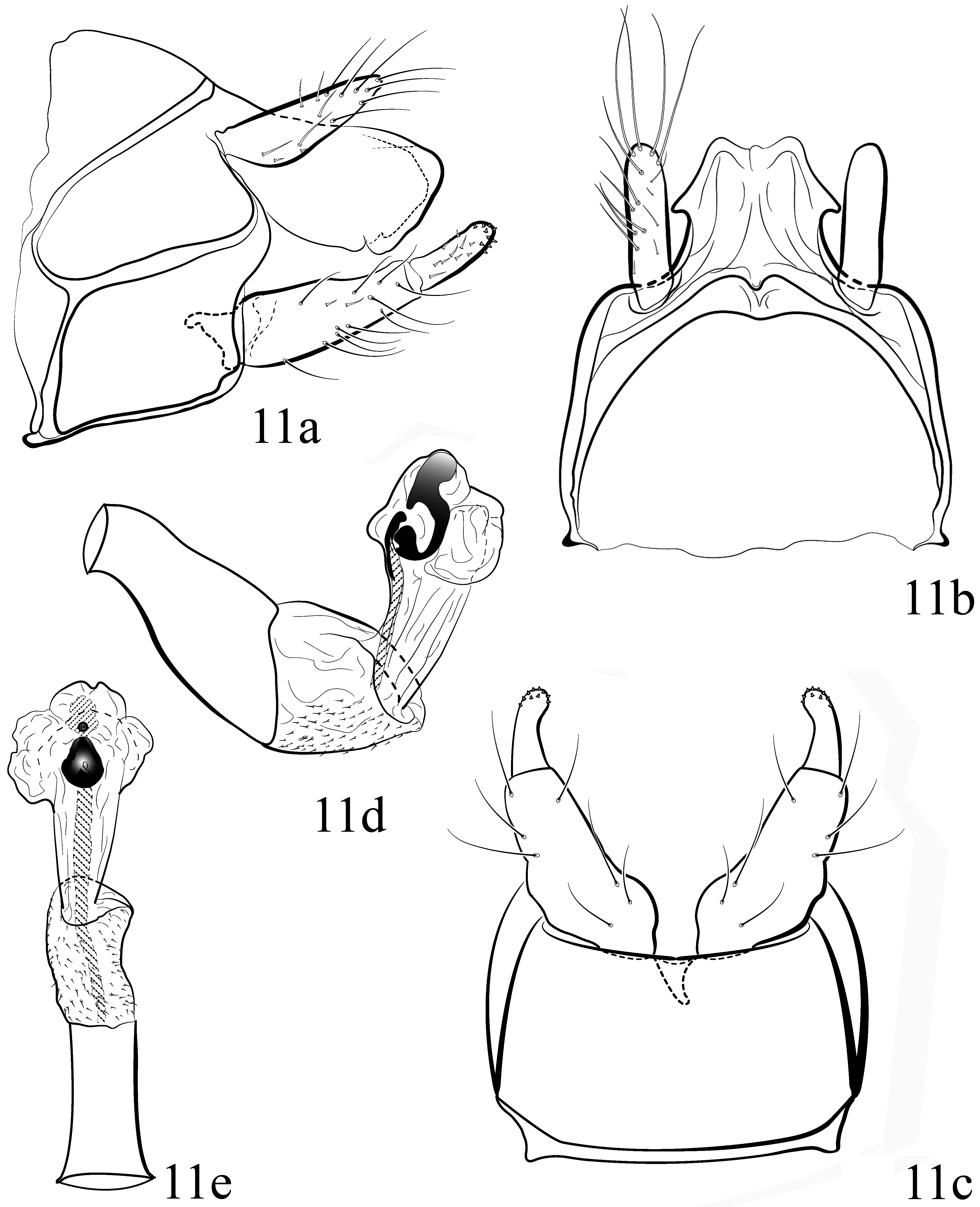
Male genitalia: Segment IX, in lateral view with anterior margin incised and posterior margin slightly projected at midheight; midlateral sutures separating each side of segment IX into 3 parts, ventral plate occupying almost half of height of segment IX ( Fig 11a View FIGURE 11 ); dorsal part not projected over segment X ( Fig 11b View FIGURE 11 ). Preanal appendages, in lateral view short, tapering to pointed apex ( Fig 11a View FIGURE 11 ); finger-like, in dorsal view, with base almost as broad as apex, with numerous setae ( Fig 11b View FIGURE 11 ). Segment X with apex subrectangular in lateral view ( Fig 11a View FIGURE 11 ); sub-quadrate, slightly broader at base, apex arrow-like and with median notch in dorsal view ( Fig 11b View FIGURE 11 ). Inferior appendages with two articles, basal one cylindrical, with base as broader than apex, curved slightly mesad; apical one short, with small conical spines apically, in ventral view curved mesad with internal margin concave ( Fig 11c View FIGURE 11 ). Phallus tubular, in lateral view curved about 30 basally ( Fig 11d View FIGURE 11 ), in ventral view straight ( Fig 11e View FIGURE 11 ); endotheca membranous, with small conical spines; phallotremal sclerite hook-shaped in lateral view ( Fig 11d View FIGURE 11 ), subtriangular in ventral view ( Fig 11e View FIGURE 11 ).
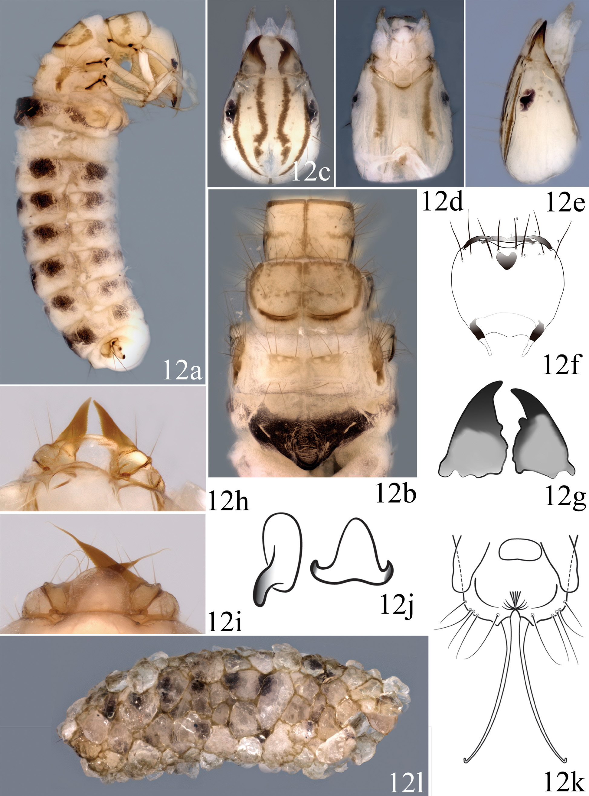
Final Instar Larva: Body length 4.5–6.3 mm (n = 4). Background color of sclerites yellow and abdomen light gray with dark spots on each segment in alcohol ( Fig 12a View FIGURE 12 ).
Head: In dorsal view quadrangular, tapered anteriorly and broader behind midlength, yellowish gray with pair of distinct black lines mesal of carinae extending from eyes to juncture of sutures, frontoclypeus with pair of dark, sinuous submesal lines but with posterior ends of frontoclypeal sutures and short coronal suture unmarked ( Fig 12c View FIGURE 12 ); in ventral view with pair of longitudinal dark brown lines not reaching posterior margins, ventral apotome 4.0 times as long as broad and separating genae entirely ( Fig 12d View FIGURE 12 ). Eyes rimmed by light areas ( Fig 12e View FIGURE 12 ). Labrum with slightly concave apical margin and convex apicolateral margins broader than respective concave basal and convex basolateral margins; setae 1 and 2 on each side robust, with setae 3, 4, 5, 6 straight and of less rigid thickness ( Fig 12f View FIGURE 12 ). Mandibles robust, acute, and asymmetrical, left mandible longer than right mandible and with mesal tooth farther from apex ( Fig 12g View FIGURE 12 ).
Thorax: Pronotum yellow, with setae on anterolateral corners, anterior margin dark and slightly concave, with pair of transverse black lines laterally at midlength of each sclerite and not reaching its midline or lateral margin, and median longitudinal black line faintly reaching posterior margin. Mesonotum divided into 3 pairs of sclerites: Anteromesal sclerites (sa 1) yellow, with dark spots at anterior setal bases; posteromesal sclerites (sa 2) yellow, each with dark narrow area on posterior margin covering one-fifth of sclerite; lateral sclerites (sa 3) yellow with many setae and spots on anterior margins. Metanotum divided into 5 sclerites: Anteromesal pair (sa 1) yellow with setae and spots on anterior margins; lateral pair (sa 3) brown with setae on anterior and lateral margins; posteromesal pair (fused sa 2 sclerites) yellow, elongate transversally, straight, with few lateral setae ( Fig 12b View FIGURE 12 ). Legs yellow.
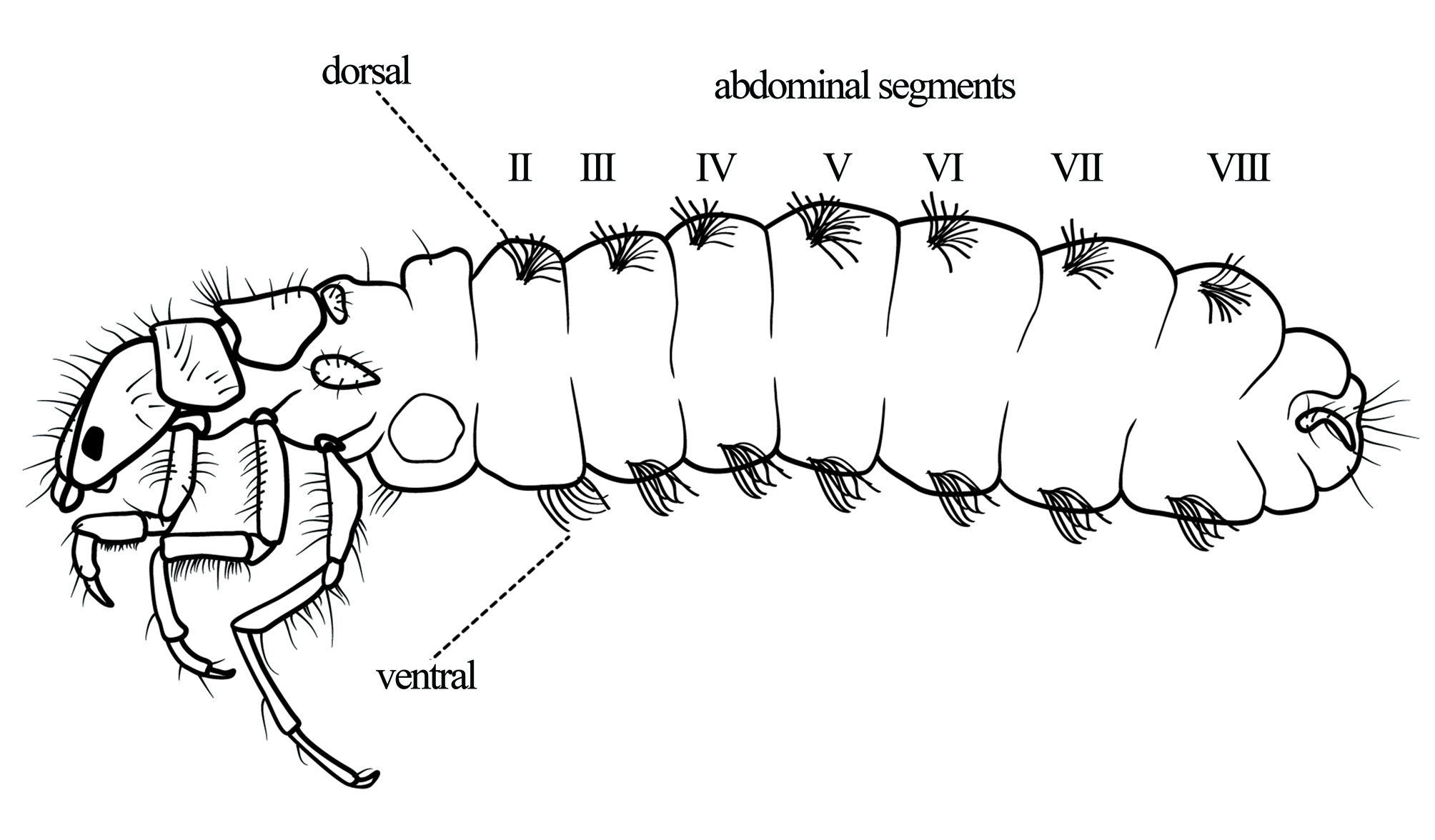
Abdomen: Abdominal gill formula as in Fig 13 View FIGURE 13 . Tergite IX subtriangular. Anal proleg without teeth on external margins of claws.
Pupa: Body length 6.4–6.5 mm (n = 2). Body brown in alcohol.
Head: Mandibles long, more than three times as long as wide, each with distal portion narrowed then extended in finely attenuated sclerotized filament ( Figs 12i View FIGURE 12 ), readily broken ( Fig 12h View FIGURE 12 ), serrate on entire internal margin. Labrum subquadrate, with lateral angles rounded ( Fig 12i View FIGURE 12 ).
Thorax: Mesotarsi each with fringe of long setae.
Abdomen: Segments III–VII each with pair of small oval anterior hook plates, each with one hook oriented posterad; segment V with pair of triangular posterior hook plates, each hook plate with 2 posterior hooks oriented anterad ( Fig 12j View FIGURE 12 ). Terminal processes long, slender, and divergent from base ( Fig 12k View FIGURE 12 ).
Case: Length 5.0– 6.6 mm (n = 4), composed of grains of coarse sand, slightly curved, nearly parallel-sided but broadening slightly from posterior to anterior ( Fig 12l View FIGURE 12 ).
Etymology: The specific epithet is a noun in apposition, referring to Cunhà-Poranga, which means “beautiful girl” in the native Nheengatú language, the most beautiful woman in the tribe, in reference to the remarkably beautiful larvae of this species.
Bionomics: This species is found mainly in pools of slowly flowing streams with widths of 1 to 5 m, depths of 0.15 to 1.5 m, with sand, leaves, and logs on the bottom. The larvae were found mostly in roots of riparian vegetation. We collected a considerable number of larvae.
Comments: The holotype is a pharate male removed from the pupal exuviae to better observe the genitalia; for this reason, we do not have illustrations of the holotype male wings. The wings were described and illustrated from the male paratype. We also observed that pupae of emerged adults had no apical attenuations on the mandibles ( Figs 12h, 12i View FIGURE 12 ), these possibly having been broken as the insects opened their cases.
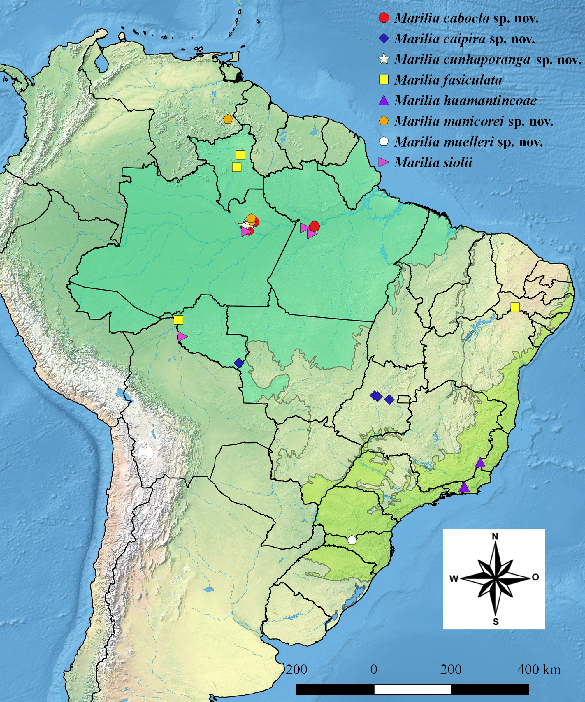
Distribution: BRAZIL: Amazonas ( Fig 29 View FIGURE 29 ).
Holotype pharate male: BRAZIL, Amazonas , Manaus: Km 18, BR 174 , 02°49’00.8”S 60°02’05.6”W, 07.iv.2009, A.M.O. Pes, J. Albino, R. Boldrini leg., pharate male and its pupal exuviae [alcohol] ( INPA-TRI 000070 ). GoogleMaps
Paratypes: Same data as holotype, except 3 pupae [alcohol] ( INPA-TRI 000071 ); Rio Preto da Eva , ZF 3, Fazenda Esteio , Igarapé do km 21, Ponte, PDBFF, 02°26’03”S 59°54’16”W, raiz correnteza, 12.xi.2003, A.M.O. Pes leg., 2 larvae [alcohol] ( INPA-TRI 000072 ). Same data as holotype, except larva collected 18.ii, molted to pupa 28.ii, emerged as adult 31.iii.2020, A.M.O. Pes & G. R. Desidério, leg. 1 male (pinned), and its pupal and larval exuviae [alcohol] ( INPA-TRI 000073 ) [wings illustrated] GoogleMaps .

| R |
Departamento de Geologia, Universidad de Chile |
No known copyright restrictions apply. See Agosti, D., Egloff, W., 2009. Taxonomic information exchange and copyright: the Plazi approach. BMC Research Notes 2009, 2:53 for further explanation.
|
Kingdom |
|
|
Phylum |
|
|
Class |
|
|
Order |
|
|
Family |
|
|
Genus |