
Medauroidea extradentata (Brunner von Wattenwyl, 1907)
|
publication ID |
https://doi.org/ 10.5531/sd.sp.55 |
|
DOI |
https://doi.org/10.5281/zenodo.7733269 |
|
persistent identifier |
https://treatment.plazi.org/id/038D8781-FF9C-20C4-FCDD-FDB5A37EF84F |
|
treatment provided by |
Felipe |
|
scientific name |
Medauroidea extradentata |
| status |
|
Medauroidea extradentata View in CoL View at ENA
“Vietnamese walking stick”
Figures 91 View FIGURE 91 , 92 View FIGURE 92 (lateral, anterior, posterior); 93 View FIGURE 93 , 94 View FIGURE 94 (dorsal, anterior, posterior); 95 View FIGURE 95 , 96 View FIGURE 96 (ventral, anterior, posterior)
Plates 55 (lateral), 56 (dorsal), 57 (ventral)
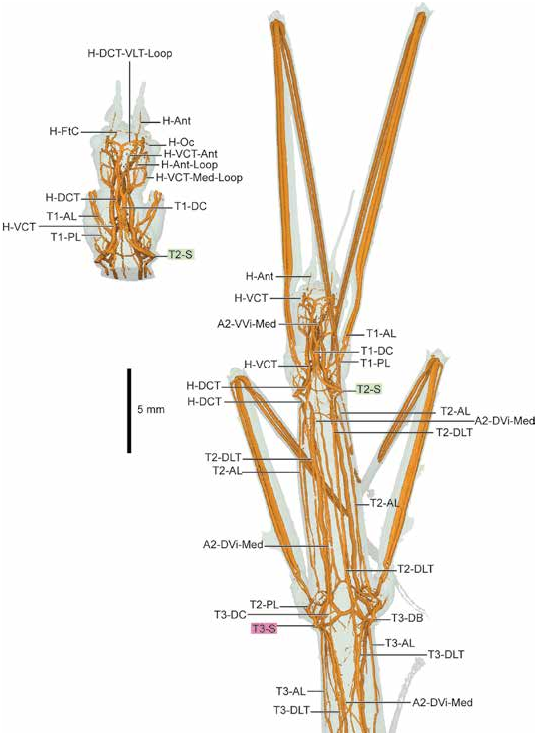
Although Strauss (2021) conducted the most recent work on stick insect tracheae, he focused on the prothorax and prolegs, concentrating on hearing. He employed terminology from Ander (1939), also incorporated here, although he left several branches unlabeled. In the Medauroidea scan here, T1-AL and T1-PL remain separate, at least as far as the distal end of the foretibia. Strauss indicates foreleg (and mid- and hind leg) adaptations for hearing; these cannot be verified from this scan.
Medauroidea is a good example of assessing homology using secondary criteria (serial homology). The T2-DB branching pattern posteriorly in Medauroidea is somewhat ambiguous, in particular the placement of T2-AL and T2-Wbr. T2-AL and T2-Wbr could be swapped; however, the decision that the dorsal one is T2-AL is based on serial homology with T3. Additionally, the shorter DB branches, such as T3-DB and T3-VB here in Medauroidea (but also applicable elsewhere), could be argued to be branching directly from the spiracle. The presence or absence of a spiracular “atrium” is not specific here; these structures are most prominent in groups such as Orthoptera , where a large “cavity” sits just inside the spiracle with multiple tracheae branching in various directions. In Medauroidea , it appears that a very short “stub” of DB or VB may have trachea branching from it. T2-CT may be present, but this stub may also be a spiracular atrium, as the closest (phylogenetically) relative with T2-CT is Plecoptera (rather distant).
The tracheal branching in first abdominal segment is very unusual and assessing its homology calls for some explanation. A1-DLT is quite clearly absent, as no tracheae arc posteriorly in the form of a DLT; T3-DLT connects directly to the dorsum of A1-S with no continuation. A1-VLT initially appears to be A1-MLT, but the ventral branch of T3-VL into the hind leg is indicative of it being a VLT, not MLT. Additionally, A1-VC branches from T3-VL, rather than A1-VB, an unusual arrangement. This branch is not a section of A1-VB, as A1-VLT connects A1-S and A2-S. The remaining branches are typical.
The second abdominal segment is also slightly modified from the remaining segments extending posteriad. A2-DB and A2-VB are basically absent—while A2-DC and A2-VC are present, they branch directly from A2-DLT and A2-VLT. The primary connection between A2-S and A1-S anteriad is A1-VLT. A2-DC extends a little anteriad of A2-S; A2-DC could arguably be A1-DC but the branching pattern is from A2-DLT, so homologizing based on connectivity or branching pattern seems more reasonable.
DESCRIPTION: HEAD: The head tracheal morphology of M. extradentata features a network of loops interconnecting both dorsoventrally and laterally. Exploration of the 3D models in the supplementary digital data is encouraged. Three sets of tracheae into head: H-DCT, H-VCT, and additional H-VLT. H-DCT dorsad, proceeding anteriorly and forming a prominent H-DCTVLT-Loop anteriad and ventrad, connecting directly with H-VLT. H-Lbm anteriad from ventral apex of H-DCT-VLT-Loop. H-VCT runs anteriad, dividing into H-Ant and two branches, one looping posteriad to connect with H-VCT, forming H-Ant-Loop; second branch looping ventrad and posteriad to join H-VLT.
THORAX: T2-S with four connections: possible T2-CT, T2-DB, T2-VB, and T1-PL. T2-CT short and running directly anteriad, bifurcating into H-DCT and H-VCT near posterior margin of prothorax; as T2-CT absent in other Phasmatodea , this T2-CT is possibly a deeper spiracular atrium rather than T2-CT. H-DCT runs anteriad, extending through prothorax into head. H-VCT anteriad, with T1-AL splitting off into foreleg; short connection to T1-VLT at this branching point. T2-DB runs posteriad, curving slightly ventrad before splitting into two pairs: T2-DLT/T2-AL dorsal branch and T2-Wbr/ T2-lvl ventral branch; T2-AWL notably absent. For dorsal T2-DLT/T2-AL pair, T2-DLT as with other specimens, positioned along dorsum with connection posteriad to T3-DB; T2-AL extending posteriad, connecting with T3-S via T2-PWB. Ventral T2-Wbr/T2-lvl pair with T2-AL posteriad with shallow arc dorsad to connect with T2-PWL; T2-lvl along venter, connecting with T3-S via T2-VLT connection just anteriad of T3-S. T2-VB short and directly ventrad, linking with T2-VLT posteriad and T1-VLT anteriorly. Both T1-VC and T2-VC present. T1-PL runs anteriad, linking with T1-VLT via short T1-VL; T1-PL extends into foreleg without joining T1-AL. T1-AL and T1-PL do not join and remain separate at least until the distal end of the foretibia. Two small, visceral medial A1-DVi-Med and A1-VVi-Med extend through mesothorax, originating at A1-S and extending into head. T3-S with four branches: T2-VLT, T3-DB, T3-VB, and T2-PL. T2-VLT from anterior, connecting T2-S to T3-S. T3-DB short and mediad, quickly branching into T3-AL posteriorly and remaining T3-DB dorsad; T3-DB joining with T2-DLT anteriorly and T3-DLT posteriorly in Y-shaped junction, linking to T2-S and A1-S. T3-AL posteriad, joining with T3-PL to form T3-L, extending into hindleg. T3-VB short, similar to T3-DB, quickly splitting into T3-VLT posteriad and remaining T3-VB ventrad. T3-VLT runs directly posteriad to A1-S; T3-DC present toward posterior margin of metathorax. T3-VB continues to T3-PVC, which forms posteriad segment of hexagonal network comprised of T3-VC anteriad and lateral sections connected to T2-VLT. T2-PL anteriad, joining T2-AL and extending into mideg. T2-VL branching from T2-VLT, also extending into midleg; T2-VL and T2-L remain separate to distal end of midleg tibia. A2-DVi-Med and A2-VVi-Med extending through metathorax, with laterally asymmetric connections: A2-DVi-Med to T2-VLT on specimen’s left side, A2-DVi-Med to T2-VLT on specimen’s right side.
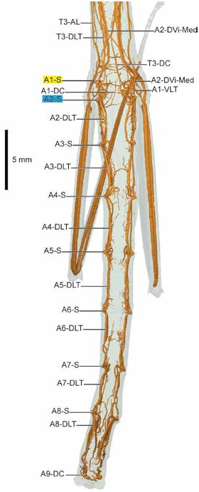
ABDOMEN: A1..8-S present. First abdominal segment approximately half as long as remaining abdominal segments. A1-S and A2-S branching patterns highly modified from remaining abdominal segments. A1-S with four branches: T3-DLT, A1-VLT, T3-VLT, and A1-PL. A1-S with additional A2-VVi-Med connection on left side only, see description of A2-VVi-Med below. T3-DLT runs dorsad, connecting in anterior arc from T3-S; A1-DLT notably absent. A1-VLT runs directly posteriad to A2-S; T3-VL directly ventrad from A1-VLT; A1-PVC branching from T3-VL. T3-VLT from anterior, with two ventral commissures A1-AVC1 and A1-AVC2. T3-PL ventrad, linking with T3-AL before arcing posteriad to extend into hind leg. A2-S with seven branches: A1-VLT, A2-DVi-Med, A2-VVi-Med, A2-DLT, A2-DVi, A2-VVi, and A2-VLT. A1-VLT runs directly anteriad from A1-S. A2-DVi-Med beginning as sinusoidal, looping branch, extending anteriorly and medially, each side combining in Y-shaped junction in metathorax and proceeding anteriorly along venter; A2-DVi-Med asymmetric, with sinusoidal form on both sides but only right side leading to Y-shaped join, with left side coming from A1-S. A2-VVi-Med similar, arranged along venter. A2-DLT runs dorsad and posteriad in arc connecting to A3-S; A2-DC present off A2-DLT. A2-DVi and A2-VVi ventrad, connecting with A3-S. A2-VLT likewise in ventral arc, also connecting with A3-S; four tracheae connect A2-S with A3-S. Remaining A3.. A8 segments similar, with varying degrees of A n -DB; A3-DB short, A4-DB not present, etc. A n -DLT and A n -VLT present on all, connecting segments longitudinally. A3..8-S with anteriad visceral tracheae generally dorsad, posteriad visceral tracheae generally ventrad. A4-Vi-VC present, formed from visceral tracheae and not from VLT as is typical.


No known copyright restrictions apply. See Agosti, D., Egloff, W., 2009. Taxonomic information exchange and copyright: the Plazi approach. BMC Research Notes 2009, 2:53 for further explanation.