
MICRONETINAE
|
publication ID |
https://doi.org/ 10.11646/zootaxa.3674.1.1 |
|
publication LSID |
lsid:zoobank.org:pub:981F80ED-96D7-40C7-8A3C-677954416A2E |
|
DOI |
https://doi.org/10.5281/zenodo.6162275 |
|
persistent identifier |
https://treatment.plazi.org/id/038D6700-FFB8-5612-118C-00C0AF0AB26A |
|
treatment provided by |
Plazi |
|
scientific name |
MICRONETINAE |
| status |
|
SUBFAMILY MICRONETINAE
Hull (1920) defined the subfamily Micronetinae by the absence of femoral and lateral spines, the absence of “auxiliary claws” and the presence of two, four or five promarginal teeth on the chelicerae. All these characters are very broad and can be found in large number of Linyphiidae . Hull (1920) included in the subfamily Micronetinae Agyneta , Microneta , Meioneta, Rhabdoria , Syedra and Aphileta .
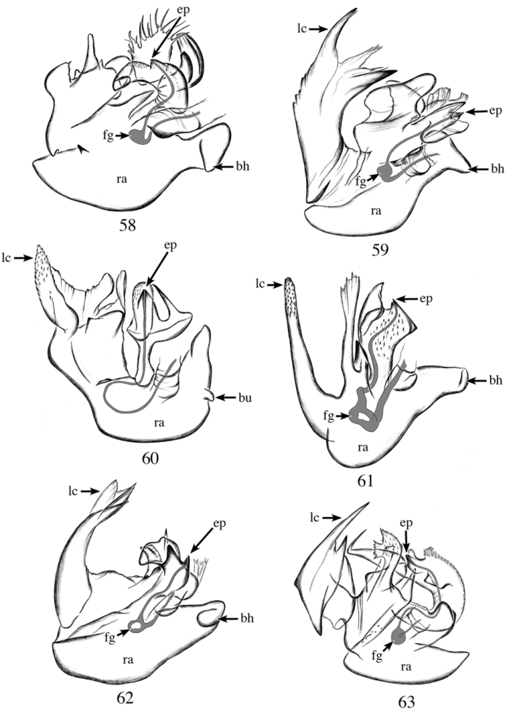
Saaristo & Tanasevitch (1996) redelimited the subfamily Micronetinae in reference to the male palp and the conformation of the epigynum. They gave a general description of the male palp based on Lepthyphantes minutus ( Blackwall 1833) to represent the conformation found in the subfamily Micronetinae . Saaristo & Tanasevitch (1996: 170) also mentioned the presence in the radix “at the site of the embolic base the sperm duct is dilated forming the so called Fickert’s gland”, and that “In certain cases this gland has been lost which sometimes is compensated by a secondary dilatation inside the embolus like for example in some Agyneta Hull, 1911 species”. I found that not all Agyneta (sensu Saaristo) have a Fickert’s gland, but when present it is always in the embolus. It seems unlikely that this corresponds to a secondary dilation, and I would argue that the Fickert’s gland has been displaced to the embolus, since its position within the radix varies greatly and it has shifted so far as to be in the base of the embolus in Poeciloneta variegata ( Blackwall 1841) ( Fig. 59 View FIGURES 58 – 63 ).
Saaristo & Tanasevitch (1996: 176) gave a short description “Micronetids may be recognized by the complex type of embolic division as described above. Also the presence of the outward pointing suprategular apophysis just below the tip of the cymbium in the unexpanded palp is often diagnostic” and included 54 genera in the subfamily Micronetinae . In studying the complex embolic division, Saaristo & Tanasevitch overlooked some important characters such as the position of the embolus in relation to the radix and the position of the embolus proper (when present). They did mention that the embolus proper is situated on the retrolateral side of the embolus in Lepthyphantes Menge 1866 , whereas it is on the prolateral side in Agyneta and Meioneta , and that in Microneta the sperm duct just ends at the tip of the embolus without an embolus proper. I re-studied some of the genera included in Saaristo & Tanasevitch’s (1996) study, based on the type species (except for Maorineta Millidge 1988 , as I did not have access to the type species Maorineta tibialis Millidge 1988 ). I examined the details of the male palp, particularly the embolus shape, morphology and its attachment to the radical division as well as the morphology of the radix.
No known copyright restrictions apply. See Agosti, D., Egloff, W., 2009. Taxonomic information exchange and copyright: the Plazi approach. BMC Research Notes 2009, 2:53 for further explanation.