
Henosferus molus, ROUGIER & MARTINELLI & FORASIEPI & NOVACEK, 2007
|
publication ID |
https://doi.org/ 10.1206/0003-0082(2007)507[1:NJMFPA]2.0.CO;2 |
|
persistent identifier |
https://treatment.plazi.org/id/03E187D0-FFA4-FFB6-FC84-FCE2FEB6FD48 |
|
treatment provided by |
Carolina |
|
scientific name |
Henosferus molus |
| status |
sp. nov. |
Henosferus molus , new species
ETYMOLOGY: From the Latin mola, meaning millstone in reference to the well-developed talonid of the lower molars.
DIAGNOSIS: As for the genus, for monotypic attribution.
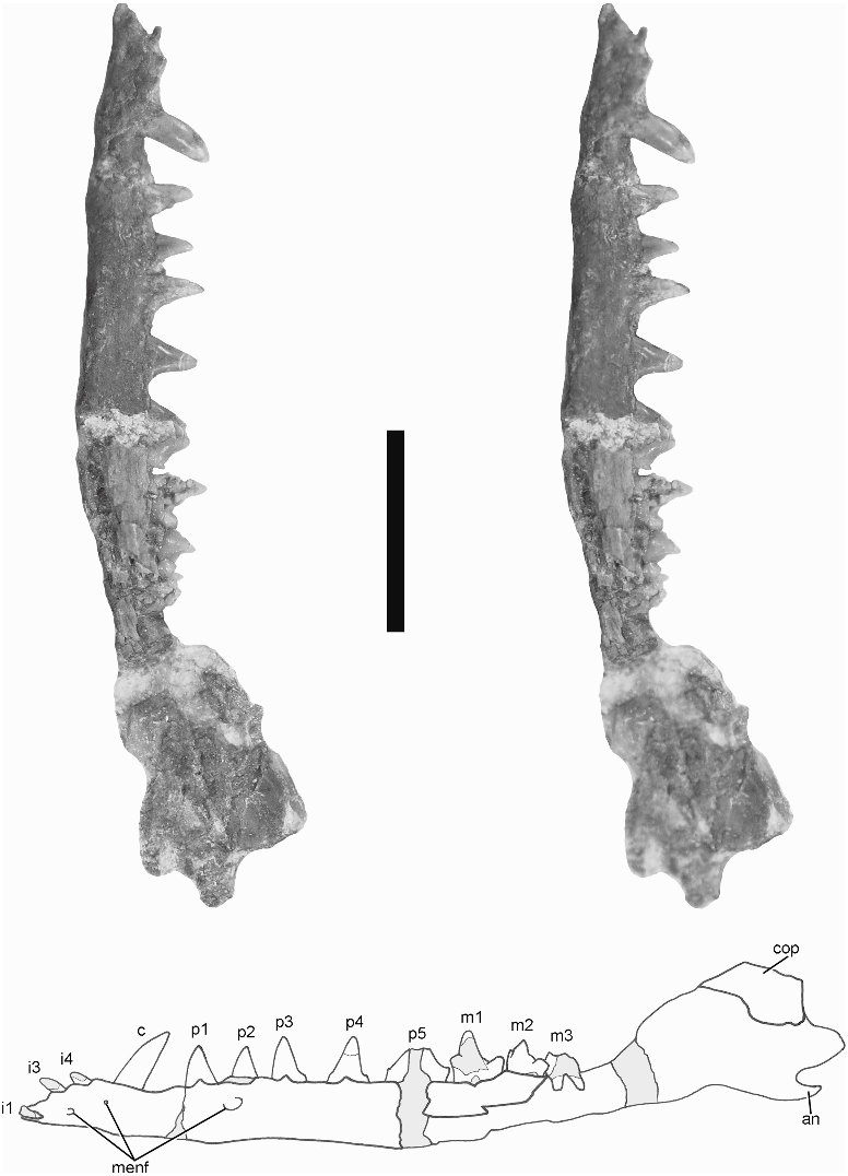
HOLOTYPE: MPEF 2353 : Right lower jaw with the dentary well preserved, bearing the distal half of p1 and p2, and almost complete m1 (fig. 2). The posterior portion of the dentary shows a deep trough and a medial flange; it also bears a deep and small coronoid facet, a remarkable Meckelian groove, and a transversely wide angular process. This specimen is chosen as the holotype because it preserves an almost complete molar that bears numerous diagnostic features, making it possible to compare with most other Mesozoic mammals.
HYPODIGM: MPEF 2354: Left lower jaw bearing the roots of the first and second incisors, the broken crown of the third and fourth incisors, a complete canine, five premolars, and three damaged molars ( fig. 3 View Fig ). Behind the level of the coronoid process, the dentary is partially broken and preserved mostly as a natural cast of the medial aspect. MPEF 2357: Left lower jaw exposed in labial view preserving the canine, four premolars, and a molar with the trigonid mostly damaged ( fig. 4 View Fig ). Floating in the matrix near the jaw are two teeth here identified as p1 and m2 (not shown in figures). The p1 is complete but the m2 is missing the protoconid and a small
Fig. 2. Henosferus molus holotype MEFP 2353 . Stereophotograph of the right lower jaw in lingual view and accompanying line drawing. Gray pattern indicates broken bone and matrix. Scale bar is 5 mm. Abbreviations: an 5 angular process; con 5 condyle; cop 5 coronoid process; cor 5 scar for the paradentary coronoid bone; dt 5 dentary trough; i1r 5 root of the lower first incisor; m1 5 lower first molar; mck 5 Meckelian groove; mf 5 mandibular foramen; mfl 5 medial flange; p1 5 lower first premolar; p2 5 lower second premolar; sym 5 mandibular symphysis. The p1 and p2 are broken, preserving only the distal halves of their crowns; the premolars appear thus to be like single-rooted but they are in fact double-rooted .
portion of the talonid. The posterior part of the dentary is complete, adding information on the lateral view of the jaw not accessible in the other specimens.
TENTATIVELY REFERRED SPECIMEN: MPEF 2355: isolated upper premolar enclosed in a small block, which also includes indeterminate fragments of bone (not figured).
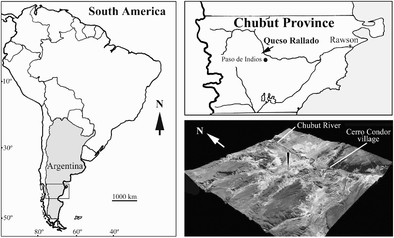
LOCALITY AND HORIZON: All specimens come from the Queso Rallado locality (43 ° 24 9 33.55 0 S/69 ° 13 9 50.1 0 W), about 3 mi west-northwest of the village of Cerro Condor, Chubut Province, Argentina ( fig. 1 View Fig ); Puesto Almada Member ( Silva Nieto et al., 2003), Cañadón Asfalto Formation, Callovian- Oxfordian ( Stipanicic et al., 1968; Tasch and Volkheimer, 1970).

No known copyright restrictions apply. See Agosti, D., Egloff, W., 2009. Taxonomic information exchange and copyright: the Plazi approach. BMC Research Notes 2009, 2:53 for further explanation.
|
Kingdom |
|
|
Phylum |
|
|
Class |
|
|
Family |
|
|
Genus |