
Multumbo dimorphicus, B, Marcio & Kury, Adriano B., 2007
|
publication ID |
https://doi.org/ 10.5281/zenodo.178288 |
|
DOI |
https://doi.org/10.5281/zenodo.6244137 |
|
persistent identifier |
https://treatment.plazi.org/id/1A51F37A-9C25-901F-DBBF-FF0107E2FC17 |
|
treatment provided by |
Plazi |
|
scientific name |
Multumbo dimorphicus |
| status |
sp. nov. |
Multumbo dimorphicus View in CoL sp. nov.
(figs. 1–9)
Type material. Santa Maria Madalena (Parque Estadual do Desengano, Serra da Rifa, 650–800 m, -21.9534 - 41.9530), A.B. Kury, A.P.L. Giupponi & M. Baptista, III/1998, 1 ɗ holotype and 2 Ψ paratypes ( MNRJ 17383); Macaé (Área de Proteção Ambiental do Sana, -22.4000 -42.1833), D. Pedroso & A. Pérez, VII/2002 1 ɗ and 2 Ψ paratypes, ( MNRJ 11353).
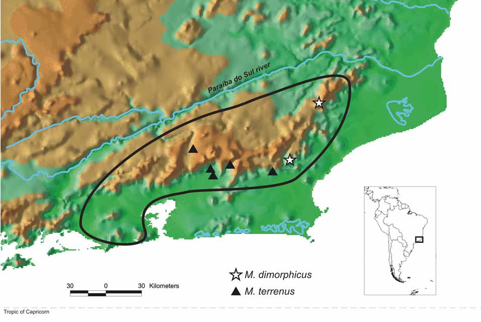
Distribution ( Fig. 10 View FIGURE 10 ). Brazil. Rio de Janeiro State. Macaé (Sana) and Santa Maria Madalena. WWF Ecoregion NT0160 (Serra do Mar coastal forests); vegetation type is Tropical & Subtropical Moist Broadleaf Forests.
Etymology. From the Greek, referring to the sexual dimorphism in leg IV, which is unusual in this genus.
Diagnosis. This species is only distinguished from Multumbo terrenus by the armed leg IV in males; females seem to be identical. Both species present similar morphological variation, as described below. Therefore, most morphological variation in the genus that is not related to secondary sexual dimorphism represents non-fixed polymorphism spread throughout the populations of both species.
Description of male holotype. Body and legs densely granular and covered by camouflage of dirt, except anterior portion of prosoma, pedipalps, chelicerae and stigmatic and genital areas. All granules and tubercles rounded, spines only present on femur IV of male ( Figs. 1, 2 and 7 View FIGURES 1 – 7 ). All setae short and strong ( Fig. 4 View FIGURES 1 – 7 ), except on pedipalps and chelicerae.
Dorsum. Anterior margin of dorsal scute with a pair of paramedian, acuminate, apophyses, which are convergent but do not meet ( Fig. 1 View FIGURES 1 – 7 ); corners of anterior margin with 2 conical apophyses larger than the central ones, more external apophysis twice as large as internal one ( Fig. 1 View FIGURES 1 – 7 ); on one side there is a more external and smaller apophysis. Ocularium with a pair of very strong parallel tubercles of same height as ocularium, slightly pointed frontward ( Figs. 1–2 View FIGURES 1 – 7 ). Anterior portion of prosoma lacking camouflage and granulation limited by a long, oblique elevation between ocularium and lateral margin of prosoma, forming a sub-anterior margin; this margin with a strong posterior tubercle and 1 or 2 smaller anterior tubercles ( Figs. 1–2 View FIGURES 1 – 7 ). Single opening of scent glands on each side. Prosoma behind ocularium with a pair of main large tubercles, those of area I further apart, and those of area II–III larger than the others; areas I–III with an additional pair of paramedian tubercles similar in size to the main prosomal tubercles ( Figs. 1–2 View FIGURES 1 – 7 ). Denser granulation close to grooves and in 2 median longitudinal rows (giving the appearance of a longitudinal sulcus) in areas of mesotergum and on prosoma behind ocularium ( Figs. 1–2 View FIGURES 1 – 7 ). Lateral margin with less dense granulation, denser on prosoma than on mesotergum; 6 larger tubercles present on widest portion of dorsal scute, close to area III, one of them very large ( Fig. 1 View FIGURES 1 – 7 ). Posterior margin of dorsal scute and free tergites with a row of tubercles and some sparse granules; larger tubercles on median portion and on posterior segments; 8 tubercles on posterior margin, 9 on free tergite I, 7 on tergite II and 5 on tergite III ( Fig. 1 View FIGURES 1 – 7 ).
Venter. Granules similar in size to those of dorsal scute, arranged in a row on free sternites and spread homogeneously on coxae, except for more proximal portion, which is smooth; stigmatic and genital areas with sparser granulation; genital operculum granular. Coxae I–III with a row of longitudinal tubercles decreasing in size from I to III. Proximal portion of coxae, stigmatic and genital areas, and genital operculum without camouflage of dirt.
Pedipalps. Coxa and trochanter with 2 ventral distal tubercles. Femur smooth, apart from a ventral basal tubercle and some sparse granules, similar in diameter to other segments. Patella almost smooth, with only some sparse minute granules. Tibia II mesal and IIi ectal setae. Tarsus II mesal and IIi ectal and 2 paramedian rows of smaller setae on distal half.
Chelicerae. Segment I with one tubercle on base of bulla and one mesal apical; II with sparse, minute granules.
Legs. All with row of sparse tubercles decreasing in size distally. Trochanters with 3 to 4 larger ventral tubercles and some sparse smaller ones. Distitarsi of legs I–II 3 -segmented (figs. 4–6). Tarsal process small. Leg I with tubercles a little stronger on venter of trochanter and femur. Basitarsus I swollen (figs. 5–6). Coxa IV with prolateral apical apophysis almost transversal and straight, with 2 distal branches, dorsal one larger, conical and curved, and ventral one rounded (fig. 1); trochanter IV with 1 prolateral sub-basal apophysis and 2 larger retrolateral apical apophyses, along with some scattered smaller tubercles (figs. 3 and 7); femur IV curved at basal third and armed with a retrolateral row of strong spines along its whole length, larger on submedian portion, and with a dorsal row of spines becoming larger towards base (fig. 7); dorsal sub-basal apophysis rounded with a small anterior process (fig. 3); patella, tibia and metatarsus similar to those of other legs.
Color. Homogeneous dark-brown with some tubercles of lateral margin and areas of mesotergum with whitish apex; legs lighter brown; pedipalps, chelicerae, trochanter I, apex of metatarsus, and tarsus yellowish.
Description of penis of male paratype ( Figs. 8–9 View FIGURES 8 – 9 ). Ventral plate widened in the region of basal setae, with slightly concave sides; distal margin concave with angles slightly pointed inward and backward; sides with 5 distal, 1 median, and 4 ventral setae; ventral face covered by bristles. Three large distal setae close together (the proximalmost seta is slightly more dorsal); 2 much smaller distal setae situated in a more ventral position; median seta a little smaller than 2 distoventrals; more ventral basal seta much smaller than other basal setae; latter 4 inserted on a lateral lobe (dorsalmost of the four is a little more distal). Glans with ventral process with flabellum bearing few projections on apex and stylus S-shaped with oblique apex (projected more dorsally).
Variation among specimens Sexual dimorphism. Males only distinguished from females by leg IV strongly armed and basitarsus I swollen. In females, coxa IV has a tubercle (instead of the apophysis of male), trochanter IV has no apophyses, femur IV has only tubercles and basitarsus I has the same diameter as the other tarsomeres.
Males. The type material includes two males, in different vials. The holotype ( MNRJ 17383) is much larger and has the secondary sexual structures, mainly of leg IV, more strongly developed than in the paratype ( MNRJ 11353). This type of male dimorphism, in which one morph has secondary sexual characters much more strongly developed than the other morph, is very common in the family Gonyleptidae (e.g., Mendes, 2005, who even refers to male polymorphism) and has also been reported from other Laniatores, such as Epedanidae , Triaenonychidae , Manaosbiidae , Cranaidae , Stygnidae , and Cosmetidae ( Forster, 1954; Suzuki, 1972; Hunt, 1985; Pérez & Vasconcelos, 2003; Gnaspini et al., 2004). These differences may either represent two different adult instars ( Gnaspini et al., 2004) or two real morphs reflecting different sexual strategies ( Tsurusaki & Fujikawa, 2004).
Males and females. Anterior margins of carapace may have two or three apophyses on corners. The number of tubercles varies on sub-anterior, lateral, and posterior margin of dorsal scute and free tergites, but maintains the general pattern described for holotype. The number of whitish tubercles on lateral and posterior margin and areas of mesotergum is highly variable.
Tarsal segmentation (number of segments of holotype in parentheses): 6, 10–12 (11), 7, 7–8 (8).
Measurements of males (those of holotype in parentheses): dorsal scute: length: 4.7–(5.9), width: 5– (6.9); legs: I: 10.5–(11.8), II: 26.9–(30.9), III: 17.9–(19.6), IV: 25.4–(28.9); femur IV: 7.1–(7.9); pedipalp: (4.8)–5.1.
Measures of females: dorsal scute: length: 5.5–5.7, width: 5.6–5.8; legs: I: 10.1–11.1, II: 25–27.4, III: 17.4–18.8, IV: 23.5–26.2; femur IV: 6.4–7.4; pedipalp: 4.7–5.4.


| MNRJ |
Museu Nacional/Universidade Federal de Rio de Janeiro |
No known copyright restrictions apply. See Agosti, D., Egloff, W., 2009. Taxonomic information exchange and copyright: the Plazi approach. BMC Research Notes 2009, 2:53 for further explanation.