
Myotis blythii longicaninus, Popov, 2004
|
publication ID |
https://doi.org/ 10.5281/zenodo.5377199 |
|
DOI |
https://doi.org/10.5281/zenodo.10543955 |
|
persistent identifier |
https://treatment.plazi.org/id/03B287E9-FF80-FF91-FF0D-61E5BADAFDCB |
|
treatment provided by |
Marcus |
|
scientific name |
Myotis blythii longicaninus |
| status |
subsp. nov. |
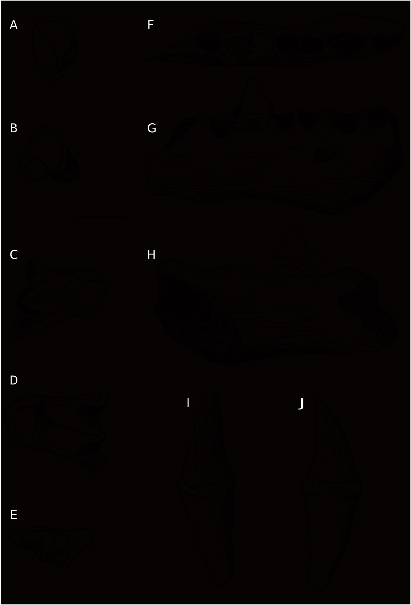
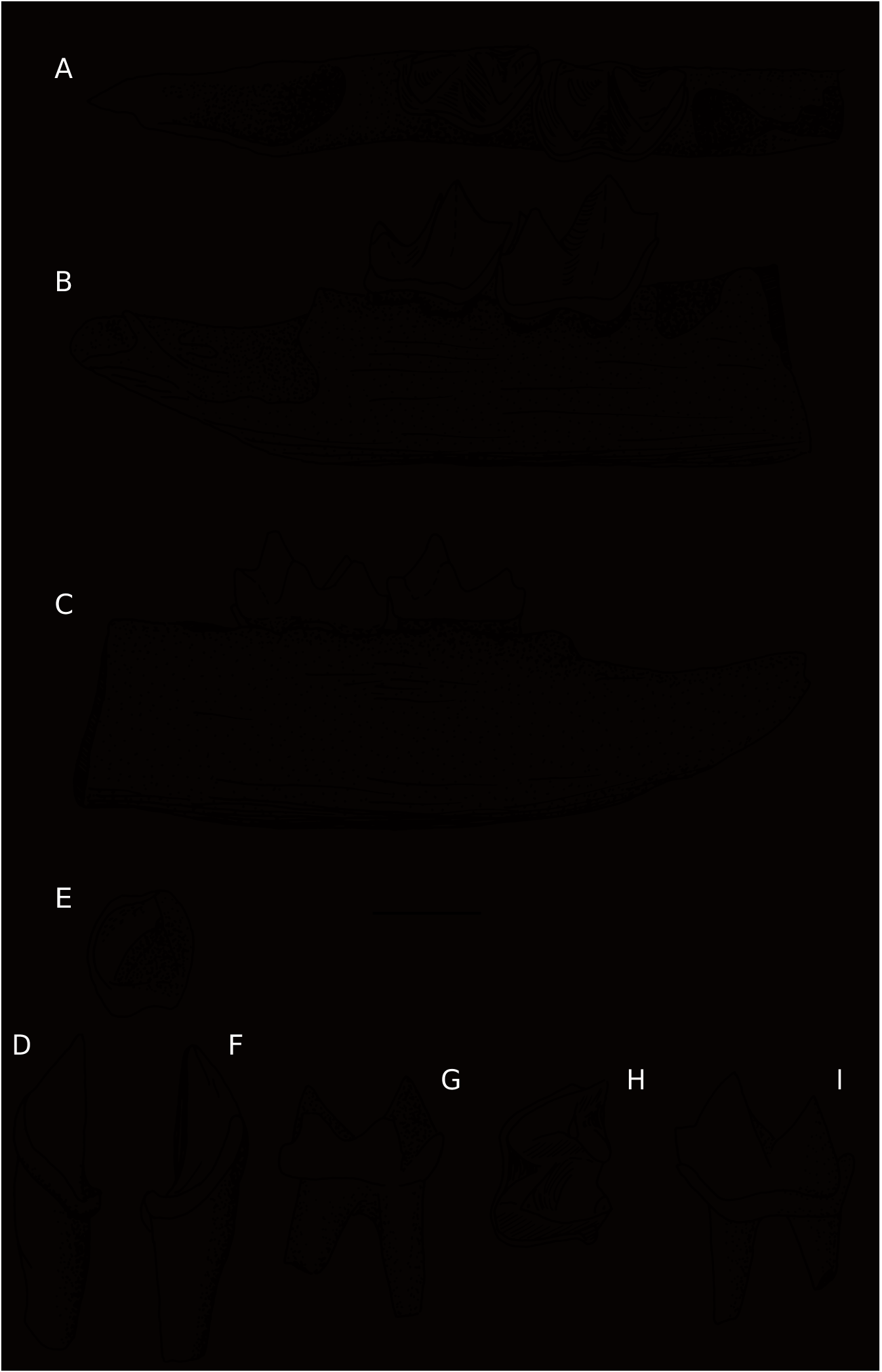
Myotis blythii longicaninus n. ssp. ( Figs 14 View FIG ; 15 View FIG )
HOLOTYPE. — 1 fragment of mandible with p4 (Ms352, Fig. 14 View FIG F-H).
FIGURED PARATYPES. — 1 fragment of mandible with m2-m3 (Ms350); 1 left C1 (Ms361-1); 1 left P4 (Ms361-2); 1 right M1 (Ms361-3); 1 left M2 (Ms361- 4); 1 right M3 (Ms361-5); 1 left c1 (Ms361-7); 1 left m1 (Ms361-8).
ETYMOLOGY. — Longicaninus because of its elongated canines.
TYPE LOCALITY. — Muselievo, Bulgaria.
OTHER MATERIAL EXAMINED. — 10 small fragments of mandible with one tooth (p4, m1, m2, m3) each
(Ms349-358, 362), 11 fragments of mandible without teeth (Ms359-360), 35 left C1 (Ms363), 38 right C1 (Ms368), 3 P4 (Ms365), 7 left and 4 right M1 (Ms366), 4 left and 2 right M2 (Ms367), 1 right M3 (Ms368), 30 right c1 (Ms369), 38 left c1 (Ms370), 3 left and 5 right p4 (Ms371), 5 right m1 (Ms372), 5 right and 4 left m2 (Ms373), 1 left and 1 right m3 (Ms374), 1 distal fragment of humerus (Ms375).
MEASUREMENTS. — For summary data on isolated teeth see Table 4.
Holotype: LSy = 3.50; HMd/m1 = 2.50; Lp4 = 1.42, Wp4 = 1.00.
Mandibular fragments: LSy = 2.87; 3.37; 3.40.
HMd/m1 = 2.62; 2.37; 2.42; 2.37; 2.50; 2.12; 2.57.
Lp2-p4 = (2.87); (2.62); (2.50); (2.30); (2.25); (2.55). HPC = 5.55.
M1 (L × W) = 2.10 × 1.45; 2.05 × 1.45; 2.07 × 1.70; 1.97 × 1.42.
M2 (L × W) = 2.00 × 1.45; 2.20 × 1.60; 2.20 × 1.42. M3 (L × trW × taW) = 1.80 × 1.42 × 1.02; 1.77 × 1.25 × 0.92; 1.87 × 1.30 × 1.02; 1.85 × 1.25 × 1.02; 1.95 × 1.50 × 1.05.
DIAGNOSIS. — A large Myotis species with elongated canines and p4s.
DIFFERENTIAL DIAGNOSIS. — Myotis blythii longicaninus n. ssp. has smaller molars than the recent M. myotis , being similar in this respect with M. blythii , but differs from the recent form in having considerably longer canines, a larger p4, more anterior position of the mental foramen, more oblique position of the symphysis, ellipsoidal basal cross-section of the upper canine (not pear-like as in the recent species), better developed mesial cingula on M1 and M2, less anteroposteriorly reduced M3, slender and elongated m1- m2, less reduced talon on m3, without interruption of the middle part of the labial cingulum on m3. The form from Muselievo differs from Myotis ghardalamensis Storch, 1974 in having more elongated p4 and m1- m2, and less reduced M3.
DESCRIPTION AND COMPARISONS
The holotype (Ms352) is a mandibular fragment with p4, which lingual margin is slightly eroded; the alveoli of the canine, p2, p3, and i3 and the symphysis are also preserved. The crown outline of p4 is rectangular (elongated antero-postereriorly) with rounded corners (occlusal view). In the recent species this tooth is shortened antero-posteriorly with a trapezoidal outline, somewhat widened out posteriorly. The anterior margin of the cingulum of the p4 overlaps the posterior rim of the alveolus of p3, in contrast to the recent species, in which the anterior end of p4 does not reach the alveolus of p3. The foramen mentale is situated under the portion between c1 and p2 (in front of the root of p2), while in the recent species it is shifted posteriorly, located on the level of the root of p2 or behind it, i.e. between p2 and p3.
c1: in general the tooth is similar to this one of the modern M. blythii , but in some specimens the posterior shelf is better developed and its distal part is curved upwardly. There is no cingular cusplet at the mesiolingual margin of the tooth.
p4: the available teeth are large, rectangular in crown outline. The labial cingulum shows two concavities, each towards a root. In labial view, the overall orientation of the ventral margin of the crown is horizontal or slightly oblique, while in the recent species it is clearly oblique, with a definitely more ventral position of the posterolabial corner of the crown than the antero-labial one.
m1 and m2: they are slender and more elongated than in the recent species. Their talonids are longer than the trigonids. In the modern species the lengths of the trigonids are nearly equal with the lengths of the talonids, or the trigonids are longer. In the fossil m1s the upper margin of the cingulum strongly ascends at the base of the protoconid. The entocristids are well developed and somewhat higher than in the recent species.
m3: the talonid shows no distinct reduction, in contrast to the recent species. The labial cingulum is equally thick along the crown, while in the recent species it is weaken or interrupted between the protoconid and hypoconid.
C1: the basal cross-section of the crown is ellipsoidal. There are two shallow grooves: one on the labial face, one on the lingual face. In the recent species these grooves are better pronounced and very often there is a trace of an antero-labial groove, which never occurs in the fossil form. The lower margin of the crown is sharp and well differentiated from the root, while in the recent species the transition between these parts of the tooth is gradual.
P4: in one unworn tooth there is an indistinct accessory tubercle on the mesio-lingual margin of the cingulum. This cingular cusplet is always missing in the recent comparative material of M. blythii . The lingual portion of the crown (talon) is better developed than in the modern species.
M1-M2: the occlusal surface of the two first upper molars is simplified: without heel, paraloph, metaloph, and metaconule, similar in these respects to the recent species. The differences concern the wider mesial cingula in the fossil teeth, which usually incorporate the parastyles. This feature reflects the loose arrangement of P4- M1-M 2 in the fossil form.
M3: the outline of the crown is transversely triangular, with a rather massive metacone protruding posteriorly. In the recent species this cusp is indistinct, transversely elongated as an extension of the premetacrista.
REMARKS
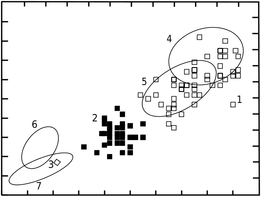
The L × W plots of the fossil molars ( Figs 11; 12 View FIG ) show that they are of approximately the same size as the teeth of the recent M. blythii . In the same time the ranges of variability of the fossil p4s and especially of the lengths of the canines are larger, covering the variability of both recent species, M. myotis and M. blythii ( Fig. 13 View FIG ). These data indicate that we deal with one species, similar in size to M. blythii but with longer canines and larger p4s.
The size of molars from Muselievo is nearly identical with the middle Pliocene material from Osztramos 9 and 13, determined as Myotis cf. blythii ( Topál 1983) . The Hungarian form has a relatively wide talonid on m3 (trWm3 = 1.22, taWm3 = 0.92; taW/trW = 83.63%), which, according to Topál (1983), “seems to be a primary and more ancient feature”. The form from Muselievo also shows a relatively wide talonid. The values of the taW/trW ratio in the two specimens from Muselievo are 73.3 and 74.8%, while in 20 recent specimens of M. blythii this ratio ranges between 63.08 and 73.08% with a mean value of 68.78%. This ratio is even lower in the recent closely related species M. myotis (62.12-71.85%). Based on this feature and on the reduced premolar rows, M. myotis , according to Topál (1963c), is more evolved than M. blythii . In this context, the differences between the fossil and the recent samples of M. blythii , presented above, indicate that the remains from Muselievo represent an unspecialized and primitive form.

No known copyright restrictions apply. See Agosti, D., Egloff, W., 2009. Taxonomic information exchange and copyright: the Plazi approach. BMC Research Notes 2009, 2:53 for further explanation.
|
Kingdom |
|
|
Phylum |
|
|
Class |
|
|
Order |
|
|
Family |
|
|
Genus |