
Myriopathes cf. stechowi ( Pax, 1932 )
|
publication ID |
https://doi.org/ 10.11646/zootaxa.4826.1.1 |
|
publication LSID |
lsid:zoobank.org:pub:1DC59C31-61D1-4458-897B-29D9CA523634 |
|
DOI |
https://doi.org/10.5281/zenodo.4448334 |
|
persistent identifier |
https://treatment.plazi.org/id/F5768787-9349-4243-FF4C-FA08FBDFFB2D |
|
treatment provided by |
Plazi |
|
scientific name |
Myriopathes cf. stechowi ( Pax, 1932 ) |
| status |
|
Myriopathes cf. stechowi ( Pax, 1932) View in CoL
Fig. 25 View FIGURE 25
Aphanipathes View in CoL ? stechowi Pax 1932, p.436 View in CoL –441, figs.16–18
Myriopathes stechowi Opresko 2001, p.349 View in CoL
Material examined. Soalara 17 m, branches from one colony, specimen INV.131335.
Depth range. 15–25 m.
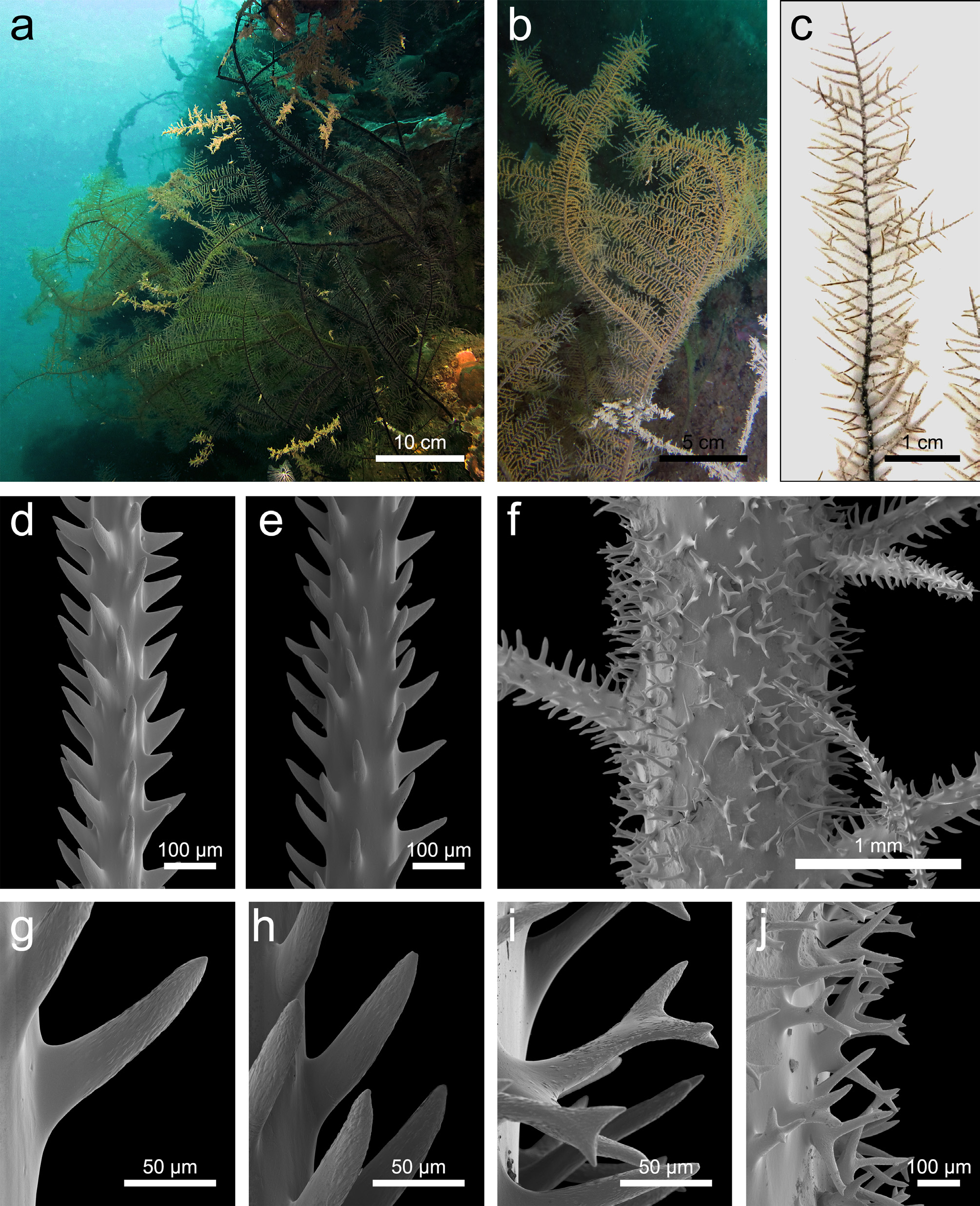
Description. The colony is branched and measures around 50 cm in width and height ( Fig. 25 View FIGURE 25 , a). The branching pattern is planar, with long branches measuring up to 30 cm in length, the whole colony appears red except for thick, dark and black branches ( Fig. 25 View FIGURE 25 , a). The branches do not tend to overlap, but rather grow by forming almost perpendicular angles to the lower order branches bearing them ( Fig. 25 View FIGURE 25 , a, b). The primary pinnules are biserial alternate and measure 0.5–1.7 cm, but mostly around 1 cm ( Fig. 25 View FIGURE 25 , c). Primary pinnules are inserted 60–80° to the branch with an average of 70° and are slightly directed towards the posterior side of the colony ( Fig. 25 View FIGURE 25 , b, c). In each row the primary pinnules are spaced 1.3–2.0 mm apart, with an average of 1.6 mm ( Fig. 25 View FIGURE 25 , c). There are generally 10–11 primary pinnules occurring along one cm of a branch, counting those in both lateral rows ( Fig. 25 View FIGURE 25 , b, c). Secondary pinnules are present but not on every primary ( Fig. 25 View FIGURE 25 , c). Secondary pinnules measure up to 5 mm in length, but mostly 2.0–3.0 mm ( Fig. 25 View FIGURE 25 , c). They are inserted out of the plane and in some places, especially on thick branches, they emerge very close to the base of the primaries, giving the appearance that they are inserted at the same point on the branch. There are usually up to three uniserial secondaries on the primaries, with a maximum of five after which they become biserial and transform into a branch ( Fig. 25 View FIGURE 25 , c). Tertiaries pinnules are rare, but when present only a single one is found on a secondary. The polyps are located on a single side of the pinnules, but on thicker branches they can be irregularly all around the axis. Polyps measure 0.6–0.8 mm and are spaced up to 0.4 mm apart, for about 10–12 polyps per cm.
The spines on the pinnules are conical and either straight-sided or slightly horn-shaped ( Fig. 25 View FIGURE 25 , d, e). They are inclined upwards, but the inclination is greater on the abpolypar side ( Fig. 25 View FIGURE 25 , d, e). Their surface is clearly papillose, with papillae elongated towards the tip of the spines ( Fig. 25 View FIGURE 25 , g, h, i). On a subpinnule measuring 0.10 mm in diameter, 5–6 longitudinal rows are seen in one aspect. Polypar spines measure 0.06–0.09 mm and abpolypar spines measure 0.08–0.10 mm, and their mutual distance is 0.09–0.16 mm. On a primary pinnule measuring 0.14 mm in diameter, polypar spines measure 0.08–0.13 mm and are spaced 0.10–0.15 mm apart, while abpolypar spines measure 0.08–0.10 mm and are spaced 0.07–0.16 mm apart. The spines become more numerous and taller, narrower and very often antler-shaped or bifid on thick branches ( Fig. 25 View FIGURE 25 , f). The bifurcation can occur at the base or near the tip of the spine ( Fig. 25 View FIGURE 25 , i, j). They are no longer inclined regularly upwards on such branches ( Fig. 25 View FIGURE 25 , f), and the longitudinal arrangement is lost. On these branches there is no distinction between polypar and abpolypar spines. They measure 0.18–0.26 mm in height on a branch 1.1 mm in diameter, and the mutual distance cannot be calculated as the longitudinal arrangement is lost.
Taxonomic remarks. Planar and flabellate myriopathids are represented by M. ulex , M. panamensis , M. stechowi , M. spinosa and M. rugosa (Opresko 2001) . Amongst the three latter species, M. spinosa ( Carter, 1880) reported by Brook (1889) from Sri Lanka at 120 m is described as having only one to three uniserial secondaries, which is different from the present species. The pinnulation is too poorly described by Brook (1889) to compare other morphological features. Myriopathes rugosa ( Thomson & Simpson, 1905) , originally reported from Sri Lanka, described as having an angular branching pattern of the main branches as seen in the present specimen, with similar angles of insertion of the primary pinnules. Again, the pinnulation is too poorly described to be confident with this identification. Finally, M. stechowi ( Pax, 1932) originally described from Japan is the closest species to the present one. They share a similar pinnulation pattern as well as pinnules and spines of similar sizes, and both have numerous antler-shaped spines. This species could be related to M. stechowi , however Pax (1932) reported smooth spines for this species. Therefore, the Malagasy specimen is at this time assigned to M. cf. stechowi .
Distribution. Japan (type locality, Pax 1932), Madagascar (present study).
No known copyright restrictions apply. See Agosti, D., Egloff, W., 2009. Taxonomic information exchange and copyright: the Plazi approach. BMC Research Notes 2009, 2:53 for further explanation.
|
Kingdom |
|
|
Phylum |
|
|
Class |
|
|
Order |
|
|
Family |
|
|
Genus |
Myriopathes cf. stechowi ( Pax, 1932 )
| Terrana, Lucas, Bo, Marzia, Opresko, Dennis M. & Eeckhaut, Igor 2020 |
Myriopathes stechowi
| Opresko 2001: 349 |
stechowi
| Pax 1932: 436 |
Aphanipathes
| Brook 1889 |