
Nanophareus araucanus, Hara, Marcos Ryotaro, Pinto-Da-Rocha, Ricardo & Kury, Adriano Brilhante, 2012
|
publication ID |
https://doi.org/ 10.5281/zenodo.212388 |
|
DOI |
https://doi.org/10.5281/zenodo.6175468 |
|
persistent identifier |
https://treatment.plazi.org/id/3C3487A8-4273-9D36-DDEB-B1CAFF95342B |
|
treatment provided by |
Plazi |
|
scientific name |
Nanophareus araucanus |
| status |
sp. nov. |
Nanophareus araucanus View in CoL sp. nov.
( Figs. 4 View FIGURE 4 , 5 View FIGURE 5 , 10 View FIGURE 10 C–D, 13)
Type material. CHILE. V Región de Valparaíso: Parque Nacional La Campana (32°58´48”S, 71°07´03”W), 16.I.2010, R. Pinto-da-Rocha, F. Cádiz L. & D. Cádiz L. leg., ma holotype (MNHNCL); idem, 1 fe paratype ( MZSP 36877); idem (32°58´52”S, 71°07´59”W, 632 m), 6.XII.2010, F. Marques, F. Cádiz L. & F. Carbayo leg., 2 ma paratypes ( MZSP 36878).
Diagnosis for males. Nanophareus araucanus sp. nov. resembles N. bosqenublado sp. nov. because of the frontal hump on dorsal scutum with an enlarged, high median tubercle, coxa IV reaching close to the posterior margin of dorsal scutum in dorsal view, prolateral apical apophysis of coxa IV surpassing the posterior margin of dorsal scutum, trochanter IV with a prodorsal apical apophysis and femur IV with a retromedian apophysis. Nanophareus araucanus sp. nov. can be distinguished from N. bosqenublado sp. nov. by: Ocularium not widened (½ of carapace width) and armed, scutal area III always with a median pair of spines (some specimens of N. bosqenublado are unarmed), prolateral apical apophysis of coxa IV bifid at the apex, prolateral apical apophysis of trochanter IV wide and truncated and patella IV with 1 proventral apical spine. Nanophareus araucanus sp. nov. can be distinguished from the other species of the genus by: Ocularium not widened; prolateral dorsoapical apophysis of trochanter IV wide and truncated; and femur IV retrolaterally with a basal and a median spine.
Diagnosis for females. Nanophareus araucanus sp. nov. can be distinguished from the other species of the genus by the combination of the following characters: ocularium not widened; ocularium and frontal hump armed with enlarged median tubercles; and undivided scutal area IV.
Etymology. In reference to the tribe of Araucanos, who lived in most part of Central and Southern Chile before they were conquered by the Spanish explorers.
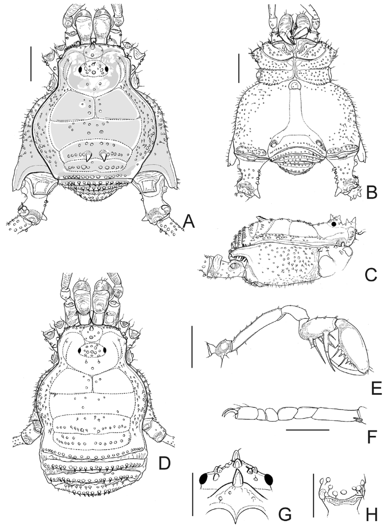
Description. Male (holotype): Dorsum ( Fig. 4 View FIGURE 4 A, C, G): Measurements: DSL 3.70; DSW 3.35; LI 7.55; LII 12.40; LIII 9.00; LIV 12.40. Anterior margin of carapace with a frontal hump bearing 2 median, enlarged, high tubercles, and 8 small tubercles on each side. Ocularium not widened (½ of carapace width), with 4–5 tubercles near each eye, 2 median enlarged ones. Carapace with sparse tubercles. Scutal area I with 3–5 tubercles on each half close to the longitudinal median groove; II with 10 tubercles concentrated in the middle; III with 2 parallel spines pointing backwards (lower than ocularium height), one anterior row of 6 tubercles, 5–6 tubercles lateral to the spines; IV undivided, with one posterior row of 9 slightly enlarged tubercles, 1 anterior small one. Lateral margin of dorsal scutum with a row of enlarged tubercles inserted among small ones, more densely distributed between grooves I and III. Posterior margin of dorsal scutum and free tergites I–III each with a row of tubercles numbering16, 17, 13, 12, respectively. Anal operculum densely tuberculate, except for smooth anterior region.
Venter ( Fig. 4 View FIGURE 4 B): Coxa I–III densely tuberculate; I with enlarged tubercles. Coxa IV with tubercles on laterals and apex. Stigmatic area smooth. Free sternites each one with a row of tubercles. Anal operculum tuberculate.
Chelicera: Segment I with 1 tubercle, bulla well-marked; movable finger with 3 teeth; fixed finger with 4 teeth.
Pedipalpus ( Fig. 4 View FIGURE 4 E): Coxa with 3 ventral tubercles. Trochanter with 2 dorsal, 2 ventral tubercles (basal largest). Femur with 2 ventral tubercles, 1 small prolateral subapical tubercle. Patella with 1 dorsal tubercle. Tibia with 3 dorsal tubercles; tibial setation: Prolateral IIi, retrolateral I[Ii] (apical bifid and longest, 1 short and 1 long setae). Tarsal setation: Prolateral IiIii, retrolateral IiIiii, iIiIiii.
Legs ( Figs. 4 View FIGURE 4 F, H, 5): Coxa IV tuberculate, more conspicuous dorso-laterally, smooth on and near apophysis, with a prolateral apical apophysis apically bifid, moderately long, directed backwards. Trochanters I–IV tuberculate; IV twice longer than wide, prolaterally with a wide median apophysis with truncate apex, a wide dorsoapical apophysis with truncate apex; retrolaterally with a moderately long, conical apical apophysis (its length ½ of trochanter IV width). Femur IV strongly sigmoid, dorsoapically unarmed; retrolaterally with a basal and a median spine, the largest of the article (its length larger than femur width); ventrally with two rows of tubercles, prolateral ones increasing in size apically (apical 3 enlarged), retrolateral ones increasing in size basally (basal 6 enlarged, pointed), 1 prolateral apical spine. Patella IV with enlarged proventral and retroventral tubercles, 1 proventral apical spine. Tibia III with two ventral rows of enlarged tubercles; IV ventrally with two rows of tubercles increasing in size subapically, becoming pointed, retrolateral ones slightly enlarged, apically with 2 spines. Basitarsus normal. Tarsal process reduced to a seta. Tarsal segmentation: 6(3); 8(3); 6; 6.
Penis ( Fig. 10 View FIGURE 10 C–D): Glans with wide sac; stylus slender, cylindrical, curved with ventral subapical scattered trichomes; ventral process slender, blunt apex directed to stylus. Ventral plate distal setae conical, placed a little far from ventral plate corner; ventral plate basal setae slightly curved on the middle (size similar to distal group).
Coloration: Mostly light brown, densely covered with small black dots, except for grooves I–V, edge of lateral margins of dorsal scutum, most part of ocularium and some patches on carapace.
Female (paratype; MZSP 36877): Dorsum ( Fig. 4 View FIGURE 4 D): Measurements: DSL 3.65; DSW 3.05; LI 6.75; LII 11.50; LIII 8.25; LIV 11.00. Scutal area III unarmed, with one anterior row of 6 tubercles and posterior row of 17 tubercles. Pedipalpus: Tibial setation: Prolateral III; tarsal setation: Prolateral IiIi, retrolateral IiIii. Legs I–IV with tubercles of similar size, unarmed. Femur IV slightly curved inwards. Coloration: Light brown with small black spots on dorsal scutum, more concentrated on carapace near groove I.
Variation in males (n=3): Measurements: DSL 3.65–3.70; DSW 3.25–3.35; LI 7.55–7.75; LII 12.40–13.50; LIII 9.00–9.10; LIV 12.40–13.30. Pedipalpus: Tibial setation: Prolateral IiI, IIi, II, III, retrolateral I[Ii]; tarsal setation: Prolateral IiIi, IiIii, retrolateral IiIii, IiIiii, iIiIii, iIiIiii. Tarsal segmentation: 6(3); 8–9(3); 6; 6.
Geographical distribution ( Fig. 13 View FIGURE 13 ): Central Chile. Valparaíso.


| MZSP |
Sao Paulo, Museu de Zoologia da Universidade de Sao Paulo |
No known copyright restrictions apply. See Agosti, D., Egloff, W., 2009. Taxonomic information exchange and copyright: the Plazi approach. BMC Research Notes 2009, 2:53 for further explanation.