
Nanophareus palpalis Roewer
|
publication ID |
https://doi.org/ 10.5281/zenodo.212388 |
|
DOI |
https://doi.org/10.5281/zenodo.6175466 |
|
persistent identifier |
https://treatment.plazi.org/id/3C3487A8-4274-9D2B-DDEB-B7DAF80B35E7 |
|
treatment provided by |
Plazi |
|
scientific name |
Nanophareus palpalis Roewer |
| status |
|
Nanophareus palpalis Roewer View in CoL
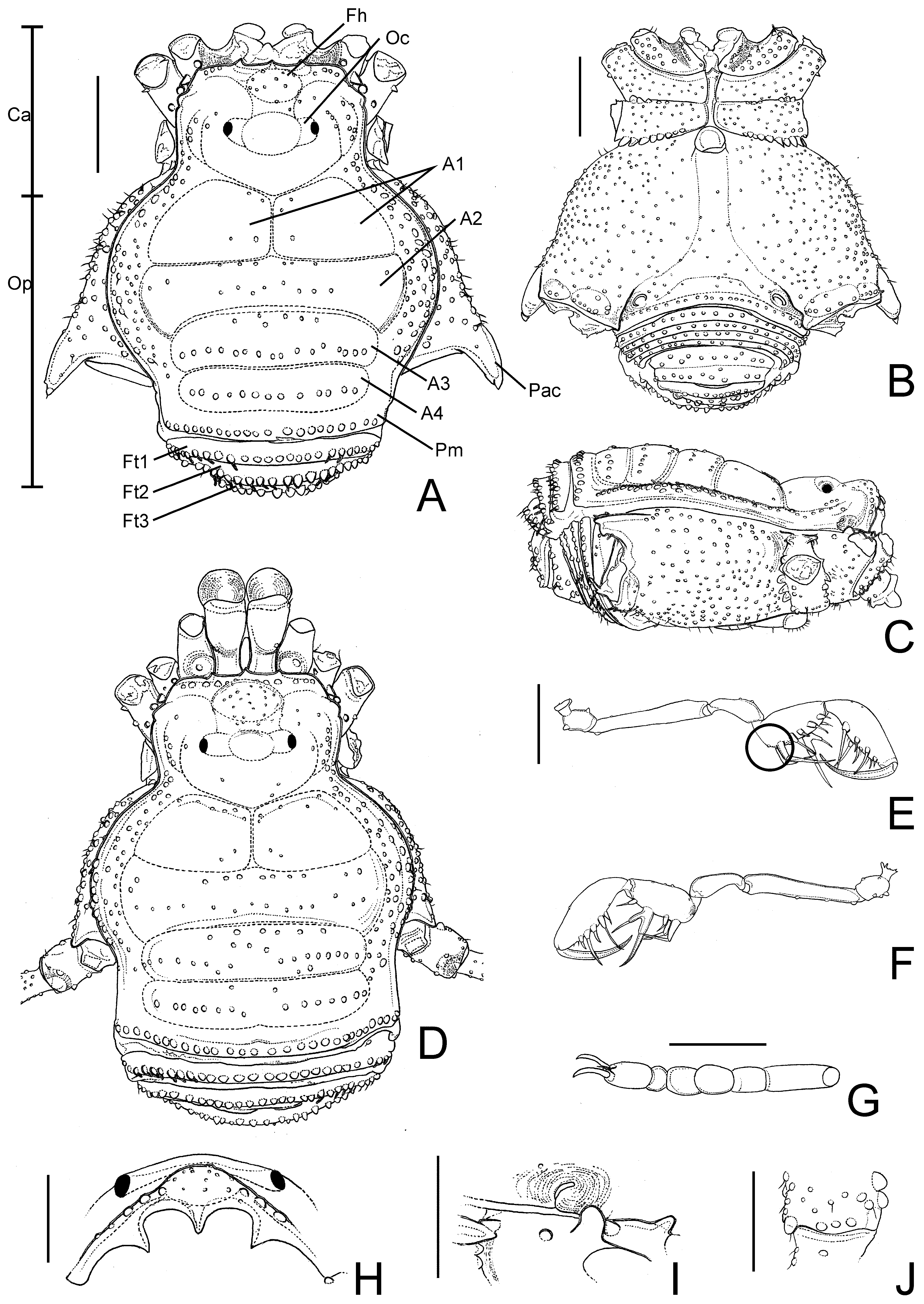
( Figs. 2 View FIGURE 2 , 3 View FIGURE 3 , 10 View FIGURE 10 A–B)
Nanophareus palpalis Roewer 1929: 281 View in CoL , fig. 46; Canals 1936: 69 (cat); Cekalovic 1985: 24 (cat); Kury 2003: 105 (cat). ( Chile; without name of more precise collection site, collector and date; 1 ma lectotype, 1 ma & 2 fe paralectotypes, indicated by Maury and designated here; SMF 986/1; examined). Lectotype lacking both LI, tarsus IV, right metatarsus and tarsus IV. Paralectotypes without most of their legs attached to the body. Therefore, we were unable to unequivocally match LI–III to the correct specimen.
Material examined. CHILE. Without further data of locality, collector name and date, 1 ma lectotype, 1 ma & 2 fe paralectotypes, indicated by Maury and designated here ( SMF 986/1).
Diagnosis for males. Nanophareus palpalis resembles N. bipartitus sp. nov. because of the unarmed frontal hump on dorsal scutum, widened ocularium, unarmed scutal area III, prolateral apical apophysis of coxa IV barely reaching the posterior margin of dorsal scutum, trochanter IV unarmed prolateral apically and femur IV without retromedian apophysis. Nanophareus palpalis can be distinguished from N. bipartitus sp. nov. by: Scutal area IV undivided and tibia IV with a retrolateral row of enlarged and pointed tubercles decreasing in size from submedian region to apex, two ventral rows of similar size tubercles, ventral apically unarmed. Nanophareus palpalis can be distinguished from the other species of the genus by: Tibia IV with a retrolateral row of enlarged and pointed tubercles decreasing in size from submedian region to apex, two ventral rows of similar size tubercles, ventral apically unarmed.
Diagnosis for females. Nanophareus palpalis can be distinguished from the other species of the genus by the combination of the following characters: Ocularium widened; ocularium and frontal hump unarmed and undivided scutal area IV.
Redescription. Male (lectotype): Dorsum ( Fig. 2 View FIGURE 2 A, C, H, I): Measurements: DSL 3.70; DSW 3.30; LII 14.10; LIII 9.05; LIV 12.10 (without tarsus IV). Median frontal hump tuberculate, with 3–4 tubercles on each side of anterior margin of carapace. Ocularium widened, low, with median eminence, smooth. Carapace with sparse tubercles. Scutal area I with few tubercles close to scutal groove I and median longitudinal groove; II with two transversal rows of 6 (anterior) and 8 (posterior row) tubercles, 1–2 tubercles on the sides; III with two rows of tubercles, anterior with 6, posterior with 16 tubercles (these larger than anterior ones); IV undivided, with one row of 13 tubercles. Lateral margin of dorsal scutum with a row of enlarged tubercles inserted among small ones, more densely distributed between grooves II and III. Posterior margin of dorsal scutum and free tergites I–III each one with a row of 21, 23, 21 and 16 tubercles, respectively. Anal operculum with 16 scattered tubercles.
Venter ( Fig. 2 View FIGURE 2 B): Coxa I–IV densely tuberculate. Stigmatic area with few small scattered tubercles. Posterior margin of stigmatic sternite and free sternites each one with a row of tubercles. Anal operculum tuberculate.
Chelicera: Segment I smooth, bulla weakly-marked; movable finger with 4 teeth; fixed finger with 5 teeth.
Pedipalpus ( Fig. 2 View FIGURE 2 E–F): Coxa with 1 ventral, 1 dorsal tubercle. Trochanter with 1 dorsal, 2 ventral tubercles. Femur with 3 small ventral tubercles. Patella with 1 dorsal tubercle. Tibia with 4 dorsal tubercles; tibial setation: Prolateral IIiIi, retrolateral Ii[Ii] (subdistal bifid and longest, 1 short and 1 long setae). Tarsal setation: Prolateral IiIii, retrolateral iIiIii.
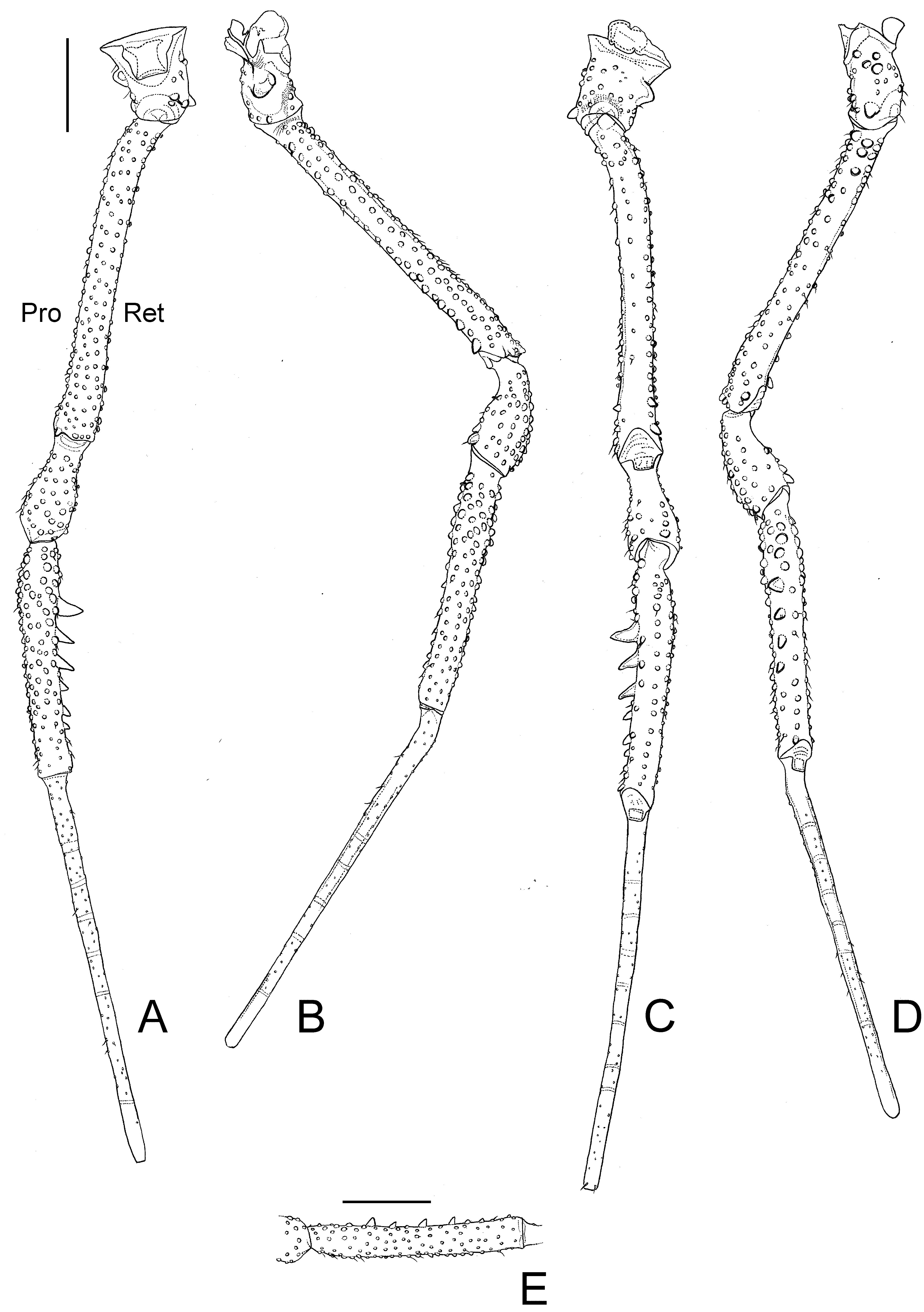
Legs ( Figs. 2 View FIGURE 2 G, J, 3): Coxa IV densely tuberculate, with prolateral apical apophysis apically bifid, moderately long, directed backwards. Trochanters I–IV tuberculate; IV 1.5 times longer than wide, prolaterally with a conical, short, blunt median apophysis, swollen in the middle; retrolaterally with 2 submedian slightly enlarged tubercles, an apical conical, short and blunt apophysis. Femur IV slightly curved basally, proventral row of tubercles increasing in size, 1 prodorsal apical enlarged tubercle, 1 proventral apical blunt spine. Patella IV tuberculate, 1 proventral apical blunt spine. Tibia IV with one retrolateral row of enlarged tubercles decreasing in size from submedian region to apex. Basitarsus I slightly swollen. Tarsal process reduced to a seta. Tarsal segmentation: 5(3); 7–8(3); 6; 6 according to Roewer description (1929) but legs I and tarsi IV missing in this specimen.
Penis ( Fig. 10 View FIGURE 10 A–B): Glans with wide sac; stylus slender, cylindrical, curved without ventral trichomes; ventral process slender, blunt apex, directed to stylus. Ventral plate distal setae conical, placed almost on ventral plate corner, slightly curved on apex; ventral plate basal setae slightly curved on apex (larger than distal group).
Coloration: As in Roewer 1929, i.e. body and all limbs pale yellow. We suppose Roewer received the type series already discolored.
Female (paralectotype): Dorsum ( Fig. 2 View FIGURE 2 D): Measurements: DSL 3.80; DSW 3.20; PI 6.40; PIV 10.60. Scutal area IV with 2 anterior tubercles, one posterior row with 15 tubercles. Trochanter IV with 1 enlarged prolateral subasal tubercle, retrolaterally with 1 median, 1 apical enlarged tubercle. Femur IV with a proventral row of enlarged tubercles on distal ¼. Tibia IV with similar sized tubercles. Tarsal segmentation: 5(3);?;?; 6.
Variation in males (n=2) ( Fig. 3 View FIGURE 3 A, E): Measurements: DSL 3.65–3.7; DSW 3.15–3.30; LI 7.00; LII 12.60–14.10; LIII 9.0; LIV 11.90–12.10 (without tarsus). Pedipalpus: Tibial setation: Prolateral IIiIi, IIiII, retrolateral Ii[Ii], Ii[Ii]i; tarsal setation: Retrolateral iIiIii, iIiIiii. Tibia IV with one retrolateral row of more or less conspicuous enlarged tubercles decreasing in size from submedian region to apex. Tarsal segmentation: 5(3); 7(3); 6; 6.
Variation in females (n=2): Measurements: DSL 3.75–3.80; DSW 3.05–3.20; PII 10.20; PIV 9.95–10.60. Pedipalpus: Tibial setation: Prolateral IIiII, retrolateral Ii[Ii]i. Tarsal segmentation: 5(3); 7(3); 6; 6.
Geographical distribution. Known only from the imprecise type locality.

| SMF |
Forschungsinstitut und Natur-Museum Senckenberg |
No known copyright restrictions apply. See Agosti, D., Egloff, W., 2009. Taxonomic information exchange and copyright: the Plazi approach. BMC Research Notes 2009, 2:53 for further explanation.
|
Kingdom |
|
|
Phylum |
|
|
Class |
|
|
Order |
|
|
Family |
|
|
Genus |
Nanophareus palpalis Roewer
| Hara, Marcos Ryotaro, Pinto-Da-Rocha, Ricardo & Kury, Adriano Brilhante 2012 |
Nanophareus palpalis
| Kury 2003: 105 |
| Cekalovic 1985: 24 |
| Canals 1936: 69 |
| Roewer 1929: 281 |