
Nesocyrtosoma guerreroi Hopp and Ivie
|
publication ID |
https://doi.org/ 10.1649/0010-065x-63.sp8.1 |
|
persistent identifier |
https://treatment.plazi.org/id/457F4C06-A84A-FFBC-E037-FA49D0CBB47C |
|
treatment provided by |
Carolina |
|
scientific name |
Nesocyrtosoma guerreroi Hopp and Ivie |
| status |
sp. nov. |
Nesocyrtosoma guerreroi Hopp and Ivie , New Species
( Figs. 10, 15 View Figs , 70 View Figs , 234–238 View Figs , 266 View Figs , 301 View Figs )
Type Material. HOLOTYPE: Male. DOM.REP:Prov. Pedernales; 26 km
N. Cabo Rojo, 915m; 09 SEP 1988,wet forest; at light and night beating;
M.Ivie, Philips and Johnson. (from WIBF, deposited NMNH). PARATYPES (21 specimens) : 1 WIBF specimen with same label data as type. DOMINICAN REPUBLIC; Barahona Prov., Filipinas; 18u 07.3399N, 71u 07.1529W; 625 m; Day beating; 7 July 2004 S. Lingafelter. (1 WIBF) GoogleMaps . DOMINICAN REP.: Prov.; Barahona, nr. Filipinas,; Larimar Mine ; 26-VI-7-; VII-1992; Woodruff and; Skelley, at light. (2 FSCA, 1 WIBF) . DOMINICAN REP.: Prov.; Barahona, nr. Filipinas; Larimar Mine ; 20–26-VI-1992; Woodruff and; Skelley; at night. (1 FSCA) . DOMINICAN REPUBLIC: Pr.; Barahona, Rd. to Polo , S.; slope, 860m 14-VII-1996; Coll. M.C. Thomas. (3 FSCA) . DOMINICAN REPUBLIC: Prov. ; La Vega, 1km NW Manabao; 6-VI-1994; coll. M.C. Thomas. (1 WIBF) . DOM.REP; LaVega Prov. ; PN. A.Bermudez, Cienaga; 19.VII-2.VIII.95, 1,100m; trop.evgrn.for.,FIT; S.+J. Peck, 95-36. (2 CMNC) . DOMINICAN REPUBLIC:; La Vega Prov., PN Armando; Bermudez , km 1–3 along trail; W of La Cienga, 900–1,100,; 19u 01.7539N, 70u 54.6549W; 6-VI-2005; Gino Nearns. (1 WIBF) GoogleMaps . DOMIN.REP: Prov. LaVega; La Cienega , 1,100m; 19u 04.079N, 70u 51.689W; 29 JULY 1999, at night; M.A.Ivie and K.A.Guerrero. (1 WIBF) GoogleMaps . DOM.REP:- Prov. Pedernales; 24 km N.Cabo Rojo, 610m; 21 AUG 1988,wet forest; at light and night beating; M. Ivie, Philips and Johnson. (1 WIBF) . DOM.REP: Prov. Pedernales; 24 km N.Cabo Rojo, 610m; 20AUG-09SEP1988,wetforest; Malaise trap, M.A. Ivie,; T. K. Philips and K.A. Johnson. (1 WIBF) . DOMINICAN REPUBLIC: Pr.; Pedernales,25kmN. Cabo; Rojo , 700m 10-VII-1996; Coll. M.C. Thomas. (1 FSCA) . DOM.REP: Prov. La Altagracia; P.N.del Este, Boca de Yuma ; entrance, 05AUG1999, at night; 18u 21.9049N, 68u 37.0879W; M.A.Ivie, beating veget. (2 WIBF) GoogleMaps . DOM.REP: Prov. La Altagracia; P.N.del Este, Boca de Yuma , entr. to Par. ; Nac. delEste, 12m, 06AUG1999; at night, 18u 21.9049N, 68u 37.0949W; M.A.Ivie beating at night. (1 WIBF) GoogleMaps . DOMINICAN REPUB- LIC: Prov.; Puerto Plata, 1km, ESE; Estero Hondo , 1-VI-1994; coll. M.C. Thomas. (1 FSCA) . DOMINICAN REPUBLIC; Peravia Prov., Cerro Gordo; south of Baní 65 meters; 18u 16.1209N, 70u 20.6079W; 28 June 2004 S. Lingafelter. (1 NMNH) GoogleMaps .
Diagnosis. This species can be distinguished by the combination of the elytral striae present as rows of small, lightly impressed, discontinuous punctures ( Fig. 234 View Figs ), hypomeral bead absent, antennomeres 6–10 transverse, ocular depression present ( Fig. 238 View Figs ), and tibiae with a weak dorsal longitudinal groove ( Fig. 37 View Figs ). It most closely resembles N. fernandoi , but can be distinguished from that species by having an antero-posteriorly broad mesoventrite ( Fig. 237 View Figs ).
Description (male). 4.0– 5.5 mm long, 2.0–3.0 mm wide. Small, body short, moderately convex ( Figs. 234, 235 View Figs ). Dark ferrugineous to purple; antennae, mouthparts, and tarsi ferrugineous. Head densely variably punctate; largest punctures subequal to a single eye facet, weakly to moderately impressed; extremely short golden seta emerging from each puncture. Antenna clavate, antennomeres 6–10 transverse, forming a loose club; apical antennomere subcircular; antennomeres 6–11 with stellate sensoria. Mentum with acute median keel raised anteriorly to a point ( Figs. 10, 12 View Figs ). Ventral portion of eye not reaching subgenal sulcus ( Fig. 8 View Figs ); ventral ocular groove present ( Fig. 238 View Figs ); postgena with distinct punctures ( Fig. 12 View Figs ). Dorsal surface of pronotum densely punctate; punctures separated by 0.5–1.5 diameters. Pronotal marginal bead complete laterally, anterior margin with marginal bead effaced medially, posterior margin lacking marginal bead; anterior angles of pronotum right, weakly produced and widely rounded apically; lateral edge of pronotum evenly curved to base; pronotum evenly convex ( Fig. 236 View Figs ). Hypomeron without distinct punctures.
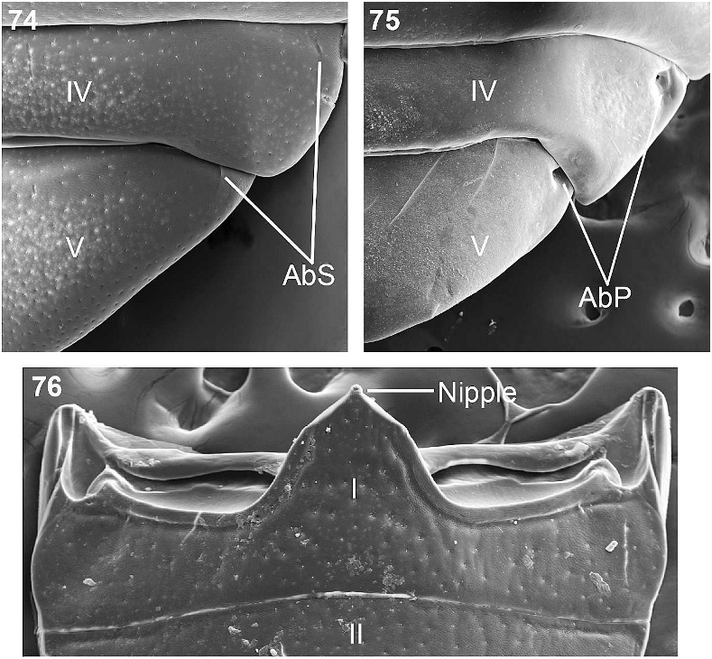
Prosternal process apically rounded with indistinct marginal grooves opposite coxae not joined apically, mediobasal portion of prosternal process raised ( Fig. 237 View Figs ). Elytral striae weakly impressed, present as rows of small punctures separated by 0.5–1.0 3 diameter; elytral interstriae flat, scarcely punctate; scutellary striae 3 punctures long; scutellum triangular, normal ( Figs. 234, 236 View Figs ). Mesoventrite broad antero-posteriorly, U-shaped, excavate, receiving prosternal process; metaventrite subequal to antero-postero length of mesocoxa ( Fig. 237 View Figs ). Metathoracic wing fully developed. Legs short, punctate; femora reaching beyond edge of elytron; tibiae with a weak dorsal longitudinal groove; metatibia straight, lacking ventral apical tooth ( Fig. 70 View Figs ). Abdominal depressions on 4th and 5th ventrites reduced to indistinct slits ( Fig. 74 View Figs ); intercoxal process of first ventrite apically rounded; ventral surface densely punctate, punctures weakly to moderately impressed ( Fig. 237 View Figs ).
Female. Identical to male.
Biology. This species has been collected at light, by beating dead branches at night in wet forest and moist tropical forest, and by beating vegetation in dry forest. It has also been taken during the day by beating secondary Acacia Mill. and Cassia L. (both Fabaceae ) thorn scrub. It has also has been taken in a Malaise trap.
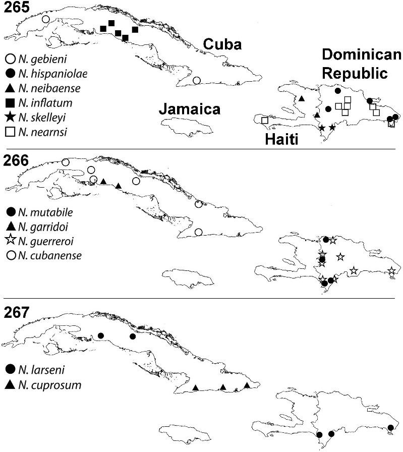
Distribution. This species is endemic to Hispaniola. It has been collected in Barahona Province at the Larimar Mines and on the road to Polo; in La Vega Province at Parque Nacional Armando Bermudez in Ciénaga; in Pedernales Province north of Cabo Rojo; in La Altagracia Province in Parque Nacional del Este Boca de Yuma; and variously in Puerto Plata Province, Peravia Province, and La Estrel Province ( Figs. 260 View Fig , 266 View Figs ).
Etymology. This species is named for Kelvin Guerrero, former student of MAI who helped collect some of the series and is infamous around our laboratory for the saying ‘‘Sistemática, no es easy!’’
Nesocyrtosoma virens (LaPorte and Brulle´), New Combination ( Figs. 12 View Figs , 37 View Figs , 71 View Figs , 80, 82 View Figs , 239–242 View Figs , 263 View Figs , 302 View Figs )
Platydema virens LaPorte and Brullé 1831: 391–392 ; Leng and Mutchler 1914:
462; Gebien 1938 – 1942 a: 538; Blackwelder 1945: 529; Marcuzzi 1984: 89. Hoplocephala flavicornis Chevrolat 1877: 170 [synonymy follows Blackwelder
1945: 529]. Neomida viridula Gundlach , nomen nudum [in Gundlach collection]. Diaperis viridula Zayas 1988: 93 , Fig. 81 View Figs . New Synonymy. Serrania viridula ; Garrido 2003: 50–51, Fig. 1 View Figs ; Peck 2005: 150. Platydema antennatum [not LaPorte and Brulle´]; Ivie 1991: 400 (misidentifica-
tion); Marcuzzi 1998: 158 (misidentification).
Taxonomic Notes. Garrido (2003) described the monotypic genus Serrania for Diaperis viridula Zayas, 1988 , placing it in the Diaperini . Diaperis viridula was described from two specimens in the Gundlach collection with the nomen nudum Neomida viridula and the number 656.
Three years after Zayas’ publication, Ivie (1991) visited the residence of Zayas and examined part of the collection and synonymized several taxa described by Zayas. Among these, Ivie incorrectly synonymized Diaperis viridula Zayas, 1988 with Platydema antennatum LaPorte and Brulle´, 1831. Garrido (2003), detecting Ivie’s mistake, attempted to resolve the taxonomic placement of this species. However, when he sent two topotypes to C. A. Triplehorn, Triplehorn identified them as Nautes Pascoe sp. in the tribe Helopini , resulting in yet another discrepancy in determination between Zayas, Ivie, and Triplehorn ( Garrido 2003). Thus, Garrido (2003) described a new genus, Serrania , with D. viridula as the type species, under the new combination, Serrania viridula (Zayas) .
While Garrido was correct that D. viridula does not fit within any of the current Diaperinae genera, he failed to recognize that it does not belong in the Diaperinae at all. This species has the clypeal membrane concealed and the metatibia does not bear a fine, crenulate, longitudinal ridge on the external (dorsal) surface. This automatically rules out this species from the Diaperinae as the Diaperinae have the clypeal membrane exposed and the metatibia bears a fine, crenulate, longitudinal ridge on the external (dorsal) surface ( Triplehorn 1965).
While dealing with the Serrania problem, a second species, Platydema virens LaPorte and Brulle´, 1831, came into the picture. Much like S. viridula , P. virens lacks the external apomorphies of the Diaperinae . Interestingly enough, after the examination of P. virens type specimens in Torino, Italy by Ivie, it was discovered that Serrania viridula (Zayas) is a synonym of Platydema virens LaPorte and Brullé New Synonymy. With this new synonymy, females of this species were dissected in order to determine its taxonomic placement.
Dissections of the female genitalia revealed the true taxonomic placement of this species in the Stenochiinae : Cnodalonini and in the genus Nesocyrtosoma . First, the defensive reservoirs are attached medially and have annular bands, which are consistent with Stenochiinae defensive reservoirs ( Fig. 82 View Figs ) ( Tschinkel and Doyen 1980). Second, the genital tube is a bursa-less vagina with an apical, smooth spermathecal accessory gland ending distally in a membranous, abruptly saccate spermatheca, which is consistent with the Stenochiinae ( Fig. 80 View Figs ) ( Tschinkel and Doyen 1980). Third, the ovipositor is consistent with the Stenochiinae with the paraprocts much shorter than the coxites, and at rest are rotated 180u so that the morphologically proximal ends lie distally beside the coxites, the proximal lobe of the coxites is longer than the distal three lobes combined ( Fig. 83 View Figs ) ( Tschinkel and Doyen 1980; Doyen 1989). Finally, the stiffened-cuticular tube is consistent with the NSCT-type, thus this species is placed in Nesocyrtosoma and recognized for the first time as Nesocyrtosoma virens (LaPorte and Brulle´) New Combination.
Type Material Examined. Diaperis viridula Zayas : HOLOTYPE: ‘‘ Col. F. de Zayas; Sierra Maestra; Turquino 6 1963; Oriente. CUBA / red circle label Tipo/ D. viridula ; Zayas’’ ( FZCM).
Serrania viridula (Zayas) : TOPOTYPES designated by Garrido (2003): 7 specimens in OHGC. Examined 5 of 7 (other two were not found in collection when visited); 1 specimen labeled ‘‘Coleccion M. Barro; Soledad; 14 VI 1933; Prov. S.C. CUBA / Tenebrionidae ; Ident. OHG 210; Serrania viridula ; Zayas, 1988;’’ 3 other specimens with the same label data but with OHG numbers 203, 207 and 1470; 1 specimen labeled ‘‘Corralillo; 14. VI.33; L. V. Cuba/ Tenebrionidae ; Ident. OHG 205; Serrania viridula ; Zayas, 1988.’’
Platydema virens LaPorte and Brulle´: LECTOTYPE of Platydema virens here designated (female): Diaperis ; virens Klug ; Is. (??) Cuba D. Schüppel. PARA- LECTOTYPE of Platydema virens here designated (sex unknown): Diaperis ; virens Klug ; Is. (??) Cuba D. Schüppel.
A second specimen with the same label data as the type is reported in the Zayas description, but was not designated as a paratype.
Other Material Examined. Coleccion M. Barro; Soledad; 14. VI 1933; Prov. S.C. CUBA. (6 OHGC —OHG 157, OHG1881, OHG 1884, OHG 1885, OHG 1889, OHG 1890,). Turquino- Ote; VI- 1967; Zayas-Alayo; Garcia. (1 OHGC — OHG 1882). Cardero-; Turquino; X-1966; I. Garcia. (7 OHGC —OHG 209, OHG 1882, OHG 1883, OHG 1886, OHG 1463, OHG 1469, OHG 1877; 3 IESC — OHG 301, OHG 302, OHG 304). Cardero-Turquino; VI-1964; I. Garcia/ Tenebrionidae ; Ident. OHG 1887; Serrania viridula ; Zayas, 1988. (1 OHGC). Turquino- Ote; VI-1967; Zayas-Alayo; Garcia/ Tenebrionidae ; Ident. OHG 1882; Serrania viridula ; Zayas, 1988. (1 OHGC). Soledad; 13. VI. 32; S.C. CUBA / Tenebrionidae ; Ident. OHG 1891; Serrania viridula ; Zayas, 1988. (1 OHGC). Soledad; 14 VI 1933; S.C. Cuba / Tenebrionidae ; Ident. OHG 1890; Serrania viridula ; Zayas, 1988. (1 OHGC). Sierra de; Rangel- P. R.; VI-1930 / Tenebrionidae ; Ident. OHG 1888; Serrania viridula ; Zayas, 1988. (1 OHGC). Cayamas; 11. 3 Cuba / E. A. Schwarz; Collector. (1 NMNH). Cayamas; 13. 3 Cuba / E. A. Schwarz; Collector. (1 NMNH). Cayamas; Cuba, Baker/ 4171/ CASEY; bequest; 1925/ Platydema ; virens ; Cast. (1 NMNH). Loma del Gato; Cobre Range, O; July 3–7 1936; about 3,000 ft. / Cuba 1936; Darlington; Collector/ Platydema ; virens Lap. and Brll. ; Det. Triplehorn ’64. (4 OSUC). [Soledad, Cuba; (Cienfuegos); May, 1936; Darlington. (1 OSUC). 454/ Cuba; Poey Coll. (2 ANSP). 454; var./ Cuba; Poey Coll. (1 ANSP). Poey; Collection/ Cuba. (1 ANSP). 285/ Poey Coll./ Cuba./ Neomida ; viridula ; M. Berl; Diaperiales. (1 ANSP). CUBA: Las Villas; Topes de Collantes; Sierra de Trinidad; 11 June 1959; M. W. Sanderson. (1 WIBF). CUBA: Oriente, Loma; (Pico) del Gato, Sierra; Maestra, 26–28 MAR 1959; M. W. Sanderson. (1 WIBF). 182; Neomida virens . (1 Gundlach Collection, IESC). 656; Neomida viridula (3 Gundlach Collection, IESC). 1128; Platydema virens . (1 Gundlach Collection– IESC). CUBA: Oriente; Yateritas; 01 June 1959; M. W. Sanderson. (5 WIBF). CUBA, 30.5.1985; Pico Turquino; S. Bilý leg. (1 NMPC). 285/ Cuba; Poey Coll./ Neomida ; viridula ; M. B.; Diaperiales. (1 ANSP).
Notes. This list of examined specimens may represent a complex of species as variation is extremely difficult to understand. If a specimen from Cuba does not fit nicely in N. cubanense , N. dentatum , or N. garridoi , it is placed in N. virens . The two specimens examined from the Gundlach Collection, 182 and 656, were listed from Cardenas in Gundlach’s record book.
Diagnosis. This species is extremely variable, but can be distinguished by the combination of weakly to strongly convex elytral interstriae ( Fig. 239 View Figs ), hypomeral bead weakly to strongly impressed ( Fig. 27 View Figs ), and males with a small ventral apical tooth on the metatibia ( Fig. 71 View Figs ).
Redescription (modified from LaPorte and Brullé 1831, Zayas 1988, and Garrido 2003) (male). 4.0– 4.5 mm long, 2.5–3.0 mm wide. Small, short, broad, slightly convex ( Figs. 239, 240 View Figs ). Shining greenish blue; antennae, mouthparts and tarsi yellow-brown to ferrugineous. Head densely punctate; punctures variable, largest punctures larger than a single eye facet and deeply impressed. Antenna clavate; antennomeres 7–10 weakly transverse, forming a loose club; apical antennomere subcircular; antennomeres 7–11 with stellate sensoria. Mentum with acute median keel raised anteriorly to a point ( Figs. 10, 12 View Figs ); ventral portion of eye not reaching subgenal sulcus ( Fig. 8 View Figs ); postgena with distinct punctures ( Fig. 12 View Figs ). Dorsal surface of pronotum punctate; punctures separated by 0.5–1.0 diameters. Pronotal marginal bead complete laterally, anterior margin with marginal bead effaced medially, posterior margin lacking marginal bead; anterior angles of pronotum obtuse, not produced and broadly rounded apically; lateral edge of pronotum evenly curved to base; pronotum evenly convex ( Fig. 241 View Figs ). Hypomeral bead weakly to deeply impressed; hypomeron with distinct punctures ( Fig. 27 View Figs ). Prosternal process with mediobasal portion of prosternal process raised, apically rounded with marginal grooves opposite coxae joined apically ( Fig. 242 View Figs ). Elytral striae lightly impressed, present as small discontinuous punctures with an impressed line through the middle, connecting the row of punctures; interstriae weakly to roundly convex; scutellary stria 4–8 punctures long; scutellum triangular, normal ( Figs. 239, 241 View Figs ). Mesoventrite broad antero-posteriorly, deeply excavate, U-shaped, receiving prosternal process; metaventrite subequal to antero-postero length of mesocoxa ( Fig. 242 View Figs ). Metathoracic wing fully developed. Legs short, stout, punctate, short golden setae emerging from each puncture; apical 1/4 of femur reaching beyond edge of elytron; tibiae with dorsal longitudinal groove ( Fig. 37 View Figs ); metatibia apically slightly curved ventrally, ventral apical tooth vestigial ( Fig. 71 View Figs ). Abdominal depressions on 4th and 5th ventrites reduced to slightly impressed slits ( Fig. 74 View Figs ); intercoxal process of first ventrite with apical nipple; ventral surface densely punctate, punctures weakly to strongly impressed ( Fig. 242 View Figs ).
Female. Identical to male, except metatibia straight without apical ventral tooth.
Biology. Unknown.
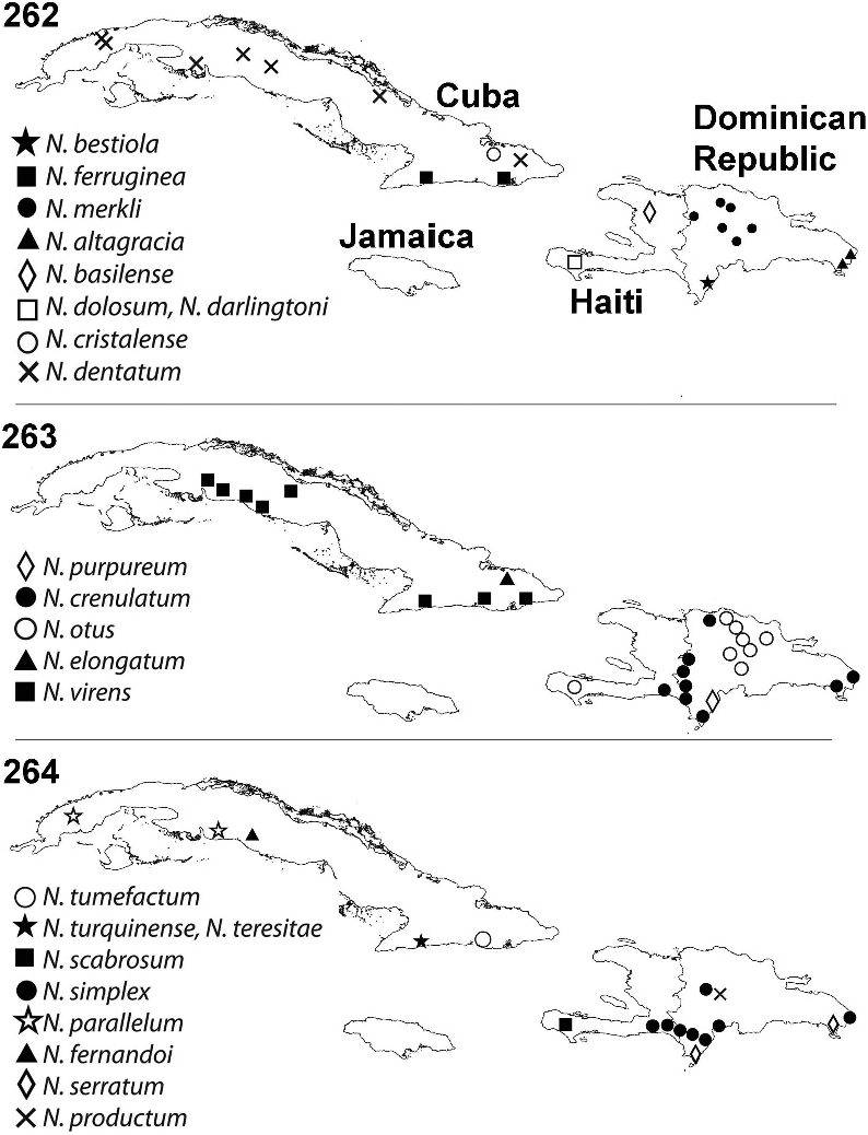
Distribution. Blackwelder (1945) synonymized Hoplocephala flavicornis Chevrolat with P. virens and states its distribution as Puerto Rico. However, Chevrolat (1877) reports H. flavicornis only from Cuba, never mentioning Puerto Rico. Thus, this species is endemic to Cuba. It has been collected primarily on Pico Turquino in the Sierra Maestra of Oriente Province. It has also been taken in Soledad, Cayamas, Loma del Gato, Coralillo, Yateritas, and Topes de Collantes ( Figs. 259 View Figs , 263 View Figs ).
Nesocyrtosoma cubanense (Kulzer) , New Combination ( Figs. 27 View Figs , 30 View Figs , 72 View Figs , 243–246 View Figs , 266 View Figs , 303 View Figs )
Apsida cubanensis Kulzer 1961: 217 ; Triplehorn 1970: 568. [Here designated correct as First Revisers]
Apsida cubaensis Kulzer 1961: 219 ; Marcuzzi 1984: 90; Ferrer and Ødegaard 2005: 640 [Here designated lapsus calami as First Revisers].
Type Material Examined. HOLOTYPE: Playa Marianao; bei Habana; 2. 6. 1932./ A. Bierig leg; Eing.Nr.129,1933./ Hapsida ; n. sp.; H. Gebein det. 1934/ =/ on white square label with red outline HOLOTYPUS; Apsida ; cubaensis mihi; det. H. Kulzer 1961. ( NHMB). ALLOTYPE: Playa Marianao; bei Habana; 2. 6. 1932./ A. Bierig leg; Eing.Nr.129,1933./ R / ALLOTYPUS; Apsida ; cubaensis mihi; det. H. Kulzer 1961. (1 NHMB). PARATYPES: Playa Marianao; bei Habana; 2. 6. 1932./ A. Bierig leg; Eing.Nr.129,1933./ PARATYPUS; Apsida ; cubaensis mihi; det. H. Kulzer 1961. (1 NHMB). Cuba,; Prov. Pinar d. Rio; Sierra d. Rosario; Rangel XII.1932 / A. Bierig leg; Eing.Nr.129,1933./ PARATYPUS; Apsida ; cubaensis mihi; det. H. Kulzer 1961. (1 NHMB).
Notes. After examination of the paratype from Pinar del Rio, it was determined that this specimen is a female of N. dentatum . However, it is not given paratype status of N. dentatum because it is a female. Kulzer (1961) used two spellings for this species, Apsida cubanensis as the first (p. 217) and second (p. 218) usage with Apsida cubaensis used in the key on p. 219. Triplehorn (1970) followed the first of these, while Marcuzzi (1984) and Ferrer and Ødegaard (2005) used the second. Under ICZN Article 32.2.1, the First Reviser [see also Art. 24.2.3] is empowered to choose between these two spellings, but in order to satisfy that provision, both names must be cited and one chosen. None of the users have satisfied this condition, so it falls to us as First Revisors to follow Triplehorn (1970) and choose Apsida cubanensis as the correct name.
Other Material Examined. Pto. Manati; Tunas- Ote 1981; L. R . Hern y; L. de Armas. (1 OHGC). Playo Giron ; C. de Zapata; VI 1962; Zayas. (1 OHGC). P. Gorda; C. de Zapata; VI 1962; Zayas. (1 OHGC). CUBA: Matanzas Prov.,; Cienaga Zapata, at; Playa Larga ; 11 and 12 Feb. 1981; P. Spangler, A. Vega / Collected in; malaise trap. (1 NMNH, 1 WIBF) .
Notes. Triplehorn (1970) published a synopsis of the genus Apsida Lacordaire and wrote a new key to the species, but did not include A. cubanensis because he had not seen specimens of this species and was therefore unable to place it in his key. However, this species does not belong in Apsida as it does not have the clypeal membrane exposed and the metatibia does not bear a fine, crenulate, longitudinal ridge on the external (dorsal) surface. The second paratype listed from Pinar del Rio is likely a female of N. dentatum , and is placed there, but not designated as a paratype as it is a female. There is an additional paratype in the series, but it was not examined.
Finally, the female specimens in the ‘‘Other Material Examined’’ section may represent a complex of species as only male specimens can be identified with certainty.
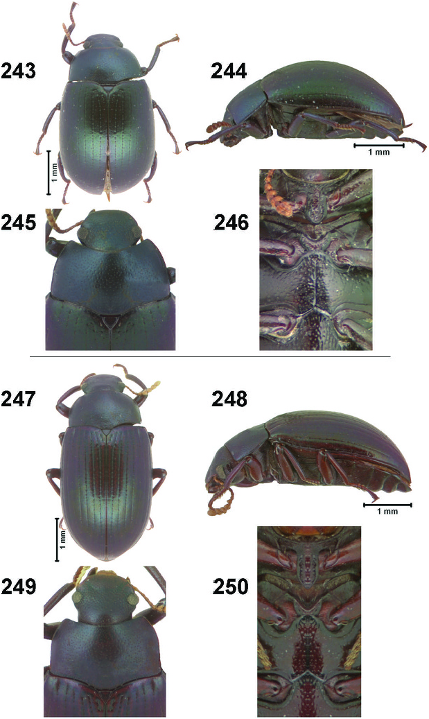
Diagnosis. Both males and females of this species can be distinguished from most other members of the Serrania species-group by the small, broad body ( Fig. 243 View Figs ), flat interstriae ( Fig. 243 View Figs ), strong hypomeral bead ( Fig. 27 View Figs ), and prosternal process raised in the mediobasal portion ( Fig. 246 View Figs ). The males of this species can be distinguished from all other species except N. dentatum by the combination of a femoral tooth on the hind margin distad the trochanter and the strongly curved metatibia with a small tooth on the apical ventral surface that flows into the apex of the metatibia ( Fig. 72 View Figs ), whereas N. dentatum males have a larger ventral apical metatibial tooth proximad apex ( Fig. 73 View Figs ).
Redescription (modified from Kulzer 1961) (male). 4.0– 4.5 mm long, 2.5–3.0 mm wide. Small, body short, broad, moderately convex ( Figs. 243, 244 View Figs ). Shining greenish blue; antennae, mouthparts, and tarsi yellow-brown to ferrugineous. Head densely variably punctate; largest punctures larger than a single eye facet, weakly to moderately impressed; extremely short golden seta emerging from each puncture. Antenna clavate; antennomeres 7–10 short, apically widened forming a loose club; apical antennomere subcircular; antennomeres 7–11 with stellate sensoria. Mentum with acute median keel raised anteriorly to a point ( Figs. 10, 12 View Figs ); ventral portion of eye not reaching subgenal sulcus ( Fig. 8 View Figs ); postgena with distinct punctures ( Fig. 12 View Figs ). Dorsal surface of pronotum punctate; punctures separated by 0.5–1.0 diameters Pronotal marginal bead complete laterally, anterior margin with marginal bead effaced medially, posterior margin lacking marginal bead; anterior angles of pronotum right, weakly produced and broadly rounded apically; lateral edge of pronotum evenly curved to base; pronotum evenly convex ( Fig. 245 View Figs ). Hypomeral bead deeply impressed; hypomeron with distinct punctures ( Fig. 27 View Figs ). Mediobasal portion of prosternal process raised; prosternal process apically rounded, with distinct marginal grooves opposite coxae joined apically ( Fig. 246 View Figs ). Elytral striae lightly impressed, present as small discontinuous punctures with a lightly impressed line through the middle, connecting the row of punctures; interstriae flat; scutellary striae 4 punctures long; scutellum triangular, normal ( Figs. 243, 245 View Figs ). Mesoventrite broad anteroposteriorly, excavate, U-shaped, receiving prosternal process; metaventrite subequal to antero-postero length of mesocoxa ( Fig. 246 View Figs ). Metathoracic wing fully developed. Legs short, stout, punctate; pro- and metafemora reaching beyond edge of elytron, mesofemur not reaching past edge of elytron; metafemur with tooth distad trochanter ( Fig. 72 View Figs ); tibiae with dorsal longitudinal groove ( Fig. 37 View Figs ); metatibia strongly curved ventrally, with small ventral apical tooth with posterior edge sloped into apex of the tibia ( Fig. 72 View Figs ). Abdominal depressions on 4th and 5th ventrites reduced to indistinct impressed slits ( Fig. 74 View Figs ); intercoxal process of first ventrite with apical nipple; ventral surface densely punctate, punctures strongly impressed ( Fig. 246 View Figs ).
Female. Identical to male, except metafemur lacking tooth distad trochanter and metatibia straight and lacking ventral apical tooth.
Biology. The only available biological data available for this species are that it has been collected in a Malaise trap.
Distribution. This species is endemic to Cuba. It seems to be widespread throughout Cuba, occurring in La Habana, Playa Giron, Playa Gorda and Playa Larga in Ciénaga de Zapata located in Matanzas Province, Manti in Las Tunas Province, and Pico Turquino in Oriente Province ( Figs. 258, 259 View Figs , 266 View Figs ).
Nesocyrtosoma dentatum Hopp and Ivie , New Species ( Figs. 73 View Figs , 247–250 View Figs , 262 View Figs , 304 View Figs )
Apsida cubanensis Kulzer 1961: 217–218 ; Triplehorn 1970: 568 [in part, one paratype].
Type Material. HOLOTYPE: Male. PinaresOri-; enteCuba’18; W M Mann. ( NMNH). PARATYPES (5 specimens): CUBA: Pinar del Rio; Aspiro-Rangel; 16 June 1959; M. W. Sanderson. (2 WIBF). Pinar del Rio; 7-12-1960 / Tenebrionidae ; Ident. OHG 316; Hapsida ; cubanensis ; Kulzer, 1961. (1 OHGC). Soledad, Cuba; (Cienfuegos); May, 1936; Darlington. (1 OSUC). CUBA: Camaguey; Monte Imias; nr. California; 07 JUN 1959; M. W. Sanderson. (1 WIBF).
Other Material Examined. Cuba,; Prov. Pinar d. Rio; Sierra d. Rosario; Rangel XII.1932 / A. Bierig leg; Eing.Nr.129,1933./ PARATYPUS; Apsida ; cubaensis mihi; det. H. Kulzer 1961. ( NHNB). PinaresOri-; enteCuba’18; W M Mann. ( NMNH).
Notes. The paratype of A. cubanensis from Pinar del Rio does not belong to that species as it is more elongate and slender and has a weakly impressed hypomeral bead. However, it is a female, so its placement here is tentative. The four recognized Cuban species of the Serrania species-group are quite variable, and even though there are some characters to use to place females within the correct species, those characters are variable and thus make it difficult to identify a female with certainty. Therefore, the female paratype of A. cubanensis from Pinar del Rio is not made a paratype of N. dentatum . The second specimen under Other Material Examined is a male and can be distinguished by the metatibia, however, the pronotum and head are missing so it is not given paratype status.
Diagnosis. Both males and females of this species can be distinguished by the elongate body form ( Fig. 247 View Figs ), weakly to roundly convex interstriae ( Fig. 247 View Figs ), weak hypomeral bead, and prosternal process not greatly raised in the mediobasal portion ( Fig. 250 View Figs ). The males of this species can be distinguished from all other species, except N. cubanense , by the combination of a tooth on the metafemur distad the trochanter, strongly curved metatibia and sharp tooth on the ventral surface proximad apex ( Fig. 73 View Figs ). It can be distinguished from N. cubanense by the males of N. dentatum having a larger ventral apical metatibial tooth proximad apex ( Fig. 73 View Figs ), whereas N. cubanense males have a small tooth on the apical ventral surface of the metatibia that flows into the apex of the metatibia ( Fig. 72 View Figs ).
Description (male). 3.5–4.5 mm long, 2.0– 2.5 mm wide. Small, body elongate, slender, slightly convex ( Figs. 247, 248 View Figs ). Shining greenish blue to purple; antennae, mouthparts, and tarsi yellow-brown. Head densely punctate; largest punctures subequal to a single eye facet, punctures moderately impressed; extremely short golden seta emerging from each puncture. Antenna clavate; antennomeres 7–10 transverse, apically widened forming a loose club; apical antennomere subcircular; antennomeres 7–11 with stellate sensoria. Mentum with acute median keel raised anteriorly to a point ( Figs. 10, 12 View Figs ); ventral portion of eye not reaching subgenal sulcus ( Fig. 8 View Figs ); postgena with distinct punctures ( Fig. 12 View Figs ). Dorsal surface of pronotum punctate; punctures separated by 0.5–1.0 diameters. Pronotal marginal bead complete laterally, anterior margin with marginal bead effaced medially, posterior margin lacking marginal bead; anterior angles of pronotum obtuse, weakly produced and broadly rounded apically; lateral edge of pronotum evenly curved to base; pronotum evenly convex ( Fig. 249 View Figs ). Hypomeral bead weakly to deeply impressed; hypomeron with distinct punctures ( Fig. 27 View Figs ). Prosternal process apically rounded with marginal grooves opposite coxae joined apically ( Fig. 250 View Figs ). Elytral striae impressed, present as small discontinuous punctures with a weakly to moderately impressed line connecting the rows of punctures; interstriae weakly convex to flat; scutellary striae 4–6 punctures long; scutellum triangular, normal ( Figs. 247, 249 View Figs ). Mesoventrite thin antero-posteriorly, deeply excavate, U-shaped, receiving prosternal process; metaventrite subequal to antero-postero length of mesocoxa ( Fig. 250 View Figs ). Metathoracic wing fully developed. Legs long, slender, punctate, short golden seta emerging from each puncture; apical 1/4 of femur reaching beyond edge of elytron; metafemur with tooth distad trochanter; tibiae with distinct dorsal longitudinal groove ( Fig. 37 View Figs ); metatibia strongly apically curved ventrally; metatibial tooth proximad apex of metatibia, sharp with posterior edge straight ( Fig. 73 View Figs ). Abdominal depressions on 4th and 5th ventrites reduced to slightly impressed slits ( Fig. 74 View Figs ); intercoxal process of the first ventrite with apical nipple; ventral surface densely punctate, punctures weakly to strongly impressed ( Fig. 250 View Figs ).
Female. Identical to male, except metafemur without tooth distad trochanter and metatibia straight without ventral apical tooth.
Biology. This species has been collected at night, but no other biological data are available.
Distribution. This species is endemic to Cuba and has been collected in Aspiro- Rangel and Pinares in Pinar del Rio Province. It has also been collected on Monte Imias in Camaguey Province, and at Ciénaga de Zapata and Topes de Collantes in Las Villas Province ( Figs. 258, 259 View Figs , 268 View Figs ).
Etymology. This species epithet comes from the Latin word dentatus meaning toothed. This species has a large tooth on the metafemur distad the trochanter, and a sharp tooth on the ventral surface proximad apex of the metatibia ( Fig. 73 View Figs ).


| WIBF |
West Indian Beetle Fauna Project Collection |
| NMNH |
Smithsonian Institution, National Museum of Natural History |
| FSCA |
Florida State Collection of Arthropods, The Museum of Entomology |
| T |
Tavera, Department of Geology and Geophysics |
| VI |
Mykotektet, National Veterinary Institute |
| V |
Royal British Columbia Museum - Herbarium |
| R |
Departamento de Geologia, Universidad de Chile |
| OSUC |
Oregon State University |
| ANSP |
Academy of Natural Sciences of Philadelphia |
| MAR |
Grasslands Rhizobium Collection |
| NMPC |
National Museum Prague |
| NHMB |
Natural History Museum Bucharest |
No known copyright restrictions apply. See Agosti, D., Egloff, W., 2009. Taxonomic information exchange and copyright: the Plazi approach. BMC Research Notes 2009, 2:53 for further explanation.
|
Kingdom |
|
|
Phylum |
|
|
Class |
|
|
Order |
|
|
Family |
|
|
Genus |
Nesocyrtosoma guerreroi Hopp and Ivie
| Hopp, Katie J. & Ivie, Michael A. 2009 |
Apsida cubanensis
| Triplehorn, C. A. 1970: 568 |
| Kulzer, H. 1961: 217 |
Apsida cubaensis
| Ferrer, J. & F. Odegaard 2005: 640 |
| Marcuzzi, G. 1984: 90 |
| Kulzer, H. 1961: 219 |
Apsida cubanensis
| Triplehorn, C. A. 1970: 568 |
| Kulzer, H. 1961: 218 |