
Neurolarthra karensharkeyae Yao, 2018
|
publication ID |
https://doi.org/ 10.11646/zootaxa.4438.3.7 |
|
publication LSID |
lsid:zoobank.org:pub:E3986654-2B9D-4DB2-88DB-2E4171966AFE |
|
DOI |
https://doi.org/10.5281/zenodo.6490305 |
|
persistent identifier |
https://treatment.plazi.org/id/922D87D0-9779-FFE9-FF71-F8CF1341EF81 |
|
treatment provided by |
Plazi |
|
scientific name |
Neurolarthra karensharkeyae Yao |
| status |
sp. nov. |
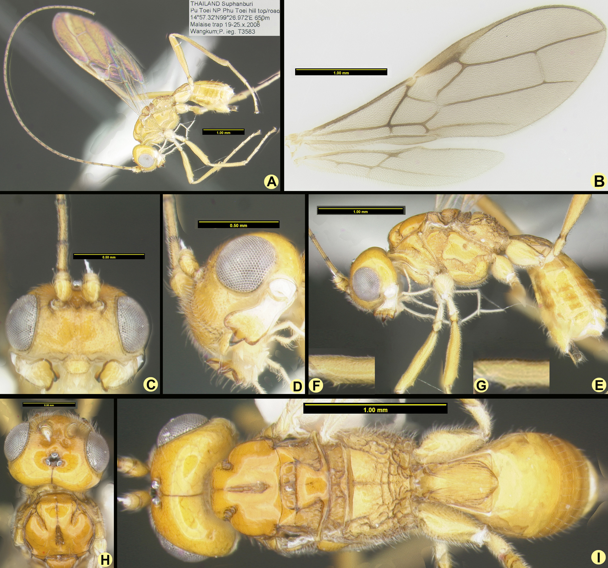
Neurolarthra karensharkeyae Yao n. sp.
Figure 2 View FIGURE 2 (A–I)
Diagnosis. Body mainly yellowish, fore and mid femora with a small ventral tooth subapically ( Fig. 2A View FIGURE 2 ); mid pit elliptical, large ( Fig. 2H View FIGURE 2 ); hind femur brownish yellow or infuscate ( Fig. 2A View FIGURE 2 ); vein m-cu of hind wing slightly antefurcal; pterostigma distinctly differentiated from vein R1 and slender subbasally ( Fig. 2B View FIGURE 2 ); vein m-cu of hind
wing comparatively close to vein 2M (thus second part of vein 1M comparatively short); vein 1m-cu of fore wing 0.5–0.7 × as long as vein 2RS ( Fig. 2A View FIGURE 2 ).
Description. Holotype, ♀, length of body 3.4 mm; length of fore wing 3.7 mm.
Head. 2.3 × as wide as long, 1.5 × as wide as mesoscutum (in dorsal view). Head at level of eyes wider than at level of temples ( Fig. 2G View FIGURE 2 ). Eyes 2.0 × as long as temples ( Fig. 2G View FIGURE 2 ). Distance between antennal sockets slightly shorter than their diameter, distance from eye to antennal socket slightly shorter than diameter of antennal socket. Excavated area anterior to ocellar area and between antennal sockets with a long groove. Distance of ocelli from each other slightly shorter than diameter of an ocellus; distance from eye to lateral ocellus longer than width of ocellar area ( Fig. 2G View FIGURE 2 ). Epicranial seam weak ( Fig. 2G View FIGURE 2 ). Face 1.8 × wider than high, almost entirely punctate, with a complete and weak medio-longitudinal carina and long and white setae. Clypeus punctate and protruding, slightly wider than long, apical margin straight, setae long and white, epistomal groove wide and broadly notched ( Fig. 2C View FIGURE 2 ). Mandible 1.75 × as long as wide, lower edge almost straight, upper up curved, apical width about 1.3 × longer than basal width. Tooth 1 wide and round, an acute incision between tooth 1 and tooth 2; tooth 2 pointed and slightly protruding; tooth 3 rounded, a right angle acute incision between tooth 2 and tooth 3; tooth 4 round and small, appearing as a lamella extending from the lower edge of tooth 3. Outer surface of mandible smooth, teeth slightly hollowed medially and slightly wrinkled laterally (externally) ( Fig. 2D View FIGURE 2 ). Maxillary palps reaching mid femur, nearly 2.3 × longer than height of head, 1.2 × longer than hind femur ( Fig. 2F View FIGURE 2 ). Antenna long and thin, apical segments missing, 46 segments remain and 2.0–2.3× as long as fore wing; F1:F2:F3=0.7:1:0.8; F1 3.5 × longer than wide, F2 6.7 × longer than wide, the remaining segments slightly shorter ( Fig. 2A View FIGURE 2 ).
Mesosoma. 1.6 × as long as high. Mesoscutum slightly longer than wide, median lobe strongly protruding, with several short setae anteriorly and along notauli, otherwise glabrous; notauli deeply crenulate, not converging, present in anterior half of mesoscutum, mid pit in posterior half of mesoscutum, crenulated, gradually widened posteriorly, and narrower than notauli ( Fig. 2H View FIGURE 2 ). Mesopleural setae dense and long, glabrous only before episternal scrobe ( Fig. 2F View FIGURE 2 ). Precoxal sulcus complete and crenulated, wide in middle, extending from anterior edge of mesopleuron reaching the mid legs, connected to epicnemial carina. Metapleuron covered with dense setae, anterior third smooth, posterior 2/3 reticulated ( Fig. 2F View FIGURE 2 ). Scutellar sulcus with four carinae. Propodeum with complete medio-longitudinal carina; setae much shorter and sparser than metapleuron; anterior third smooth and remainder reticulated ( Fig. 2H View FIGURE 2 ).
Wings. Fore wing: pterostigma rather narrow, vein r rising from middle of pterostigma, length of vein r as long as half width of pterostigma width, forming an obtuse angle with vein 3RSa, vein 3RSb as long as vein 3RSa, vein 3RSa 1.5× longer than vein 2RS, and vein 3RSb extending to wing tip, vein 1m-cu postfurcal. 2nd submarginal cell large and parallel, vein 1cu-a slightly postfurcal ( Fig. 2B View FIGURE 2 ). Hind wing: vein r almost absent, vein M+CU slightly longer than vein 1M, vein m-cu antefurcal ( Fig. 2B View FIGURE 2 ).
Legs. Hind leg 1.4× as long as the body, inner side of fore and mid femora with a sharp small tooth ( Fig. 2A, 2E, 2F, 2G View FIGURE 2 ). Width of fore femur 7.0 × longer than tooth width of fore femur ( Fig. 2F View FIGURE 2 ), same as middle femur ( Fig. 2G View FIGURE 2 ). Fore femur nearly as long as middle femur, 0.67× as long as hind femur; hind femur 0.8× longer than hind tibia, hind femur 1.8× longer than hind basitarsus.
Metasoma. T1 1.4 × longer than apical width, apical width 1.7 × longer than basal width, lateral longitudinal carinae converging and disappearing in striations in apical third. Area between lateral longitudinal carinae smooth, remainder of T1 regularly longitudinally striate ( Fig. 2I View FIGURE 2 ). Ovipositor short, barely exerted. ( Fig. 2E View FIGURE 2 ).
Color. All yellow, except eye brown, teeth margins reddish brown, apical segments of antenna brown, hind femur, tibia and tarsus dark yellow.
Male. Unknown.
Material examined. Holotype ♀ (H20142), THAILAND Suphanburi Pu Toei NP, Phu Toei hill top/road, 14°57.32'N, 99°26.972'E, elevation 650m, Malaise trap 19–25.x.2008, Wangkum ; P. leg., ( QSBG). For a map of examined material, see http://bit.ly/2o78jwe. GoogleMaps
Comparative diagnosis. This new species is similar to N. ultima , and shares the following characters: fore and mid femora with a small ventral tooth subapically; body mainly brownish or yellowish; mid pit elliptical, large; hind femora brownish yellow or infuscate; vein m-cu of hind wing slightly antefurcal, second part of vein 1M of hind wing shorter than vein 1r-m.
Neurolarthra karensharkeyae differs from N. ultima as follows: pterostigma slender subbasally and abruptly narrowed apically, well differentiated from vein R1 (wide subbasally, gradually narrowed apically in N. ultima and not well differentiated from R1); vein m-cu of hind wing comparatively close to vein 2M (far removed in N. ultima ).
Host. Unknown.
Etymology. Named in honor of Karen Sharkey, who has given me (JY) lots of warmth and help during my tenure at the University of Kentucky.
No known copyright restrictions apply. See Agosti, D., Egloff, W., 2009. Taxonomic information exchange and copyright: the Plazi approach. BMC Research Notes 2009, 2:53 for further explanation.
|
Kingdom |
|
|
Phylum |
|
|
Class |
|
|
Order |
|
|
Family |
|
|
Genus |