
Odontozona ensifera ( Dana, 1852 )
|
publication ID |
https://doi.org/ 10.11646/zootaxa.4044.3.1 |
|
publication LSID |
lsid:zoobank.org:pub:83D58648-447D-442D-B9BD-A36F192E1D3A |
|
DOI |
https://doi.org/10.5281/zenodo.5469812 |
|
persistent identifier |
https://treatment.plazi.org/id/03A66345-821D-FFC4-1BA8-FADDFAE85E36 |
|
treatment provided by |
Plazi |
|
scientific name |
Odontozona ensifera ( Dana, 1852 ) |
| status |
|
Odontozona ensifera ( Dana, 1852) View in CoL
( Figs. 15–17 View FIGURE 15 View FIGURE 16 View FIGURE 17 )
Stenopus ensiferus Dana, 1852a: 27 View in CoL .— Dana, 1852b: 607.— Dana, 1855.— Bate, 1888: 210.— Herrick, 1893: 352.— Lo Bianco, 1903: 252.—A. Milne Edwards & Bouvier, 1909: 264.
Odontozona ensifera View in CoL — Holthuis, 1946: 34, pl. IV, fig. d.—Burukovskij, 1974: 93.— Gore, 1981: 158.— Goy, 1981: 850.— Wicksten, 1982: 134.— Burukovsky, 1983: 131.— Dounas & Koukouras, 1989: 345.— Pretus, 1990: 349.— Manning & Chace, 1990: 31 — Nomura et al., 1996: 9.— Okuno, 2003: 175.— Saito & Fujita, 2009: 124.— Goy, 2010: 217. — De Grave & Fransen, 2011: 252.— Anker & Tavares, 2013: 429.— Hendrickx & Ayón-Parente, 2014: 345.
Odontozona View in CoL sp.— Bruce, 1982: 191.
Non Odontozona View in CoL ensifera— Hayashi, 1986: 24.
Material examined. New Caledonia. Lagon Sud-Ouest, Campaign No. 2, Stn 101, Ile Ouen, Baie du Prony, 23°31’S, 166°34’E, 18 m, XIII.1984, 1 female ov. (MNHN-NA).—MUSORSTOM 4, stn DW 222, 22°57.60’S, 167°33’E, 410–440 m, 30.IX.1985, 1 female ov. (MNHN-NA 11992).
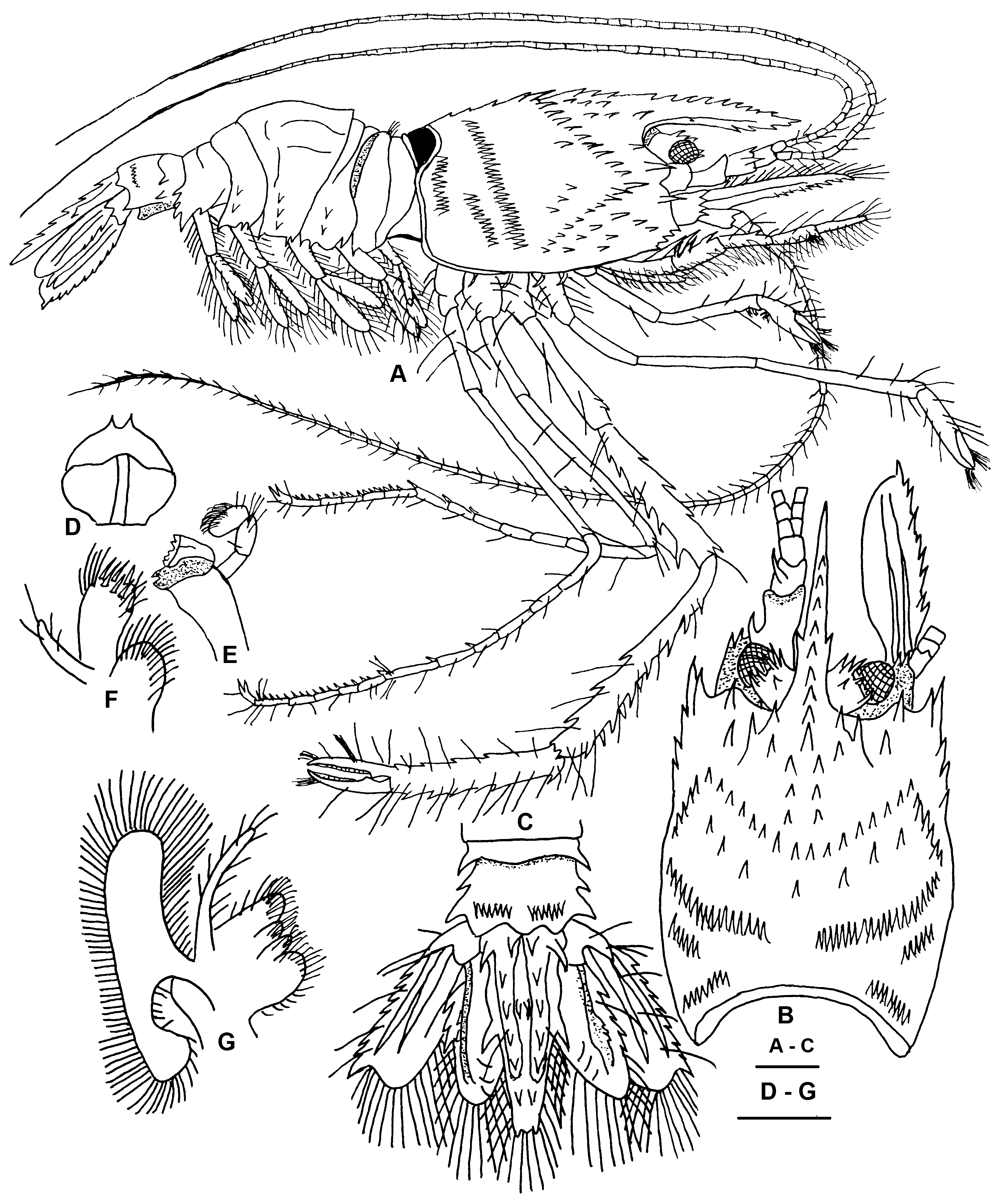
Redescription. The two females collected in New Caledonia agree rather well with the extensive description given by Holthuis (1946) based on 3 specimens from Indonesia. The only illustrations of this species are Dana (1852; pl. 40, fig. 9) who gave a lateral view of the type that lacked third pereiopods and that of Holthuis (1946) showing the spination of the third pereiopods. Therefore, a description and illustrations of whole animal, mouthparts and appendages for the specimen from Ile Ouen are presented here ( Figs. 15 View FIGURE 15 , 16 View FIGURE 16 ).
Rostrum ( Figs. 15 View FIGURE 15 A, B) compressed, slender, reaching past antennular peduncle, with 9 dorsal, 4 ventral, and small lateral spines.
Carapace ( Figs. 15 View FIGURE 15 A, B) with distinct cervical groove with cincture of 18 spines; postcervical groove with cincture of 67 spinules. Short tranverse row of 9 spinules between cervical and postcervical grooves with row of 3 spinules above tranverse row. Short tranverse row of 6–9 spinules posterior to postcervical groove, short tranverse row of 12 spinules near posterior margin. Near anterior margin; anterolateral angle few scattered spinules. Strong antennal, branchiostegal spines; 4 smaller pterygostomian spines.
First abdominal pleomere ( Fig.15 View FIGURE 15 A) with transverse carina, dorsally with row of setae, pleura ends with posterior tooth. Second abdominal pleomere with transverse carina ending midway; 2 spines laterally; posterior pleural margin ending in 1 anterior, 2 posterior teeth. Third pleomere with top broadly rounded; dorsal surface with 2 longitudinal carinae; tranverse carina posteromedially; 3 spines laterally; posterior pleural margin ending in 2 separated teeth. Fourth pleomere without carinae; 2 spines laterally; posterior pleural margin ending in 1 large posterior, 2 small anterior teeth. Fifth pleomere with short transverse carina ending about midway; posterior pleural margin with 3 large teeth; pleura laterally with 3 spines. Sixth pleomere with short transverse carina in anterior median part; 2 spines laterally; posterior part of pleomere with two rows of 7 spinules.
Eyes ( Figs. 15 View FIGURE 15 A, B) pigmented, cornea equal peduncle length. Ophthalmic peduncle robust, dorsally and medially with 6 strong spines.
Telson ( Fig. 15 View FIGURE 15 C) long, lanceolate, median longitudinal carinae with 6 strong spines. Telsonal base with pair of large outer spines; 2 spines about midway between longitudinal carinae. Lateral margins with strong spine at midlength; posterior marigin with pair of small rounded spines.
Uropods ( Fig. 15 View FIGURE 15 C) well developed with rami not exceeding telsonal length; protopodite stout, with strongly projecting lateral edge. Exopodite with 8 teeth on its outer margin, including slightly larger terminal tooth; dorsal surface bears 2 longitudinal ridges. Endopodite lobate, outer margin with 3–5 teeth; dorsal surface with one longitudinal ridge bearing 2–3 small spines on outer margin.
Antennular peduncle ( Fig. 15 View FIGURE 15 B) extending to middle of scaphocerite; proximal segment longest with strong stylocerite on outer margin; middle segment less than 0.5 proximal segment length with large spine distomedially; distal segment slightly shorter than middle segment, unarmed. Upper and lower flagella reaching nearly to end of telson; first 8 or 9 segments bear aesthetascs.
Antenna ( Fig. 15 View FIGURE 15 B) with scaphocerite reaching slightly beyond rostrum, outer margin with 9 teeth, upper surface with 2 distinct longitudinal carinae; basicerite with 3 distal spines. Flagellum well developed extending beyond tip of telson.
Epistome ( Fig. 15 View FIGURE 15 D) rounded anteriorly with 2 median spines; labrum normally developed; paragnath with median fissure.
Mandible ( Fig. 15 View FIGURE 15 E) robust, with short, fused molar and incisor processes; incisor with 7 small teeth; palp well developed, 3-segmented; segments equal in length; proximal segment glabrous, middle and distal segments setose.
Maxillule ( Fig. 15 View FIGURE 15 F) with slender undivided endopodite with 4 plumose setae; proximal endite broad, truncate distally, with numerous distal compound spinose and plumose setae; distal endite rounded with numerous plumose setae.
Maxilla ( Fig. 15 View FIGURE 15 G) with numerous plumose setae on both lobes of coxal and basal endites. Endopodite long, slender, exceeding anterior margin of scaphgnathite with distal and outer marginal plumose setae. Scaphognathite long, narrow, fringed with numerous plumose setae.
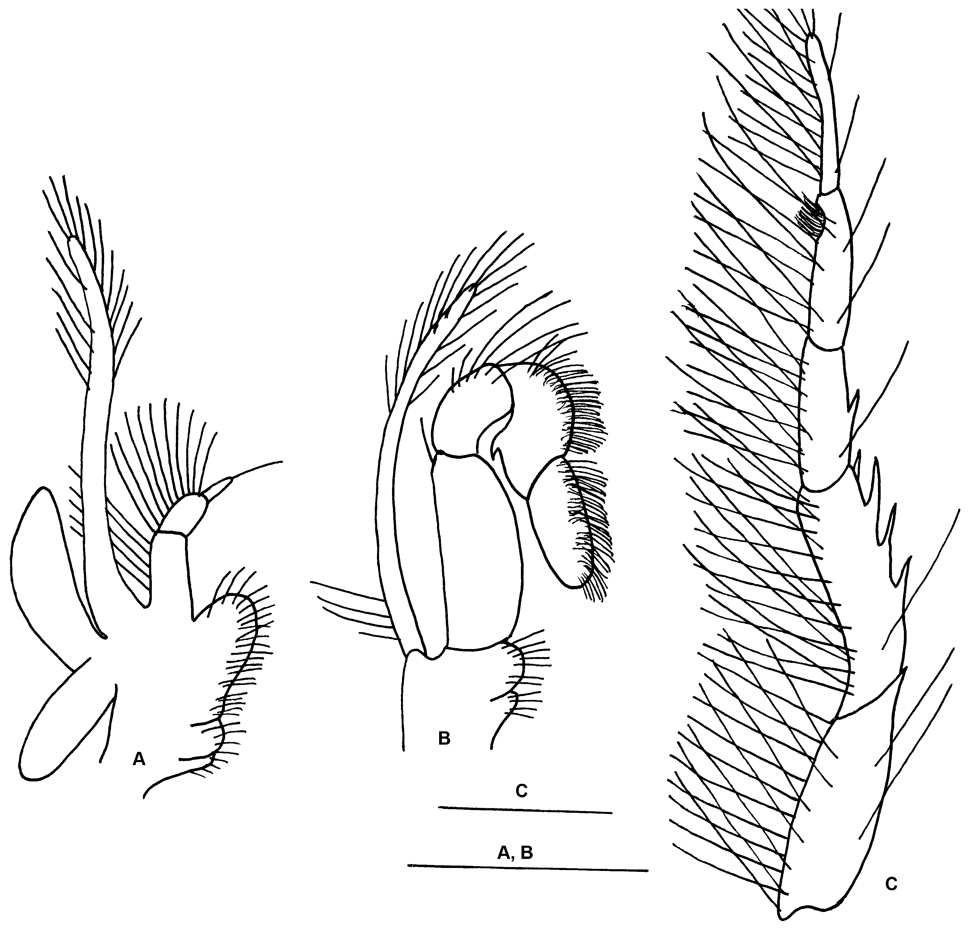
First maxilliped ( Fig. 16 View FIGURE 16 A) with 3-segmented endopodite; proximal segment longest with 6 long plumes setae on outer margin; middle segment 0.5 proximal with 10 long plumose setae on outer margin; distal segment equal middle segment in length with simple terminal seta. Basipodite large, rounded anterior, slightly concave middle, rounded posterior, bearing dense fringe of plumose setae; coxopodite bilobed with few plumose setae. Exopodite long with 16 long plumose distolateral setae. Large epipod with proximal and distal lobes.
Second maxilliped ( Fig. 16 View FIGURE 16 B) with 4-segmented endopodite; dactylus elongate with dense fringe of short setae along distodorsal margin; propodus equal dactylar length, densely setose on dorsal margin, ventral margin bearing acute proximal tooth; carpus equal propodal length with 2 short, 4 longer setae at distodorsal edge; merus 2.5 dactylar length, single distodorsal seta, ventral margin slightly convex with some long simple setae (not shown); ischium fused to basis, both lobate with few short setae. Exopodite long, slender with 4 proximal dorsal seate and 14 plumose setae distally.
Third maxilliped ( Fig.16 View FIGURE 16 C) with 7-segmented endopodite, dactylus, propodus, and carpus equal in length, with numerous long plumose setae; propodus with setiferous organ at distomesial angle; merus, ischium equal in length, merus with outer midlength spine; ischium with 3 large outer margin spines, smaller spine at distomesial angle; basis and coxa fused, unarmed; exopodite long, with distal half bearing numerous plumose setae (not shown).
First pereiopod ( Fig 15 View FIGURE 15 A) with segments without spines; fingers bearing small tufts of setae; distoventral part of carpus and distoproximal part of propodus with weak setiferous organ.
Second pereiopod ( Fig.15 View FIGURE 15 A) longer than first, segments without spines, fingers bearing small tufts of setae.
Third pereiopod ( Fig. 15 View FIGURE 15 A) strongest, slender; ischium with 2 anterodorsal spines; merus with 4 dorsal, 6 ventral spines; carpus with 7 dorsal, 3 dorsomesial and 3 ventral spines, anterior ending in 3 small spines; propodus with 6 dorsal, 4 ventral spines; dactylus unarmed. Cutting edges with large proximal tooth on propodus, dactylus, distally with rows of small peglike teeth separated by chitinous laminae.
Fourth and fifth pereiopods ( Fig. 15 View FIGURE 15 A) long, slender, carpi divided into 6 segments with 3 ventral movable spines; propodi divided into 5 segments with 15–16 ventral movable spines; dactylar unguis and corpus not separated.
Measurements (mm). PCL: 3.9, 4.0; RCL: 5.5, 5.6; TL: 12.7, 13.2. Eggs in both specimens were few in number and in very early stages of embryonic development, measuring 0.15 × 0.18 mm.
Distribution. Fiji, North Celebes, Moluccas, Indonesia, Zanzibar, Madagascar, Kenya, New Hebrides Islands, Australia, and New Caledonia (see remarks).
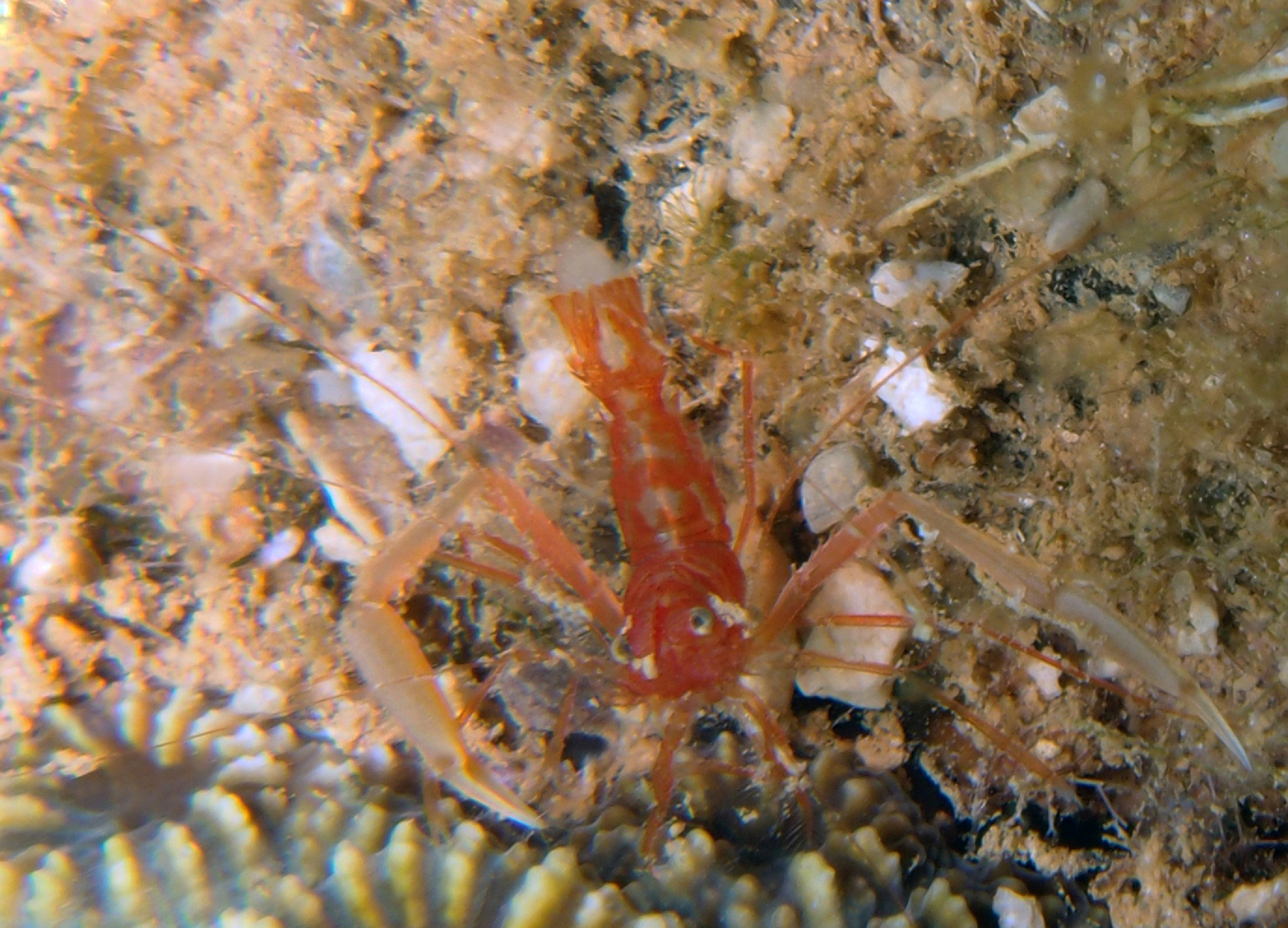
Coloration. ( Fig. 17 View FIGURE 17 ) Body and appendages generally light red, semitransparent. First to sixth abdominal pleomeres with transverse red band posteriorly; rostrum, carapace and antennae red. Eyes brown pigmented inside, whitish outside. First three pereiopods with fixed finger and dactylus white; third pereiopod propodus, carpus light orange, merus and ischium light red. Edge of telson and uropods red.
Hosts. Hayashi (1986) collected a female O. ensifera from the Solomon Islands that was associated with a crinoid, which was later identified as O. crinoidicola Saito & Fujita, 2009 . However, one specimen of O. ensifera from Indonesia (NTM Cr 009999) examined in this study was found associated with a crinoid. Bruce (1982) mentions three specimens of Odontozona sp. (no longer extant, A. J. Bruce, personal communication) from Zanzibar and Indonesia found in association with crinoids. Comparisons of Bruce’s fig. 2H with all the specimens examined here, leads this author to believe that these three specimens of Bruce belong to O. ensifera .
Remarks. An additional 26 specimens were examined throughout this species’ range of Indonesia (USNM 315621, NTM Cr 0 0 994, Cr 009999), Zanzibar (MCZH 5504), Madagascar (MNHM-NA 3908), Kenya ( FSM KE- 5), Espirtu Santos, New Hebrides Islands (AHF), off Elliot Point, Darwin (NTM Cr 007213), Arafura Sea (NTM Cr 0 0 9651, Cr 009859), and Northwest Shelf of Australia (NTM Cr 0 0 116, Cr 0 0 3738, Cr 0 0 3740, Cr 0 0 3743, Cr 0 0 3744, Cr 0 0 3747, Cr 0 0 3750, Cr 003754). This has allowed the summation of the variation seen in this small species of Odontozona . The rostrum bears 6–9 dorsal, 0–4 ventral, and 0–2 lateral spines. The cervical groove bears 10–18 marginal spines, while there are 41–70 spinules along the postcervical groove. The outer margin of the scaphocerite bears 0–1 spine at the base and 5–9 teeth distally. There are 1–5 meral teeth on the third maxilliped. The outer margin of the uropodal exopodite bears 5–9 teeth while this margin has 1–4 teeth on the endopodite. The fourth and fifth pereiopods bear 6–16 propodal movable spines. All examined specimens have carinate and grooved pleomeres. The segmentaion of the carpus and propodus of the fourth and fifth pereiopods are unreliable characters to differentiate species of Odontozona because in most cases the divisions are rather indistinct.
No known copyright restrictions apply. See Agosti, D., Egloff, W., 2009. Taxonomic information exchange and copyright: the Plazi approach. BMC Research Notes 2009, 2:53 for further explanation.
|
Kingdom |
|
|
Phylum |
|
|
Class |
|
|
Order |
|
|
Family |
|
|
Genus |
Odontozona ensifera ( Dana, 1852 )
| Goy, Joseph W. 2015 |
Odontozona
| Bruce 1982: 191 |
Odontozona ensifera
| Hendrickx 2014: 345 |
| Anker 2013: 429 |
| De 2011: 252 |
| Goy 2010: 217 |
| Saito 2009: 124 |
| Okuno 2003: 175 |
| Nomura 1996: 9 |
| Pretus 1990: 349 |
| Manning 1990: 31 |
| Dounas 1989: 345 |
| Burukovsky 1983: 131 |
| Gore 1981: 158 |
| Goy 1981: 850 |
| Holthuis 1946: 34 |
Stenopus ensiferus
| Edwards 1909: 264 |
| Lo 1903: 252 |
| Herrick 1893: 352 |
| Bate 1888: 210 |
| Dana 1852: 27 |
| Dana 1852: 607 |