
Oncodopus brongniarti, Ünal, Mustafa & Beccaloni, George W., 2017
|
publication ID |
https://doi.org/ 10.11646/zootaxa.4341.2.2 |
|
publication LSID |
lsid:zoobank.org:pub:05152B19-56AA-4CCD-A3C6-53EA3369A54C |
|
DOI |
https://doi.org/10.5281/zenodo.5611851 |
|
persistent identifier |
https://treatment.plazi.org/id/533F87C4-FF82-7329-26E2-FD18FF6ACCB0 |
|
treatment provided by |
Plazi |
|
scientific name |
Oncodopus brongniarti |
| status |
sp. nov. |
Oncodopus brongniarti View in CoL sp. nov.
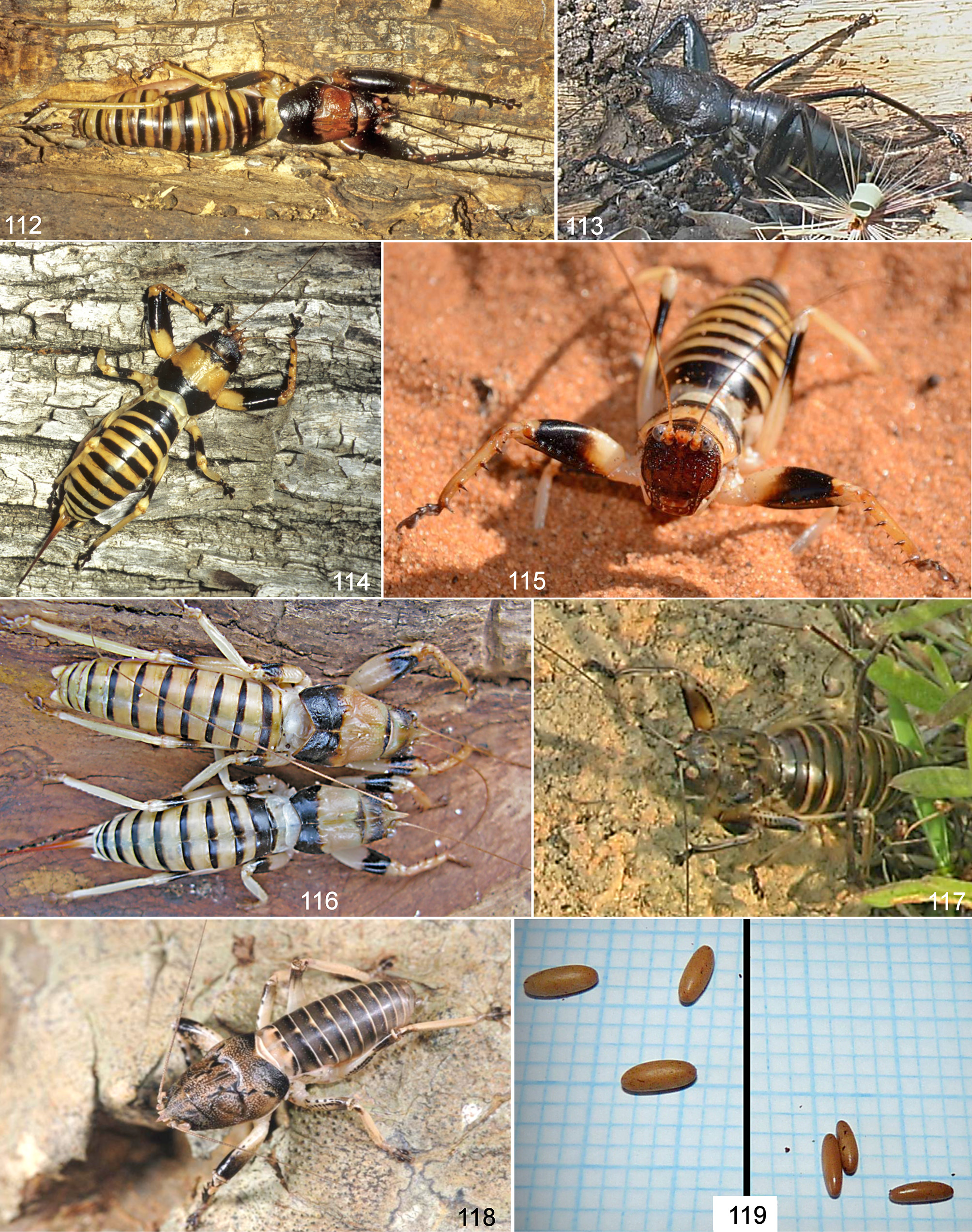
( Figs. 30–39 View FIGURES 30 – 39 , 110 View FIGURES 109 – 111 , 114–116 View FIGURES 112 – 119 )
http://lsid.speciesfile.org/urn:lsid:Orthoptera.speciesfile.org:TaxonName:499686
Type locality. Madagascar, Toliara Province, S. Morondava, Belo Sur Mer. Holotype male ( NHMUK).
Description. Male (Holotype): Body size average for genus ( Tab. 2 View TABLE 2 ) (in one male body size quite large). Fastigium of vertex ( Figs. 30, 31 View FIGURES 30 – 39 ) simple, conical, forming a short horn, upper surface flattened with a narrow longitudinal median furrow, 2.3 times longer than diameter of eye, basal part 1.4 times wider than antennal scape. Fastigium of frons ( Fig. 31 View FIGURES 30 – 39 ) with a spiniform slightly downcurved tooth. Face flattened, strongly granulated; each tubercle like a small tooth, of which the lower two are larger. Pronotum (30, 31) long, 3.2 times longer than its height and 1.2 times longer than its width; anterior margin convex; posterior margin with an angular, large median incision; upper surface slightly concave in lateral view; prozona 1.9 times longer than metazona. Prothoracic auditory spiracle large, oval; a small portion of its opening concealed under pronotum. Mesothoracic auricle small, short; its opening almost triangular; ventral lobe short. Prosternum with two long, V-shaped spines, reaching to level of ventral margin of fore coxa; widely separated towards to apex. Fore coxa with a short spine projecting forward. Femora without dorsal spines. Fore femur ( Fig. 30 View FIGURES 30 – 39 ) with a large inner spine on anterio-lateral margin; with 6 inner spines ventrally. Fore tibia ( Fig. 30 View FIGURES 30 – 39 ) with 5 inner and 5 outer spines ventrally. Mid femur with 2 ventral spines on outer margin. Hind femur with 2 outer spines ventrally (1–4 spines in other males). Fore and mid tibia without dorsal spines. Hind tibia with 7 dorsal spines on inner margin (other males with 5–6 spines); with 4 apical spurs ventrally, inner 2 slightly smaller than outer ones; without apical spurs dorsally. All tibia with ventral spines on both margins. Tegmina ( Figs. 30, 31 View FIGURES 30 – 39 ) concealed under pronotum, its apex visible from the posterior incision of pronotum in dorsal view, reaching to half of metanotum. Last tergite ( Fig. 33 View FIGURES 30 – 39 ) wider than long, posterior margin with a shallow, wide, rounded median incision. Cercus ( Fig. 34 View FIGURES 30 – 39 ) stout, apical part with a very small tooth (which is not spiniform) on its ventral margin; inner arm very long, its apex slightly bent backward, pointed with a distinct tooth. Subgenital plate ( Fig. 35 View FIGURES 30 – 39 ) almost as long as wide, its styli distinctly longer than depth of median incision.
Female: Face as in male. Fastigium of vertex ( Fig. 36 View FIGURES 30 – 39 ) short, 1.8 times longer than diameter of eye, its basal part 1.5 times wider than antennal scape. Pronotum ( Figs. 36, 37 View FIGURES 30 – 39 ) as wide as long; posterior margin with a large rounded incision; pronotum 2.9 times longer than its height; prozona 3 times longer than metazona. Tegmina ( Fig. 37 View FIGURES 30 – 39 ) strongly reduced, scale like laterally, reaching to half of mesonotum. Prosternal spines V-shaped, slightly shorter than that of male. Fore femur ( Fig. 36 View FIGURES 30 – 39 ) with 6–7 ventral spines on inner margin and a large inner spine on anterio-lateral margin. Tibial spines as in male. Mid femur with 2 outer spines ventrally, in other females with 2–4 spines. Hind femur with 2 ventral spines on outer side. Last tergite very short and wide, its posterior incision distinct. Cercus stout, basal part broad, apical third strongly and sharply narrowed, its apex slightly incurved and pointed. Subgenital plate ( Fig. 39 View FIGURES 30 – 39 ) almost triangular with a median carina, its apex with a small rounded incision. Ovipositor ( Fig. 38 View FIGURES 30 – 39 ) clearly longer than hind femur, slightly upcurved.
Colour (dry specimens). Body milky brown with distinct transverse dark reddish-brown bands. Head reddish brown. Eye, fastigium of vertex, labrum, maxillary and mandibulary palps slightly lighter. Anterior part of pronotum milky brown, posterior part (metazona) very dark with reddish-brown. In female metazona dark brown. All tibiae milky brown. Apical part of each femur dark, with a large black or reddish-black band. Posterior half of first abdominal tergite in male and the whole of it in the female dark, black or reddish brown; posterior part of each abdominal tergite with transverse dark band, black or reddish brown. All sternites, cerci and subgenital plate yellowish milky brown. Ovipositor milky brown in basal 1/3, reddish brown in remaining part.
Diagnosis. Based on the colour pattern (with distinct transverse dark bands), the male cercus with a long inner arm, and the shape of the ovipositor this new species appears to be most closely related to O. zonatus . It differs from it by the absence of a distinct apical spine on the male cercus, the larger median incision of the last tergite in both sexes, the more pronounced tubercles of the face and the female cercus. It is recognized by the shape of the male cercus and the last tergite.
Material examined. Madagascar, Toliara Province, Belo Sur Mer ( South of Morondava ), 20 ° 44'53.63" S, 43 ° 59'36.97" E, 22.11.2007, Coll. G.W. Beccaloni, 2 males (including Holotype) GoogleMaps , 2 females, under bark of dead tree; Madagascar, S. Centr . Madagascar, 1 male [no more data] ; Toliara province, Forest near village of Mangily , c. 27 km N. of Toliara, (-23.15, 43.60) [23 ° 8'60'' S, 43 ° 36'0'' E], 11.11.2004, Coll. G.W. Beccaloni, 1 female, under bark of dead tree ; Madagascar, Foret de Zombitsy [Zombitse-Vohibasia N.P.], near Sakaraha , 650 m, 16.12.1959, 1 female (leg. E. S. Ross) (all in NHMUK).
Measurements (mm). Holotype (male): Body length: 40.3; pronotum length: 14.2; width of pronotum: 12.1; fore femur length: 14.5; width of fore femur: 5.7; fore tibia length: 13.3; hind femur length: 15. See Table 2 View TABLE 2 for the range of measurement of the other specimens including females.
Etymology. This species name is dedicated to Charles Brongniart who was the first to discover this group of insects in 1897. He is the author of the genus Oncodopus and the species Oncodopus zonatus and Colossopus redtenbacheri .
Distribution. South-west and south-central Madagascar ( Fig. 110 View FIGURES 109 – 111 ). Toliara Province: Toliara, Mangily, Foret de Zombitsy [Zombitse-Vohibasia N.P.], Morondava.
Habitat. This species is known from spiny forest, dry costal scrub and dry deciduous forest. It has been found under the bark of dead trees.
Phenology. Adults have been found in November and December.
Remarks. The body size is variable as in above 2 species (see under the Remarks section for O. zonatus and Table 2 View TABLE 2 ).


| NHMUK |
Natural History Museum, London |
No known copyright restrictions apply. See Agosti, D., Egloff, W., 2009. Taxonomic information exchange and copyright: the Plazi approach. BMC Research Notes 2009, 2:53 for further explanation.
|
Kingdom |
|
|
Phylum |
|
|
Class |
|
|
Order |
|
|
Family |
|
|
Genus |