
Monarea Szépligeti, 1904
|
publication ID |
https://doi.org/ 10.11646/zootaxa.3795.4.2 |
|
publication LSID |
lsid:zoobank.org:pub:86D99803-D8AE-47B0-B3EF-EA66A66C9CFA |
|
DOI |
https://doi.org/10.5281/zenodo.6133743 |
|
persistent identifier |
https://treatment.plazi.org/id/03B8B527-1966-8F41-FF0E-FDB76A65DC60 |
|
treatment provided by |
Plazi |
|
scientific name |
Monarea Szépligeti, 1904 |
| status |
|
Monarea Szépligeti, 1904 View in CoL View at ENA
Szépligeti, 1904: 68; Enderlein, 1912: 35; Shenefelt & Marsh, 1976: 1320; Fischer, 1981a: 243; Yu et al., 2012. Type species: Osmophila fasciipennis Szépligeti, 1902 .
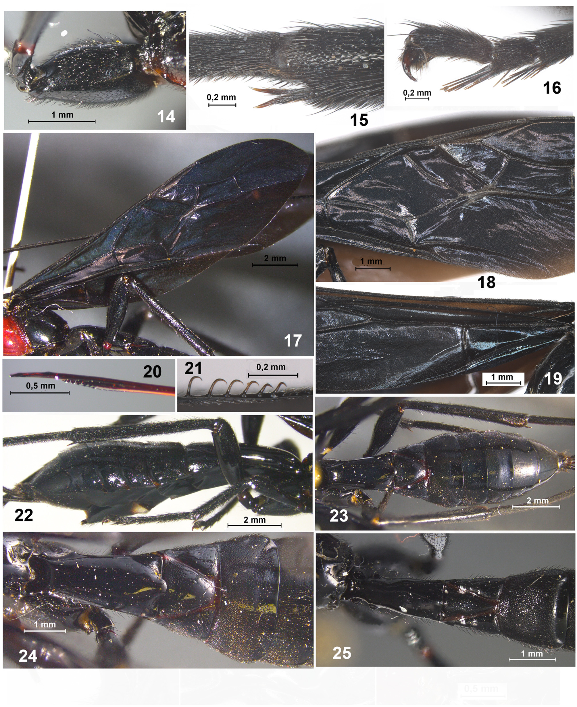
Diagnosis. Members of this genus can be distinguished from other doryctine taxa by having the following combination of characters: frons distinctly concave and with high median keel ( Figs 7, 8 View FIGURES 2 – 13 ); propleuron with additional lower lateral lobes slightly before its anterior margin ( Fig. 11 View FIGURES 2 – 13 ); sternaulus very shallow ( Fig. 10 View FIGURES 2 – 13 ); most part of body smooth ( Fig. 2 View FIGURES 2 – 13 ); discoidal (first discal) cell of fore wing sessile anteriorly ( Figs 17, 18 View FIGURES 14 – 25 ); brachial (first subdiscal) cell of fore wing closed apico-posteriorly by very short brachial (2cu-a) vein ( Fig. 18 View FIGURES 14 – 25 ); recurrent (m-cu) vein of fore wing strongly antefurcal; submedial (subbasal) cell of hind wing long ( Fig. 19 View FIGURES 14 – 25 ); recurrent (m-cu) vein of hind wing completely absent; medial (basal) cell of hind wing not widened in apical half, narrow and long; hind coxa without basoventral corner and tubercle ( Fig. 14 View FIGURES 14 – 25 ); hind basitarsus long; second metasomal tergite with subtriangular areas delineated by deep furrows ( Figs 23–25 View FIGURES 14 – 25 ).
Monarea Szépligeti is morphologically similar to Megaloproctus Schulz, 1906 , but differs from the latter genus by having the following combination of main characters: frons concave and with distinct median keel, propleuron with additional lower lobes slightly before its anterior margin, discoidal (discal) cell of fore wing almost sessile, recurrent vein (m-cu) of hind wing absent, medial (basal) cell of hind wing not widened from middle towards apex, and second metasomal tergite with median area delineated by deep furrows. Within the Holcobracinini, Monarea is similar to Nervellius Roman , but differs from it in having the medial (basal) cell of hind wing narrow, not widened towards apex; radial (marginal) cell of hind wing without transverse (r) vein; propleuron with additional lower lobes slightly before its anterior margin; second metasomal tergite with median area delineated by deep furrows; propodeum and all metasomal tergites entirely smooth, and in lacking the recurrent vein (m-cu) of hind wing.
Description. Head. Head not depressed, transverse. Vertex smooth. Ocelli arranged in triangle with base distinctly larger than its sides ( Fig. 4 View FIGURES 2 – 13 ). Frons distinctly concave, with distinct, flat, wide and high median longitudinal keel in its anterior 0.5–0.7, without lateral protuberances and lateral longitudinal carina ( Figs 7, 8 View FIGURES 2 – 13 ). Eyes glabrous. Occipital carina dorsally complete, though largely fine or interrupted in some areas, concavely curvedly below and fused with hypostomal carina. Malar suture absent. Clypeus low, semi-round, delineated from face by distinct furrow, with fine and short lower flange. Hypoclypeal depression large and round ( Fig. 3 View FIGURES 2 – 13 ). Postgenal bridge narrow. Maxillary palpus of medium length, 6-segmented, three basal segments thickened, third segment distinctly widened in apical half, sixth segment about as long as fifth segment or shorter. Labial palpus short, 4-segmented, two basal segments thickened, third segment more or less shortened ( Fig. 6 View FIGURES 2 – 13 ). Scape of antenna ( Fig. 5 View FIGURES 2 – 13 ) wide and short, apically without flange and lobe, without basal constriction; ventral margin of scape distinctly shorter than dorsal margin (lateral view). First flagellar segment subcylindrical, not curved, slightly longer than second segment. Flagellar segments entirely covered with short and very dense black setae. Apical segment pointed apically, without apical spine.
Mesosoma ( Figs 9, 10 View FIGURES 2 – 13 ). Mesosoma not or (male) slightly depressed, long. Neck of prothorax short. Pronotum slightly convex dorsally (lateral view), with short and up-curved anterior flange; pronotal carina absent. Pronope absent. Sides of pronotum mainly smooth, with distinct subvertical groove situated closely to its posterior margin.
Propleuron with a pair of additional lower lateral lobes immediately before its anterior margin ( Fig. 11 View FIGURES 2 – 13 ). Propleural dorsoposterior flange long and narrow. Mesonotum highly and roundly elevated above pronotum ( Figs 2, 9 View FIGURES 2 – 13 ), entirely smooth. Median lobe of mesonotum without median longitudinal furrow, its anterolateral corner wide and obtuse. Notauli more or less complete, mainly shallow, deeper anteriorly, wide, entirely smooth or finely crenulate anteriorly. Tegula distinctly widened distally, concave along outer margin ( Fig. 12 View FIGURES 2 – 13 ). Scuto-scutellar (transscutal) suture distinct and complete. Prescutellar depression long medially, usually narrow laterally, more or less shallow, with several high carinae. Lateral longitudinal flanges on level of prescutellar depression low. Scutellum slightly convex, not shorter than anterior width, without lateral carinae. Subalar depression deep and narrow. Mesopleural pit fine and small. Sternaulus shallow, narrow, long, almost straight, oblique. Prepectal carina distinct, high below, laterally before sternaulus, strongly curved towards apex of prepectus. Postpectal carina absent. Metanotum without median tooth. Metapleural flange short or very short, subpointed or rounded apically, with carina prolongated posteriorly along lower margin of metasoma ( Fig. 13 View FIGURES 2 – 13 ). Propodeum without areas, mostly smooth; lateral tubercles and propodeal bridge absent ( Figs 9, 10 View FIGURES 2 – 13 ). Propodeal spiracles small and bean-shaped. Metapleuron slightly convex, entirely smooth, indistinctly separated from propodeum.
Wings ( Figs 17–19 View FIGURES 14 – 25 ). Pterostigma of fore wing narrow and long. Radial (r) vein arising almost from middle of pterostigma ( Fig. 18 View FIGURES 14 – 25 ). Radial (marginal) cell not shortened. Both radiomedial veins (RS and r-m) present. Second radiomedial (submarginal) cell short or long, narrow. Recurrent (m-cu) vein distinctly antefurcal. Nervulus (1cu-a) slightly postfurcal or interstitial. Discoidal (first discal) cell sessile or subsessile anteriorly. Basal (M) and recurrent (m-cu) veins subparallel. Parallel (2CUb) vein not interstitial, arising from posterior 0.15–0.20 of apical margin of brachial (first subdiscal) cell. Brachial (first subdiscal) cell closed postero-apically, brachial (2cu-a) vein short, complete and sclerotised. Transverse anal veins (1a and 2a) absent. Hind wing with at least seven hamuli ( Fig. 21 View FIGURES 14 – 25 ). First abscissa of costal vein (C+Sc+R) shorter than second abscissa (SC+R). Radial (RS) vein arising from costal (R) vein closely to basal (r-m) vein, or from basal (r-m) vein closely to costal (R) vein. Radial (marginal) cell subparallel-sided or slightly narrowed apically, without transverse (r) vein. Medial (basal) cell ( Fig. 19 View FIGURES 14 – 25 ) not widened from middle towards apex, narrow, 0.5–0.6 times as long as hind wing. Nervellus (cu-a) present, perpendicular to mediocubital vein. Submedial (subbasal) cell long. First abscissa of mediocubital vein (M+CU) slightly longer than second abscissa (1M). Recurrent vein (m-cu) completely absent ( Fig. 19 View FIGURES 14 – 25 ).
Legs. Fore tibia on inner surface with several short and strong spines arranged in almost vertical line or narrow vertical stripe. Fore tarsus 2.5–3.0 times longer than fore tibia. Middle tibia with a few or single spines on lateral surface. Middle tarsal segments long. Hind coxa short and wide in female, long and narrow in male ( Fig. 14 View FIGURES 14 – 25 ), without basoventral angle and tubercle, dorsally without spines or teeth ( Fig. 14 View FIGURES 14 – 25 ). Fore and middle femora with low but distinct dorsal protuberances. Hind femur slender, elongate-oval. Hind tibia with area of dense numerous setae on its inner distal part. Hind tibial spurs short, slightly curved, glabrous apically ( Fig. 15 View FIGURES 14 – 25 ). Basitarsus of hind tarsus almost as long as second-fifth segments combined. Claws short, simple, coarse, strongly curved ( Fig. 16 View FIGURES 14 – 25 ).
Metasoma ( Figs 22–25 View FIGURES 14 – 25 ). First tergite not petiolate, medium size or elongated, wide, convex dorsally (lateral view). Acrosternite of first segment not elongate, 0.15–0.20 times as long as first tergite, finishing before spiracular level. Dorsope of first tergite large and deep; basolateral lobes short and subpointed; spiracular tubercles distinct, situated in basal 0.3 of tergite; tergites subbasally without semi-circular transverse carina, almost without dorsal carinae. Second tergite with deep and convergent but usually not united posteriorly furrows fusing with second suture, furrows and second suture delineated almost in “V” or “U” shape, but (rarely) suboval area (with usually rounded posterior corner) ( Figs 24, 25 View FIGURES 14 – 25 ). Second suture deep, complete, almost straight or slightly sinuate. Third tergite without transverse furrow. Second to fourth or fifth tergites with separate laterotergites. Tergites behind second one mainly cover by dense, short and almost erect setae, only their posterior narrow areas bare. Hypopygium protruding posteriorly, with short and wide median process and pair lateral cuttings on posterior margin ( Fig. 22 View FIGURES 14 – 25 ). Ovipositor subapically with two low and widely separated nodes. Ovipositor sheath longer than metasoma.
Distribution. Neotropical Region ( Brazil, Peru, British Guiana, Mexico).

No known copyright restrictions apply. See Agosti, D., Egloff, W., 2009. Taxonomic information exchange and copyright: the Plazi approach. BMC Research Notes 2009, 2:53 for further explanation.