
Paracharon caecus Hansen, 1921
|
publication ID |
https://doi.org/ 10.1206/4000.1 |
|
persistent identifier |
https://treatment.plazi.org/id/043487D1-2C5D-B64B-FE25-C58D210D4AF2 |
|
treatment provided by |
Felipe |
|
scientific name |
Paracharon caecus Hansen, 1921 |
| status |
|
Paracharon caecus Hansen, 1921 View in CoL
Figures 2 View FIGURE 2 , 4C View FIGURE 4 , 5C View FIGURE 5 , 6C, 7E, F, 8E, F, 10A, 11A, 13A, B, 14A, B, 15A, B, 16A, B, 17A, B, 19A; table 1
Paracharon caecus Hansen, 1921: 11 View in CoL , 12, pl. 1, fig. 2A–E; 1930: pl. 14, fig. 7A; Mello-Leitão, 1931: 53; Werner, 1935: 470; Turk, 1964: 239; Cloudsley-Thompson, 1968: 156; Lawrence, 1968: 2; Mullinex, 1975: 28; Quintero, 1981: 163; 1983: 50; Delle Cave, 1986: 158; Quintero, 1986: 206, figs. 8, 28; Weygoldt, 1994: 242; 1996: 188, 191, 193–198, figs. 1, 12, 21, 34, 39, table 2; 1999: 104, fig. 1; 2000a: 14, 17, 23, 50, 86, 135, 139, figs. 14, 40–42, 103, 185; 2000b: 340, 346, fig. 2; Harvey, 2002b: 364; 2003: 31; Shultz, 2007: 249; Dunlop et al., 2008: 165, 166, 167, 172, 174, fig. 7B; Fahrein et al., 2009: 456; Penney, 2009: 72; Santer and Hebets, 2011: 3; Engel and Grimaldi, 2014: 1, 2, 13; Réveillon and Maquart, 2015: 190, 196; Beron, 2016: 484; Giupponi and Miranda, 2016: 7; Maquart and Réveillon, 2016: 27, 33; Garwood et al., 2017: 1, 3, 4, 7, 10, figs. 1C, D, 2A–F; Beron, 2018: 95, 469, 682, table 8.4; McArthur et al., 2018: 62; Miranda et al., 2018a: 24; 2018b: 35, figs. 14, 15A; Miranda and Reboleira, 2019: 9; Nadein and Perkovsky, 2019: 603; Deharveng and Bedos, 2019: 125; Haug and Haug, 2021: 406–409, table 1; Schmidt et al., 2022: 164; Miranda et al., 2022: 161.
Paracharon coecus: Fage, 1939: 153 , 160; 1954: 182.
Pracharon caecus: Weygoldt, 1999: 106 View in CoL , fig. 1.
TYPE MATERIAL: The original series comprised 17 syntypes with the following data: GUINEA BISSAU: Bolama [11°34.629′N 15°29.237′W], vi–xii.1899, 1 ex. [sex indet.] GoogleMaps ; Rio Cassine [11°07.687′N 15°01.317′W], i–ii.1900, L. Fea, 1 ex. GoogleMaps ; same data, except ‘ iv.1900,’ 15 ex. ( ZMUC 24556 View Materials ). Only 2 ♀♀ ( ZMUC 24556 View Materials ), hereby designated lectotype and paralectotype GoogleMaps of P. caecus , could be located and examined for the present investigation; all other specimens are presumed lost. The specimen selected as lectotype is in better condition than the specimen selected as paralectotype.
DIAGNOSIS: As for genus.
DESCRIPTION: Based on adult female lectotype (fig. 4C). Measurements (mm) in table 1.
Coloration: Body and appendages orange-brown, carapace darker (fig. 4C).
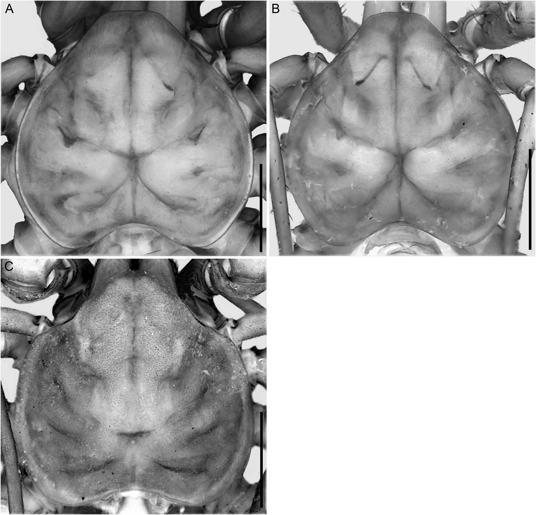
Carapace: Shape cordate (fig. 5C), as wide as long, with bumpy area anteromedially; anterior margin rounded, markedly projecting, with lateral margins curved, width of projection half width of carapace; posterior margin with shallow median notch. Two median and four pairs of dorsosubmedian transverse sulci, all distinct; anteromedian sulcus extending from anterior margin to 0.4 of carapace length, not connected to fovea; posteromedian sulcus extending from posterior margin to fovea; in anterior-posterior axis, first and third dorsosubmedian pairs of sulci not connected to fovea, second and fourth pairs connected to fovea. Median ocular tubercle, and median and lateral ocelli absent (fig. 5C). Frontal process projecting and recurved, 1.5× wider than long (fig.
6C). Cuticle strongly sclerotized, pigmented,
apodemes barely visible.
Sterna: Tetra segmented (fig. 7E, F). Tritosternum vestigial, not projecting, consisting of single, flat, subtriangular plate, with single anteromedian microseta, pair of lateral setae and pair of posterosubmedian macrosetae. Tetrasternum comprising single plate with rounded apex bearing two setae, and with two setae on surface of plate. Pentasternum comprising single plate, projecting slightly ventrally and terminating in rounded apex bearing two setae, and with two setae on surface of plate. Metasternum comprising single subtriangular plate, anterior margin narrow, posterior margin wider, with two ventrosubmedian setae in posterior half.
Chelicerae: Basal segment, retrolateral margin without teeth but with vestigial eminence
(fig. 8F); prolateral margin with three teeth (fig.
8E), first tooth (ventralmost) ca. 1.5× longer and FIGURE 6. Paracharontidae Weygoldt, 1996 , cara- broader than other teeth, second tooth absent, pace, anterior aspect. A, B. Jorottui ipuanai , gen. et third and fourth teeth similar in size, fourth sp. nov. A. Paratype Ƌ (ICN-Am 187). B. Holotype tooth (dorsalmost) bicuspid, dorsal cusp slightly ♀ (ICN-Am 180). C. Paracharon caecus Hansen, 1921 , lectotype ♀ (ZMUC 24556). Scale bars: A, C, larger than ventral cusp; prolateral surface with 0.1 mm; B, 0.2 mm. transverse row of ca. 10 setae in basal third; proventral and retroventral margins densely covered with fringe of setae (fig. 8E, F). Movable finger (claw) with nine denticles (four apparently broken) on ventral margin; prolateral surface densely covered with fringe of setae (fig. 8E).
Pedipalps: Oriented perpendicular to body in vertical plane (figs. 10A, 11A). Trochanter with moderately elongated anteroventral apophysis (AVA), ca. 1.2× longer than trochanter, terminating in small spine (fig. 13A); prolateral margin of AVA with small dorsal setiferous tubercles and row of microsetae ventral to that (fig. 13A); two dorsal primary spines (Tr1 = Tr2; fig. 13B), small setiferous tubercle distal to Tr1, two small setiferous tubercles between Tr1 and Tr2, and three small setiferous tubercles proximal to Tr2. Femur with four ventral primary spines (FII> FI> FIII> FIV; fig. 14A) and small setiferous tubercle at base of FI; femur ca. 3.8× longer than FII, FII ca. 1.45× longer than FI, ca. 2.4× longer than FIII, and ca. 4.2× longer than FIV; two dorsal primary spines (F1 = F2; fig. 14B) and small setiferous tubercle near proximal margin of segment; femur ca. 12× longer than F1 and F2. Patella with four ventral primary spines (PII> PIII> PI> PIV; fig. 15A); patella ca. 3.2× longer than PII, PII ca. 3.7× longer than PI, ca. 3.4× longer than PIII, and ca. 10× longer than PIV; three dorsal primary spines (P2> P3> P1; fig. 15B), small setiferous tubercle proximal to
P3, and small setiferous tubercle between P2 and
P3; patella ca. 3.7× longer than P2, P2 ca. 1.75×
longer than P1 and 1.4× longer than P3 (ratios based on sinistral pedipalp). Tibia with three ventral primary spines (TiI> TiII> TiIII; fig. 16A)
(only two ventral spines on sinistral pedipalp),
small setiferous tubercle proximal to TiII (only on sinistral pedipalp), and two small setiferous tubercles between TiII and TiI; tibia 2× length of
TiI, TiI ca. 2.4× longer than TiII, and ca. 7× longer than TiIII (on sinistral pedipalp); three dorsal primary spines (Ti1> Ti2> Ti3; fig. 16B), tibia ca. 2.2× longer than Ti1, Ti1 1.9× longer than Ti2
and ca. 6.5× longer than Ti3. Tarsus with one ventral primary spine (TaI; fig. 17A); tarsus ca. 2.8×
longer than TaI; three dorsal primary spines (Ta1
> Ta2> Ta3; fig. 17B), tarsus ca. 2× length of Ta1,
Ta1 ca. 1.5× longer than Ta2 and ca. 3.2× longer than Ta3. Cleaning brush organ situated along distal two-thirds of tarsus (fig. 17A); ventral setal row comprising imbricate, sickle-shaped (falcate)
setae; dorsal setal row comprising curved setae;
granular area densely covered with rectangular squamiform structures, each with several lipped,
major glands; clavate setae, situated near cleaning organ, long and lanceolate. Claw 1.1× longer than tarsus (fig. 17A, B), curved, terminating in acute apex (fig. 17A).
Legs: Legs II–IV femora each with rounded
FIGURE 8. Paracharontidae Weygoldt, 1996 , chelic- process projecting distally. Leg I tibia and tarerae, prolateral (A, C, E) and retrolateral (B, D, F) sus lost but, according to Weygoldt (1996), aspects. A–D. Jorottui ipuanai , gen. et sp. nov. A, B. comprising 16 and 28 articles, respectively. Leg Paratype Ƌ (ICN-Am 187). C, D. Holotype ♀ (ICNAm 180). E, F. Paracharon caecus Hansen, 1921 , lec- IV basitibia comprising two articles, distal third totype (ZMUC 24556). Scale bars: A, B, 1.5 mm; C, of second article with bt trichobothrium (fig. D, 1 mm; E, F, 0.5 mm. 19A); distitibia with 10 trichobothria: single proximal bf trichobothrium near articulation with basitibia, three trichobothria in sc series, four trichobothria in sf series, and single tc and
tf trichobothria, all situated distally, near articulation with tarsus.
Opisthosoma: Barely translucent (contents not visible), ca. 1.8× longer than wide (fig. 4C);
each tergite with pair of dorsosubmedian depressions (internal apodemes) in anterior half.
Male Genitalia: Male unknown.
Female Genitalia: Based on Weygoldt (1999: 106, fig. 1), gonopods oval, globose, and cushionlike, not situated near posterior margin of genital operculum; each with soft projections, forming unsclerotized, membranous flaps, covering atrial opening; lateral flap as wide as median flap, with rounded posterior terminus and without projections on anterolateral surface; atrial opening barely visible in dorsal aspect.
VARIATION: Total length, 8.1 and 7.7 mm (table 1). The lectotype possesses two primary ventral spines on the sinistral pedipalp tibia and three on the dextral pedipalp tibia.
DISTRIBUTION: Paracharon caecus is known from only two localities, Bolama and Rio Cassine, in Guinea Bissau (fig. 2).
NATURAL HISTORY: According to Hansen (1921), the type specimens of P. caecus were collected inside termitaria, except for a single specimen with no information on the label. No new specimens have been collected since 1900, hence the conservation status of this species is unknown.


No known copyright restrictions apply. See Agosti, D., Egloff, W., 2009. Taxonomic information exchange and copyright: the Plazi approach. BMC Research Notes 2009, 2:53 for further explanation.
|
Kingdom |
|
|
Phylum |
|
|
Class |
|
|
Order |
|
|
Family |
|
|
Genus |
Paracharon caecus Hansen, 1921
| Moreno-González, Jairo A., Gutierrez-Estrada, Miguel & Prendini, Lorenzo 2023 |
caecus: Weygoldt, 1999: 106
| Weygoldt, P. 1999: 106 |
Paracharon coecus:
| Fage, L. 1939: 153 |
Paracharon caecus
| Schmidt, M. & R. R. Melzer & R. D. Bicknell 2022: 164 |
| Miranda, G. S. de & A. P. Giupponi & N. Scharff & L. Prendini 2022: 161 |
| Haug, C. & T Haug 2021: 406 |
| Miranda, G. S. de & A. S. P. S. Reboleira 2019: 9 |
| Nadein, K. S. & E. E. Perkovsky 2019: 603 |
| Deharveng, L. & A. Bedos 2019: 125 |
| Beron, P. 2018: 95 |
| McArthur, I. W. & G. S. de Miranda & M. Seiter & K. J. Chapin 2018: 62 |
| Miranda, G. S. de & A. P. L. Giupponi & L. Prendini & N. Scharff 2018: 24 |
| Miranda, G. S. de & A. B. Kury & A. P. L. Giupponi 2018: 35 |
| Garwood, R. J. & J. A. Dunlop & B. J. Knecht & T. A. Hegna 2017: 1 |
| Beron, P. 2016: 484 |
| Giupponi, A. P. L. & G. S. de Miranda 2016: 7 |
| Engel, M. S. & D. A. Grimaldi 2014: 1 |
| Santer, R. D. & E. A. Hebets 2011: 3 |
| Fahrein, K. & S. E. Masta & L. Podsiadlowski 2009: 456 |
| Penney, D. 2009: 72 |
| Dunlop, J. A. & G. R. S. Zhou & S. J. Braddy 2008: 165 |
| Shultz, J. W. 2007: 249 |
| Harvey, M. S. 2003: 31 |
| Harvey, M. S. 2002: 364 |
| Weygoldt, P. 1996: 188 |
| Weygoldt, P. 1994: 242 |
| Delle Cave, L. 1986: 158 |
| Quintero, D. J. 1986: 206 |
| Quintero, D. J. 1983: 50 |
| Quintero, D. J. 1981: 163 |
| Mullinex, C. L. 1975: 28 |
| Cloudsley-Thompson, J. L. 1968: 156 |
| Lawrence, R. F. 1968: 2 |
| Turk, F. A. 1964: 239 |
| Werner, F. 1935: 470 |
| Mello-Leitao, C. 1931: 53 |
| Hansen, H. J. 1921: 11 |