
Paraphasma sooretama Chiquetto-Machado, 2022
|
publication ID |
https://doi.org/ 10.11646/zootaxa.5122.1.1 |
|
publication LSID |
lsid:zoobank.org:pub:EC13A69D-D6FA-4926-AC59-648A5626C9B9 |
|
DOI |
https://doi.org/10.5281/zenodo.6399556 |
|
persistent identifier |
https://treatment.plazi.org/id/03B587AA-FFFF-FFE5-FF2A-FB8EFF1BF5F3 |
|
treatment provided by |
Plazi |
|
scientific name |
Paraphasma sooretama Chiquetto-Machado |
| status |
sp. nov. |
Paraphasma sooretama Chiquetto-Machado View in CoL sp. nov.
Figs 30–34 View FIGURE 30 View FIGURE 31 View FIGURE 32 View FIGURE 33 View FIGURE 34 , Table 7 View TABLE 7 .
http://zoobank.org/ urn:lsid:zoobank.org:act:5F2C1B2A-5E3A-4105-8B99-B706BE04D0F0
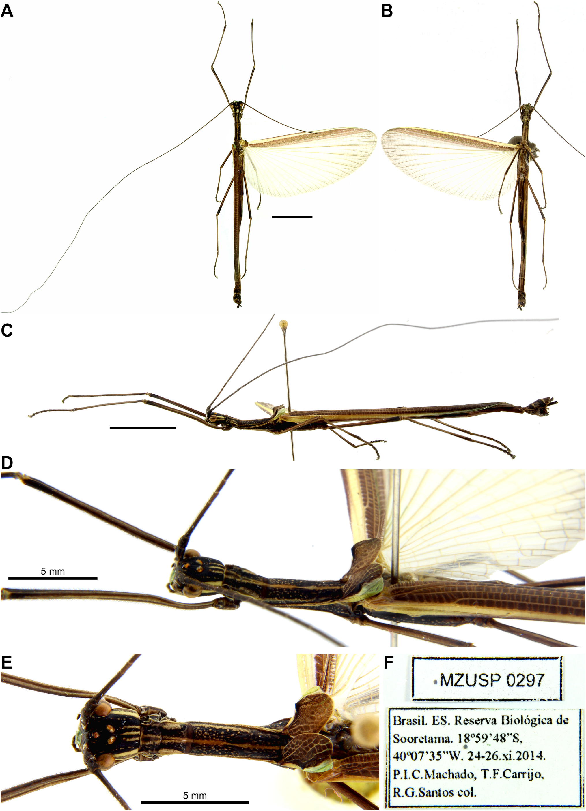
Paraphasma sp. 1 , Chiquetto-Machado & Cancello, 2021: 4, 26, figs 7, 14E, 19C, 20A, 25, 26. Holotype: ♂, Brazil, Espírito Santo, Reserva Biológica de Sooretama GoogleMaps , 18º59’48”S, 40º07’35”W, 24–26.xi.2014, P. I. C. Machado, T. F. Carrijo , R . G. Santos ( MZUSP 0297 View Materials ) ( Fig. 30 View FIGURE 30 ) . Paratypes: 1♂, same data as holotype ( MZUSP 0294 View Materials *); 2♀, Brazil, Espírito Santo, Reserva Biológica de Sooretama , 19º03’15”S, 40º08’48”W, 24–26.xi.2014, P. I. C. Machado, T GoogleMaps . F. Carrijo , R . G. Santos ( MZUSP 0300 View Materials , 0309 View Materials ); 2♀, Brazil, Espírito Santo, Santa Tereza , 5.ii.1967, C. T . & C. Elias ( DZUP 380045 View Materials , 380046 View Materials ); 1♀, Brazil, Espírito Santo, Santa Tereza , 22.xi.1967, C. T . & C. Elias ( DZUP 380003 View Materials ) ( Fig. 33A–D,H View FIGURE 33 ); 1♀, 1 egg, Brazil, Espírito Santo, Sta. Tereza , 2–6.i.68, C. & C. T . Elias ( DZUP 380029 View Materials ); 1♂, Brazil, Espírito Santo, Sta. Tereza , 22–31.i.68, C. & C. T . Elias ( DZUP 380012 View Materials *); 1♀, Brazil, Espírito Santo, Sta. Tereza , 1–3.ii.68, C. & C. T . Elias ( DZUP 380025 View Materials ); 1♂, Brazil, Minas Gerais, Marliéria, Parque Florestal Rio Doce, 12.xii.72, Grisi ( ESALQ *) .
Etymology. Noun in apposition after the type locality of the species, the Reserva Biológica de Sooretama (Sooretama Biological Reserve), a protected Atlantic Forest area in the state of Espírito Santo, Southeast Brazil. This reserve serves as an important refuge for many species endemic to the Atlantic Forest.
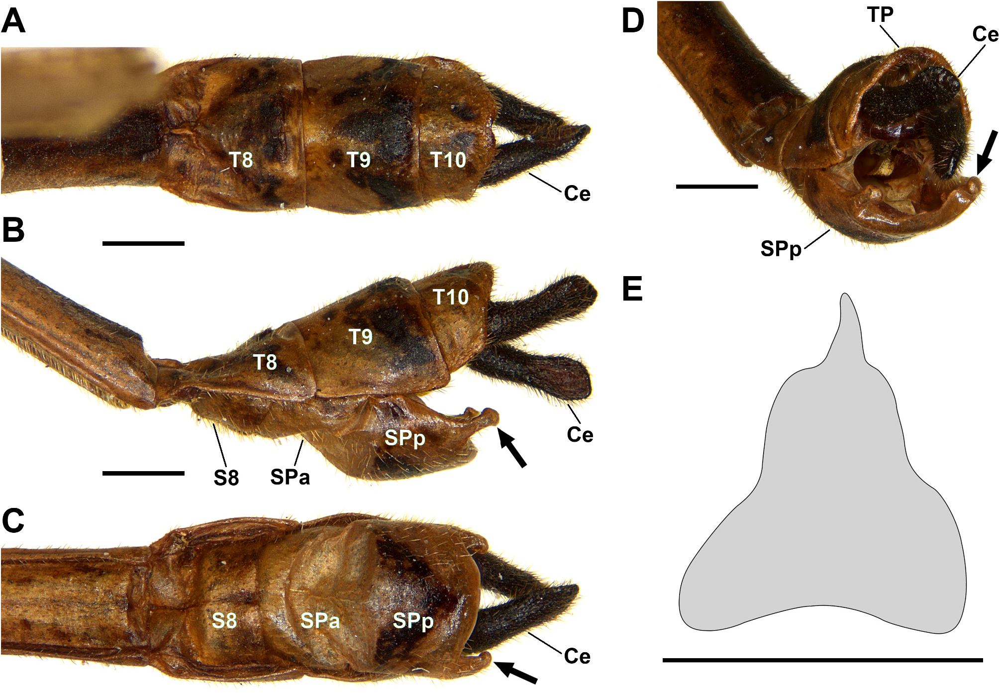
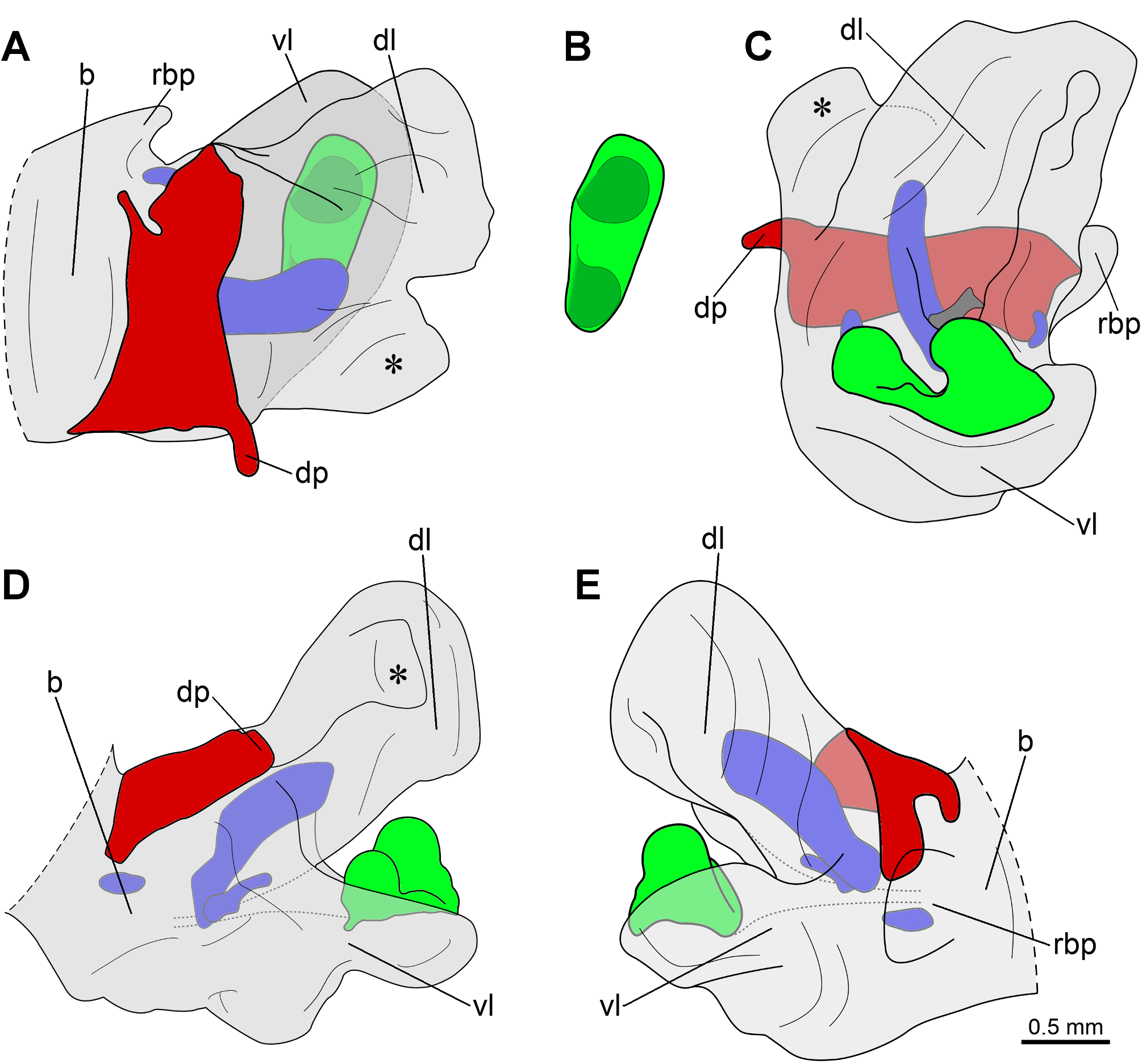
Diagnosis. Similar to P. conspersum , P. indistinctum sp. nov., P. laterale , P. marginale and P. minus , but clearly distinguishable by the phallic organ, with a “small-simple type ” sclerite of the ventral lobe ( Fig. 32 View FIGURE 32 , in green), which is oblong in dorsal view and has two rounded protuberances positioned side by side and projected towards each other. Although not exclusive of Paraphasma sooretama sp. nov., the following features may also be useful for the identification of this species: male cerci with rounded and internally concave apex (a feature that is shared only with P. laterale ) ( Fig. 31A–D View FIGURE 31 ); vomer longer than wide ( Fig. 31E View FIGURE 31 ); male subgenital plate with posterolateral margins forming a pair of approximately triangular expansions with malleable aspect ( Fig. 31 View FIGURE 31 : arrows); female sternite VII with a rounded indentation on the posterior margin.
Description of male. Color ( Figs 30 View FIGURE 30 , 31 View FIGURE 31 ): Live specimens present either a cryptic pattern, with body mostly brown or pale orange and a pair of pale yellow lateral stripes extending along head, prothorax, mesothorax and costal region of tegmina and hindwings, or a more vivid pattern, with body mostly shiny black and lateral stripes in a brighter yellow. Head, pro- and mesonotum with yellow or light brown dorsomedian line; pro- and mesonotum black or brown, with stains or spots in the same color as dorsomedian line. Legs brown or black, usually darkening towards apex of femora and tibiae. Anal region of hindwing bright yellow surrounded by a brownish stripe running along apical and posterior margins. Body ventrally light brown; subgenital plate black-stained. Dried specimens usually retain general color pattern, but less vivid and with anal region of hindwing becoming colorless. Head ( Fig. 30A–E View FIGURE 30 ): Smooth; slightly longer than wide; sub-rectangular in dorsal view; vertex weakly convex. Compound eye very prominent, large, covering nearly half of head length, almost round in lateral view. Ocelli well-developed; median one distinctly separated from lateral pair.Antennae filiform, very long, distinctly surpassing end of abdomen; scape compressed dorsoventrally; pedicel cylindrical, slightly shorter than scape; first flagellomere about 3x longer than pedicel. Thorax ( Fig. 30A–E View FIGURE 30 ): Prothorax smooth; slightly longer and distinctly narrower than head; weakly convex dorsally and ventrally, laterally flat. Pronotum sub-rectangular, with slight constriction on anterior third; anterolateral corners with rounded indentations, outlining openings of paired defensive glands; posterior margin convex; pair of gentle dorsolateral carinae originating posterior to defensive glands and extending until nearly posterior margin. Mesothorax slightly rugose, approximately 1.5x longer than prothorax; about as wide as prothorax on anterior half and gradually widening on posterior half. Mesonotum with weak longitudinal carina extending along each lateral margin; mesepisternum with more pronounced carina extending along ventral margin. Metathorax and median segment smooth; parallel-sided, as wide as posterior region of mesothorax; dorsally convex, laterally flat, weakly convex ventrally; metathorax about 3x longer than median segment; both combined almost 2x longer than mesothorax. Metepisternum with longitudinal carina extending along ventral margin. Legs ( Fig. 30A–E View FIGURE 30 ): Fairly long and slender. Profemur slightly longer than combined length of mesothorax, metathorax and median segment; curved and compressed basally; approximately trapezoidal in cross-section, with carinate edges and distinct ventromedian carina; anterodorsal carina weakly raised. Mesofemur about as long as pro- and mesothorax combined; 0.6x length of profemur. Metafemur slightly shorter than profemur. Meso- and metafemur sub-rectangular in cross-section, with dorsal and ventral faces slightly convex; edges weakly carinate; ventromedian carina absent. Tibiae slightly shorter than corresponding femur, 1.5–2x longer than corresponding tarsus; rectangular or trapezoidal in crosssection; ventromedian carina absent; with conspicuous area apicalis. Pro- and metabasitarsus slightly longer than following three tarsomeres combined; mesobasitarsus about as long as following three tarsomeres combined. Wings ( Fig. 30A–E View FIGURE 30 ): Tegmina short, not reaching median region of metanotum; in dorsal view about 2.5x longer than wide; posterior margin gently rounded, apical margin rounded or weakly acuminate; shoulder pad very prominent, sometimes developed into a sharp spine; anal region with conspicuous reticulate venation. Hindwing well-developed, reaching abdominal tergite VII. Abdomen ( Figs 30A–C View FIGURE 30 , 31 View FIGURE 31 ): As in Paraphasma conspersum , but apex of cerci rounded in lateral view and internally concave ( Fig. 31A–D View FIGURE 31 ). Phallic organ ( Fig. 32 View FIGURE 32 ): Dorsal sclerite distinctly wider than long ( Fig. 32 View FIGURE 32 , in red); distal process short and fairly wide, directed to the left, perpendicular in relation to longitudinal axis of organ ( Fig. 32 View FIGURE 32 : dp). Dorsal and ventral lobes partially fused on left side. Dorsal lobe ( Fig. 32 View FIGURE 32 : dl) subdivided into main body on the right and a dorsal smaller pouch on the left ( Fig. 32 View FIGURE 32 : asterisks); inner face with a small sclerotized region ( Fig. 32C View FIGURE 32 , dark gray). Sclerite of the ventral lobe of “small-simple type ” ( Fig. 32 View FIGURE 32 , in green), covering only central region of inner face of ventral lobe; oblong in dorsal view, distinctly wider than long; with two rounded protuberances projected towards each other; right protuberance larger and more strongly projected. One of base apodemes ( Fig. 32 View FIGURE 32 , in blue) projecting into dorsal lobe as a spatulate expansion.
Description of female. Color ( Fig. 33 View FIGURE 33 ): As in male, but subgenital plate brown, usually lighter than preceding sternites. Head and thorax ( Fig. 33A–D View FIGURE 33 ): As in male. Legs ( Fig. 33A–D View FIGURE 33 ): As in male, but slightly shorter in relation to body, with profemur not surpassing combined length of mesothorax, metathorax and median segment; and anterodorsal carina of profemur distinctly raised medially. Wings ( Fig. 33A–D View FIGURE 33 ): As in male, but hindwing reaching abdominal tergite VIII. Abdomen ( Fig. 33A–C,E–G View FIGURE 33 ): Approximately 1.7x longer than the combined length of head, thorax and median segment; dorsally and ventrally smooth, but tergite X gently carinate longitudinally. Segments II and III the longest; then gradually shortening from III to VII. Tergites VIII–X ( Fig. 33E,F View FIGURE 33 ) distinctly shorter and slightly narrower than II–VII; tergite IX slightly shorter than VIII and about 1.3x longer than X. Tergite X slightly longer than wide; tectiform; posterior margin slightly emarginate. Cerci ( Fig. 33E–G View FIGURE 33 ) short, about as long as tergite X; straight and cylindrical; apex blunt. Epiproct rounded; hardly visible under tergite X. Sternite VII with small but conspicuous praeopercular organ ( Fig. 33G View FIGURE 33 ), developed into an elongate, shiny protuberance, extending until rounded indentation on posterior margin of sternite VII. Subgenital plate ( Fig. 33F,G View FIGURE 33 ) lanceolate, extending until posterior margin of tergite X; apex somewhat sharp; inner face longitudinally carinate. Cerci, tergite X and subgenital plate densely pilose.
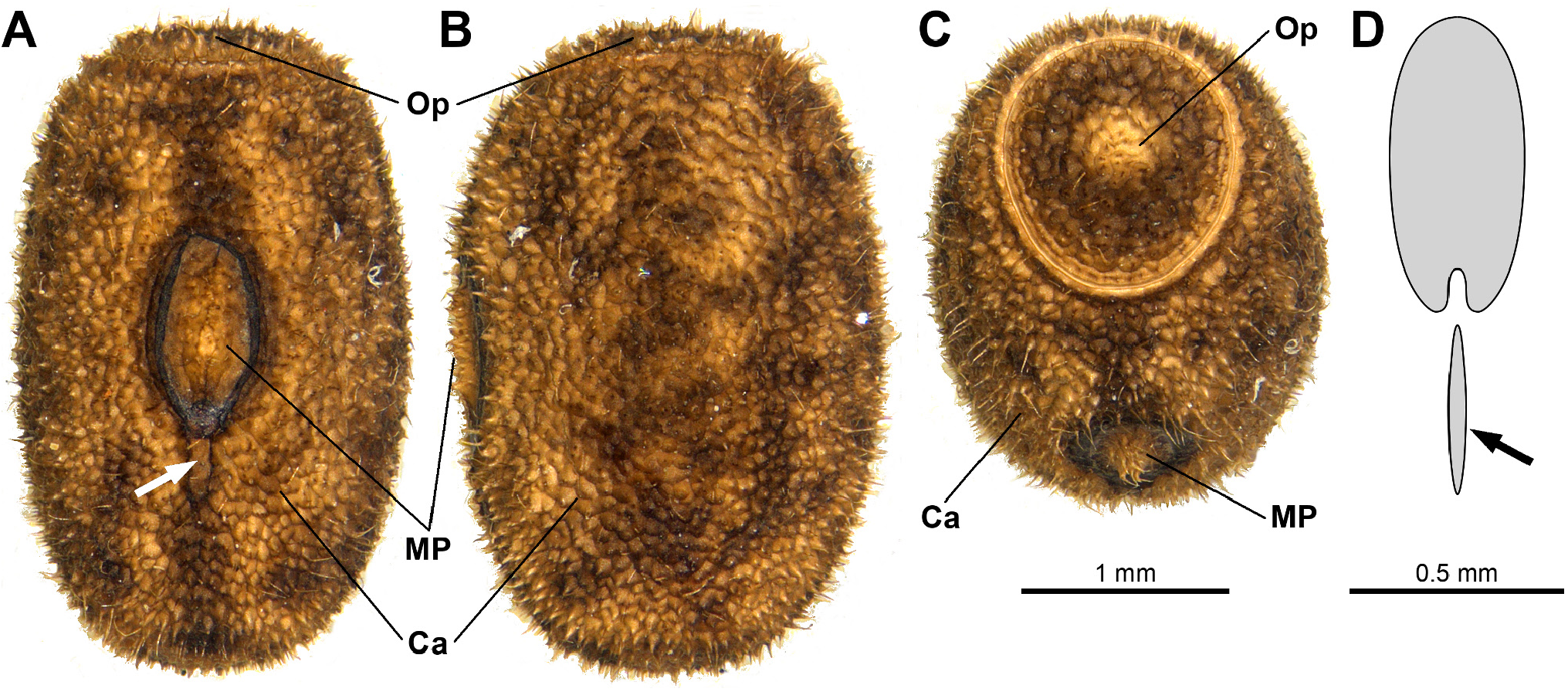
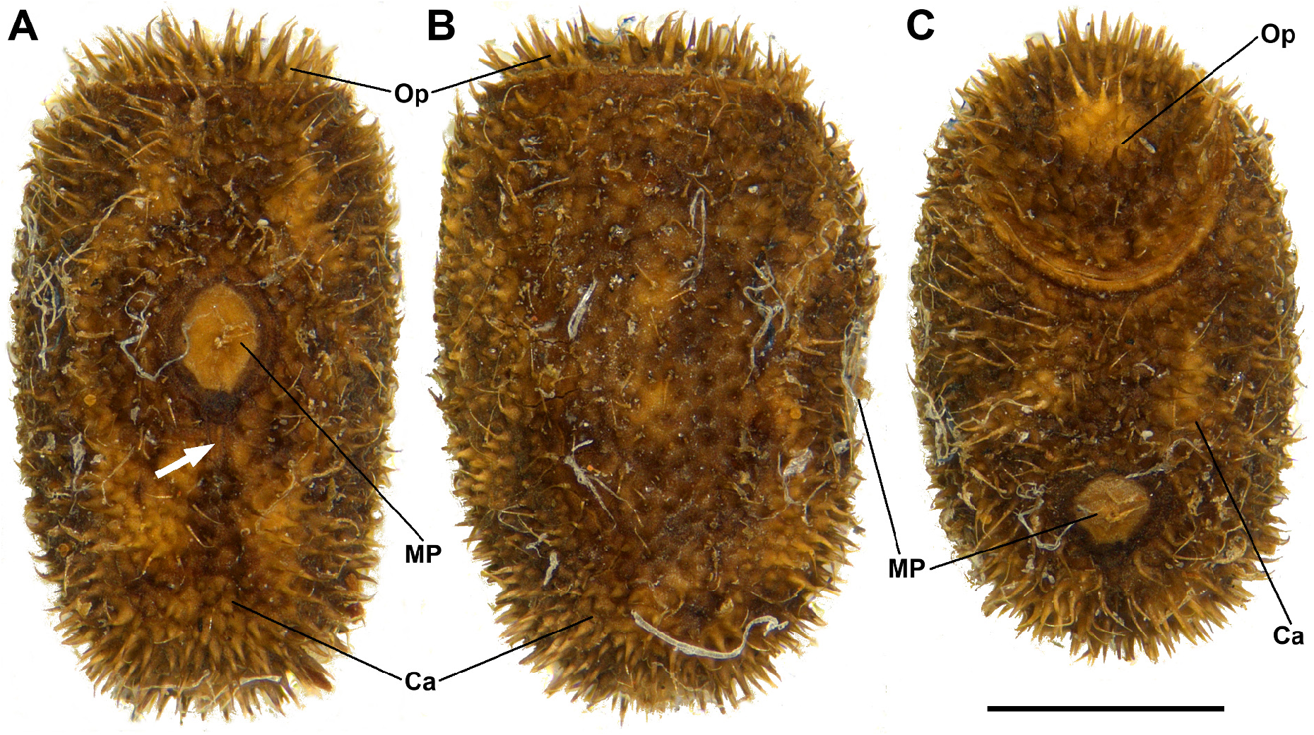
Description of egg ( Fig. 34 View FIGURE 34 ). Capsule somewhat elongate, approximately barrel-shaped, higher than wide on median region, slightly narrowing towards operculum and polar area, almost flat on polar area; densely covered with uniformly distributed granules, each one originating a fairly long bristle, longer on polar area. Operculum approximately oval, slightly higher than wide, perpendicular in relation to longitudinal axis of capsule; bearing long bristles. Micropylar plate rounded but slightly elongate, positioned medially on capsule; with a small central hump. Median line ( Fig. 34A View FIGURE 34 : arrow) very short and inconspicuous. Internal micropylar plate not examined. Egg light brown with sparse darker regions. Measurements (mm, n = 1): capsule length, 2.5; capsule width, 1.6; capsule height, 1.7; operculum width, 1.1; operculum height, 1.4; micropylar plate length, 0.7; micropylar plate width, 0.5.
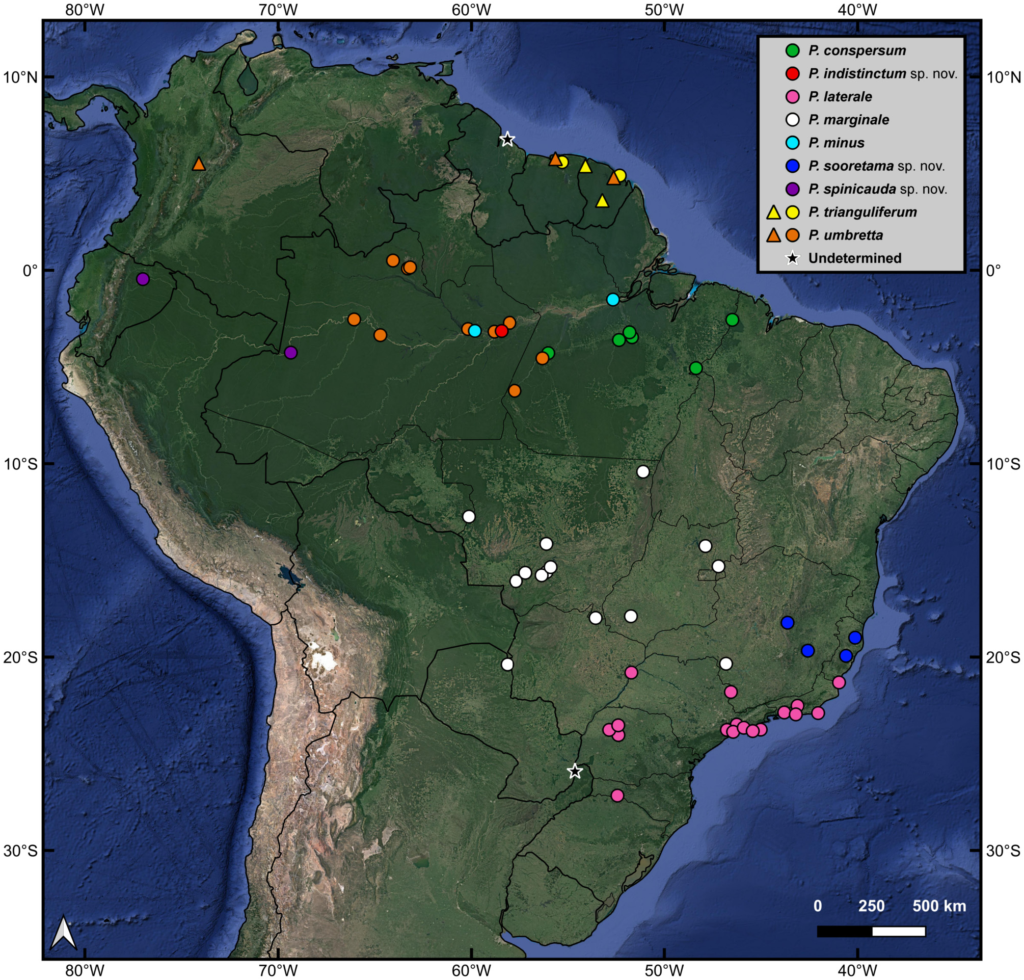
Distribution ( Fig. 1 View FIGURE 1 : dark blue circles). Paraphasma sooretama sp. nov. is recorded from the Brazilian Atlantic Forest in the states of Espírito Santo and eastern Minas Gerais and also from a locality in the Cerrado of central Minas Gerais.
Remarks. The only egg of Paraphasma sooretama sp. nov. examined ( Fig. 34 View FIGURE 34 ) is similar to the eggs of Paraphasma laterale ( Figs 16 View FIGURE 16 , 17 View FIGURE 17 ) but has a slightly more rounded micropylar plate. However, in a large series of eggs of P. laterale (more than 50 eggs examined), the micropylar plate varied from fairly elongate to almost as rounded as in the egg of P. sooretama sp. nov. It is possible that some variation also occurs in the shape of the plate of P. sooretama sp. nov., and for this reason the rounded micropylar plate is not regarded as a diagnostic feature of this species.
Additional material examined. BRAZIL. Espírito Santo: 1 egg, same data as paratype DZUP 380029 View Materials (extracted from the female terminalia) . Minas Gerais: 6♂, 2♀, Diamantina, Serra dos Cristais, atrás da R . das Rosas , campo rupestre arbustivo, 25–26.x.2018, V . M. Ghirotto ( MZUSP 1160 View Materials , 1161 View Materials , 1162 View Materials , 1163 View Materials , 1164 View Materials , 1188 View Materials , 1246 View Materials , 1355 View Materials *) .


| R |
Departamento de Geologia, Universidad de Chile |
| T |
Tavera, Department of Geology and Geophysics |
| V |
Royal British Columbia Museum - Herbarium |
No known copyright restrictions apply. See Agosti, D., Egloff, W., 2009. Taxonomic information exchange and copyright: the Plazi approach. BMC Research Notes 2009, 2:53 for further explanation.
|
Kingdom |
|
|
Phylum |
|
|
Class |
|
|
Order |
|
|
Family |
|
|
Genus |