
Paraulax perplexa Kieffer, 1904
|
publication ID |
https://doi.org/ 10.5281/zenodo.189597 |
|
publication LSID |
lsid:zoobank.org:pub:AC41ACF9-2D19-45A2-96DE-16470E7D9C7F |
|
DOI |
https://doi.org/10.5281/zenodo.5681152 |
|
persistent identifier |
https://treatment.plazi.org/id/03ADE415-FFC6-FF92-FF58-9202FB29B8A1 |
|
treatment provided by |
Plazi |
|
scientific name |
Paraulax perplexa Kieffer, 1904 |
| status |
|
Paraulax perplexa Kieffer, 1904
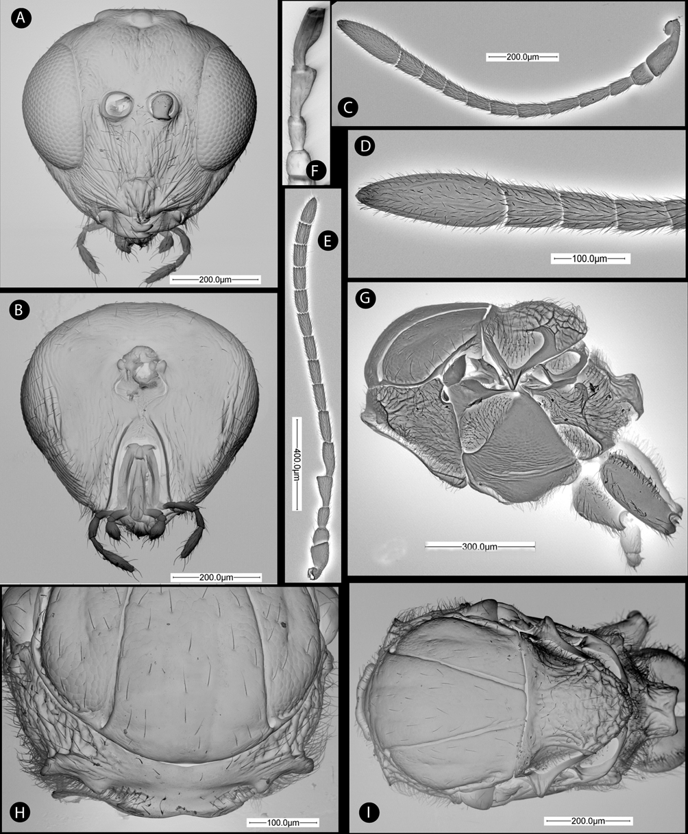
( Figs. 2 View FIGURE 2 , 3 View FIGURE 3 & 10A–B)
Paraulax perplexa Kieffer, 1904
P. perplexus Kieffer in: Bull. Soc. Metz, ser 2 v. 11 p. 60 (female and male). Bisexual.
Type material. Neotype Ƥ here designated: CHILE, El Maule, VII Región, Cauquenes, Reserva Nacional Los Queules, 35º59´10´´S, 72º42´30´´O, 420 m; caught with a Malaise trap operating in a fragment of native forest , 27.ix/ 25.x.2006. J.L. Nieves-Aldrey & A. Grez leg. Deposited in Museo Chileno de Historia Natural, Santiago de Chile, card mounted. Other material from type series: 43 same data as Neotype, except 23 collected on 22.viii/ 22.ix. 13 in MCHN, remaining exemplars in Museo Nacional Ciencias Naturales, Madrid ( Spain). Additional material (13, 1Ƥ from the type series were dissected for SEM observation); one additional female preserved in ethanol (extracted DNA). Non-type material: 1Ƥ, Chile, Ñuble, Ese Recinto. 1330 m, 29/XII/1982, Newton & Thayer leg ( AEI).
Diagnosis. P. p e r p l e x a differs from P. queulensis and P. ronquisti by a not elongate body (Figs. 10A), in the female only 3 times longer than high. The males of this species are readily distinguished of the other species of Paraulax by the strongly broadened and distally truncate second flagellomere ( Fig. 2 View FIGURE 2 F).
Redescription. Body length (measured from anterior margin of head to posterior margin of metasoma) 1.8 mm (range 1.7–2.0; N = 2) for females; 1.6 mm (range 1.5–1.8; N = 4) for males. Coloration: female body entirely black; antennal flagellum, tarsi, pro and mesotibia and apex of femora dark brown. Forewing hyaline, veins brown. Male similar in coloration to female, but antenna and fore legs slightly paler.
Female. Head. In dorsal view 2.1 times as wide as long. Gena not expanded behind compound eye. POL 1.6 times as long as OOL, posterior ocellus separated from inner orbit of eye by about 2 times its diameter. In anterior view ( Fig. 2 View FIGURE 2 A) head more or less rounded, 1.1 times as wide as high. Face with some sparse setae, more abundant in lower face; facial strigae present, radiating from clypeus, strong, laterally reaching ventral margin of eye and medially almost reaching ventral margin of torulus; vertical median carina absent ( Fig. 2 View FIGURE 2 A). Frons and vertex with coriaceous sculpture; ocellar plate slightly raised; malar space 0.38 times height of compound eye. Clypeus indistinct, more or less rectangular; ventral margin slightly projecting over mandibles. Subocular impression present though not well marked. Ventrolaterally on gena 5–7 regular vertical carinae present. Anterior tentorial pits visible; epistomal sulcus and clypeo-pleurostomal lines indistinct. Torulus situated at mid-height of compound eye; transfacial line 0.9 times height of eye; distance between antennal rim and compound eye 0.5 times width of antennal socket including rim. Occiput dorsally pubescent with coriaceous-alutaceous sculpture, without dorsal occipital carina; some strong longitudinal rugae on lateral margins of head, but without distinct genal occipital carina ( Fig. 2 View FIGURE 2 B). Posterior tentorial pits narrow, arched. Hypostomal sulci meeting slightly before hypostoma ( Fig. 2 View FIGURE 2 B). Distance between occipital and oral foramina as long as height of occipital foramen.
Mouthparts ( Fig. 2 View FIGURE 2 B). Mandibles exposed; right mandible with three teeth; left with two teeth. Cardo of maxilla visible, maxillary stipes 5.8 times longer than wide. Maxillary palp with five segments; last segment 3.3 times longer than wide. Labial palp with three segments.
Antenna ( Fig. 2 View FIGURE 2 C). 0.7 times length of body, with 12 segments; flagellum widening towards apex; antennal segments with coriaceous sculpture and setae no longer than width of a segment. Placodeal sensilla v i s i b l e o n l y o n F7 –F 1 0 (F i g. 2 D). R e l a t i v e l e n g t h s o f a n t e n n a l s e g m e n t s: 25:12:15:20:20:20:17:15:15:16:17:45; pedicel 0.9 times its width; 0.8 times length of F1; F1 1.9 times longer than wide. Ultimate flagellomere spindle-shaped, 3 times longer than wide, 1.2 times wider than penultimate, not truncate at apex.
Mesosoma. Pronotum, anterior view, almost glabrous medially, strongly pubescent laterally ( Fig. 2 View FIGURE 2 H). Ratio of median to lateral length of pronotum 0.3. Pronotal plate distinct, 5.5 wider than long; dorsal part distinctly set off, anterolateral margin marked and somewhat projecting laterad; some strong longitudinal rugae visible in anterior and lateral view between margin of pronotal plate and surface of pronotum laterally. Admedian pronotal depressions separated by more than median length of pronotum. Posterior pronotal plate more or less rectangular, bare and smooth, ventral and lateral margins marked. Lateral surface of pronotum coriaceous, some strong, short rugae running from the lateral margin of pronotal plate ( Fig. 2 View FIGURE 2 G).
Mesonotum. Mesoscutum ( Fig. 2 View FIGURE 2 I) 1.2 times wider than long; with weak coriaceous sculpture, more distinct at lateral lobe, with few setae. Median mesoscutal impression absent. Notauli percurrent, straight and narrow, converging posteriorly. Separation of notauli posteriorly at transscutal fissure relatively narrow, 0.3 times width of separation at anterior margin of mesoscutum. Anteroadmedian signa visible. Mesoscutum and scutellum separated by narrow transscutal fissure. Scutellar foveae indistinct, visible only as shallow depression with some rugae ( Fig. 2 View FIGURE 2 I). Scutellum, in dorsal view with strong rugae also present medially; in lateral view convex, with prominent rugose sculpture. Posterodorsal and posterior margins of axillula distinct. Mesopleuron ( Fig. 2 View FIGURE 2 G) ventral to mesopleural triangle with a prominent longitudinal mesopleural impression, more or less complete, ending at margin of mesopleural triangle. Some irregular longitudinal striae and coriaceous sculpture dorsal to furrow. Mesopleuron smooth ventral to mesopleural impression. Mesopleural triangle distinctly impressed and densely pubescent; dorsal margin diffuse anteriorly, not meeting area near prepectus but meeting posterolateral margin of pronotum well below prepectus.
Metanotum ( Fig. 3 View FIGURE 3 A). Metascutellum with distinct median constriction. Ventrally divided in two parts by a median vertical bar. Median width wider than metanotal trough. Metanotal trough smooth, pubescent.
Metapectal-propodeal complex. Metapleural sulcus ( Fig. 2 View FIGURE 2 G) meeting posterior margin of mesopectus at about mid height of metapectal-propodeal complex. Lateral propodeal carinae narrow, parallel, subdivided into irregular carinae near nucha ( Fig. 3 View FIGURE 3 A). Lateral and median propodeal areas smooth, pubescent. Nucha dorsally with some irregular longitudinal rugae.
Legs. Profemur with ventral swelling in basal third, wearing 4–5 rows of sharp closely spaced, deep costulae ( Figs. 3 View FIGURE 3 B & 3C). Protarsi 1.2 times longer than protibia. Metatarsal claw with a acute basal lobe or tooth, about ¼ of length of apical tooth ( Fig. 3 View FIGURE 3 D).
Forewing ( Fig. 3 View FIGURE 3 E). 1.2 times longer than body. Radial cell closed along anterior margin, 3.7 times longer than wide; R1 slightly depigmented along margin of radial cell; radius (Rs) straight, reaching anterior margin of wing. Areolet absent; vein Rs+M and M almost invisible, directed towards lower half of median vein. Fringe of long setae along apical margin of wing.
Metasoma. Metasoma ( Fig. 3 View FIGURE 3 F) shorter than head plus mesosoma; in lateral view 1.4 times longer than high; laterally compressed. Abdominal petiole smooth dorsally, ventrally with deep longitudinal grooves; about as long as high. T2 smooth and shining, covering about 2/3 of metasoma; anteromedian area of T2 with only 4–5 long setae. Projecting part of hypopygial spine 4 times longer than high; apical pubescence of hypopigial spine projecting beyond apex of spine, subapical setae longer than apical hairs, together forming a small tuft.
Male. Similar to female except as described below (size and coloration already discussed). Head as wide as high. Antenna ( Fig. 2 View FIGURE 2 E) with 13 flagellomeres. Flagellum not widening towards apex. F2 abruptly expanded from base towards apex, apex 2.5 times wider than base. F3 slightly curved at basal third, slightly wider towards apex ( Fig. 2 View FIGURE 2 F); F4 and following flagellomeres cylindrical, not modified. Relative length of antennomeres: 15:10:13:20:21:20:17:15:14:15:14:14:14:13:17. Placodeal sensilla present on all flagellomeres, except F1, arranged in row of 4–5 sensilla on each flagellomere. Metasoma ( Fig. 3 View FIGURE 3 H); T2 covering ¼ of length of metasoma. Anteromedian area of T2 with group of only 3 setae.
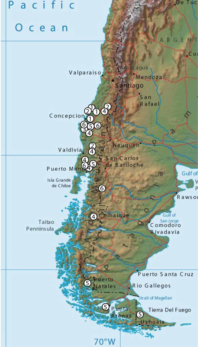
Distribution. Chile, Concepción and Los Queules (IX and X Regiones) ( Fig. 15 View FIGURE 15 ).
Biology. Unknown. One potential host is Nothofagus galls induced by species of Espinosa Gahan , and maybe also Aditrochus , both ormocerine chalcids ( Pteromalidae : Ormocerinae ). In the collecting area of Los Queules we sampled two potential Espinosa host galls, both on Nothofagus obliqua ; Espinosa nothofagi Gahan ( Fig. 12 View FIGURE 12 G) and Espinosa sp. ( Fig. 12 View FIGURE 12 H), identified according to De Santis et al. (1993). Collection data indicate a flight period in late winter and early spring (from August to October).
Remarks. Weld (1952) stated that Kieffer´s types of perplexa were specimens captured near Concepción, Chile, Pablo Herbst leg., with unknown habitat. The material was sent to Kieffer in different years from different localities. Hence, Kieffer inferred that the species must be abundant. The location of the types is unknown.
A neotype is here designated with the purpose of clarifying the taxonomic position of this taxon.
We further justify this nomenclatural act by the close resemblance of this species with the original, albeit somewhat short, description. Of special importance in the original description is the mention of the following diagnostic characters: “face irrégulierement ridée” but without mention of a median vertical carina; 12 antennal segments as long as the three preceding segments; 4 antennal segments of the male apically truncate; the “mesopleures finement striées”; the toothed tarsal claws. All these characters fit with the characters presented for the neotype, and does not apply to the species reared from Aditrochus galls that in this paper have been included in the new genus Cecinothofagus .
Also, the collecting locality of the neotype material is near the original locality, and the two localities (Concepción and los Queules) share the same vegetation type described as Bosque Maulino, with Nothofagus obliqua forest as the predominant habitat.
The neotype is deposited in the Museo Chileno de Historia Natural (Santiago de Chile).


No known copyright restrictions apply. See Agosti, D., Egloff, W., 2009. Taxonomic information exchange and copyright: the Plazi approach. BMC Research Notes 2009, 2:53 for further explanation.