
Ricanula peronata, Zhang & Wang & Stroiński & Qin, 2021
|
publication ID |
https://doi.org/ 10.11646/zootaxa.5047.3.7 |
|
publication LSID |
lsid:zoobank.org:pub:2FDA4955-4718-4A34-8053-09ACA9BB0241 |
|
DOI |
https://doi.org/10.5281/zenodo.5540965 |
|
persistent identifier |
https://treatment.plazi.org/id/038FEA2D-9A40-FFA7-19AA-4993FD10377D |
|
treatment provided by |
Plazi |
|
scientific name |
Ricanula peronata |
| status |
sp. nov. |
Ricanula peronata sp. nov.
( Figs 7–9 View FIGURE 7 View FIGURE 8 View FIGURE 9 , 11 View FIGURE 11 )
Etymology. The name is derived from the Latin word ‘ peronatus ’, referring to the periandrium with boot-shaped ventral processes in ventral view.
Diagnosis. The species is similar to Ricanula curva sp. nov., but differs from the latter by having periandrium with boot-shaped ventral processes in ventral view ( Fig. 9F View FIGURE 9 ); ventral processes of aedeagus oriented ventrally in lateral view ( Fig. 9G View FIGURE 9 ) (apical part of ventral processes of periandrium strongly curved laterally in dorsal and ventral view; ventral processes of aedeagus S-curved in lateral view in Ricanula curva sp. nov.).
Description. Measurements. Length (inc. tegmen): male 6.5–7.4 mm, female 7.3–8.8 mm.
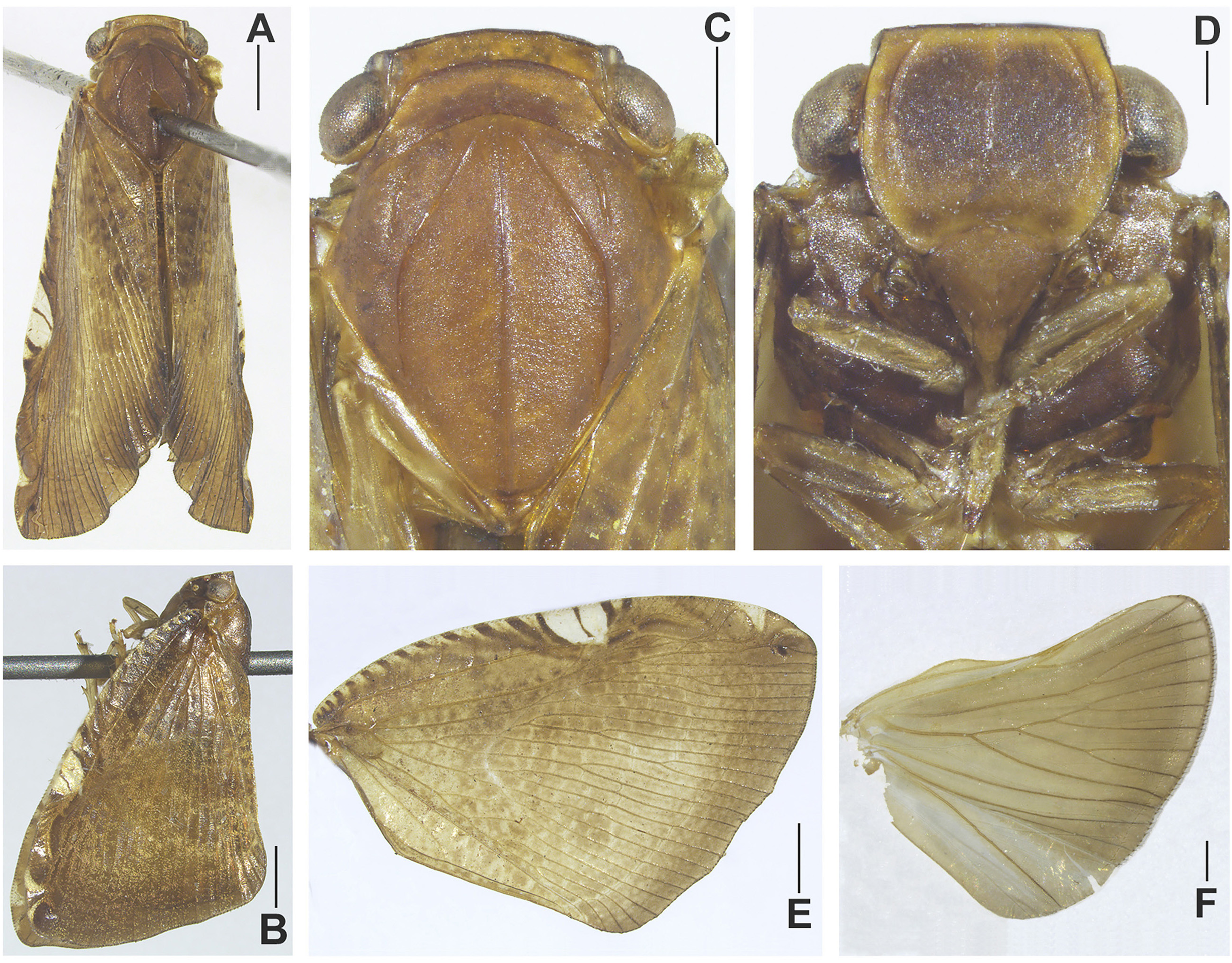
Head. Vertex ( Figs 7A, C View FIGURE 7 ) without median carina. Frons: median and lateral carinae of frontal disc surpassing half of disc, ending about the level of antennae; apical parts of median carina weakly visible.
Thorax. Pronotum ( Figs 7A, C View FIGURE 7 ) with small round depressions submedially on each lateral side. Mesonotum: lateral carinae ( Figs 7A, C View FIGURE 7 ) not reaching posterior margin; antero-lateral carinae not connected with anterior margin.
Tegmen: postero-apical part of tegmen with two eye-spot black cells, posterior margin arcuate. Longitudinal veins ScP+RA and RP, MP arising as short common stem from basal cell. Claval veins Pcu and A 1 fused on midlength of CuP vein. Hind wing ( Fig. 7F View FIGURE 7 ) without transverse veinlets.
Hing legs: Basitarsomere of metatarsus with 8 apical teeth. Metatibiotarsal formula 2/6/8.
Male terminalia. Anal tube (in dorsal view, Fig. 8B View FIGURE 8 ) nearly square, posterior margin strongly concave, basal margin slightly convex, lateral margins straight; anus placed before midlength, paraproct surpassing the posterior margin. Pygofer (in lateral view, Fig. 8A View FIGURE 8 ) with dorsal posterior angle without process. Genital styles (in lateral view, Fig. 8A View FIGURE 8 ) broadly triangular; ventral margin weakly sinuate; dorsal margin weakly convex, with small concavity before spine-like process.
Phallic complex ( Figs 9A–C View FIGURE 9 ): Periandrium ( Figs 9D–F View FIGURE 9 ) with boot-shaped ventral processes in ventral view, apical part of ventral processes straight; dorsal periandrium with U-shaped structure with membranous apical part sclerotized base in dorsal view; lateral margin of periandrium with small rod-shaped processes in lateral view, rodshaped processes hidden in the periandrium (in ventral view); ventral periandrium distinctly convex.
Aedeagus ( Figs 9G–I View FIGURE 9 ) apically with two pairs of processes. Median split asymmetrical: ventral split present only in 1/5; dorsal split very deep, reaching almost basal part. All processes single armed: lateral processes longer than ventral processes, about two thirds of aedeagus; ventral processes oriented ventrally in lateral view.
Female terminalia ( Figs 8C–I View FIGURE 8 ). Pregenital sternite ( Fig. 8I View FIGURE 8 ): posterior margin medially with two prominent processes, margin between processes with wide and shallow incision.
Anal tube (in dorsal view, Fig. 8C View FIGURE 8 ) ovoid, with widest part medially, basal margin weakly convex, posterior margin widely concave, lateral margins arcuate; anus placed after midlength, paraproct surpassing the posterior margin.
Gonoplac ( Fig. 8H View FIGURE 8 ): posterior margin with two rows of small teeth.
Coloration. General color brown ( Figs 7A–B View FIGURE 7 ). Median part of frons ( Fig. 7D View FIGURE 7 ) black brown. Eyes brown ( Figs 7B–C View FIGURE 7 ), ornamented with irregular brown patches. Gena ( Fig. 7B View FIGURE 7 ) black brown with two yellowish spots. Tegmen ( Figs 7B, E View FIGURE 7 ) brown, costal margin with about 14 transverse black brown stripes from base to a little beyond middle, between the transverse brown stripes filled with light yellow stripes, sub-medially of tegmen with a large flavescent spot marked by 2 central transverse back lines. Wings brown, each side of A 2 with a longitudinal grayish narrowed band ( Fig. 7F View FIGURE 7 ). Abdomen and terminalia brown.
Type material. Holotype, male, China: 20 Apr. 1964, Yunnan, Xishuangbanna, Menglun , 650 m, coll. Baolin Zhang.
Paratypes (8 males, 17 females, China) : 1 male: 13 Jul. 1958, 650– 700 m, coll. Chunpei Hong; 2 males, 4 females: 21/ 30 Apr. 1974, coll. Yao Chou, Feng Yuan et Yinyue Hu; 1 female: 22 May 1982, 1 female: 24 May 1982, coll. Qinmei Wang et Jingruo Zhou; 1 male, 1 female: 18 May 1991, 1 female: 20 May 1991, 1 male, 1 female: 22 May 1991, 2 females: 26 May 1991, 3 males, 6 females: 31 May 1991, coll. Yinglun Wang et Wanzhi Cai; Yunnan, Xishuangbanna, Menglun .
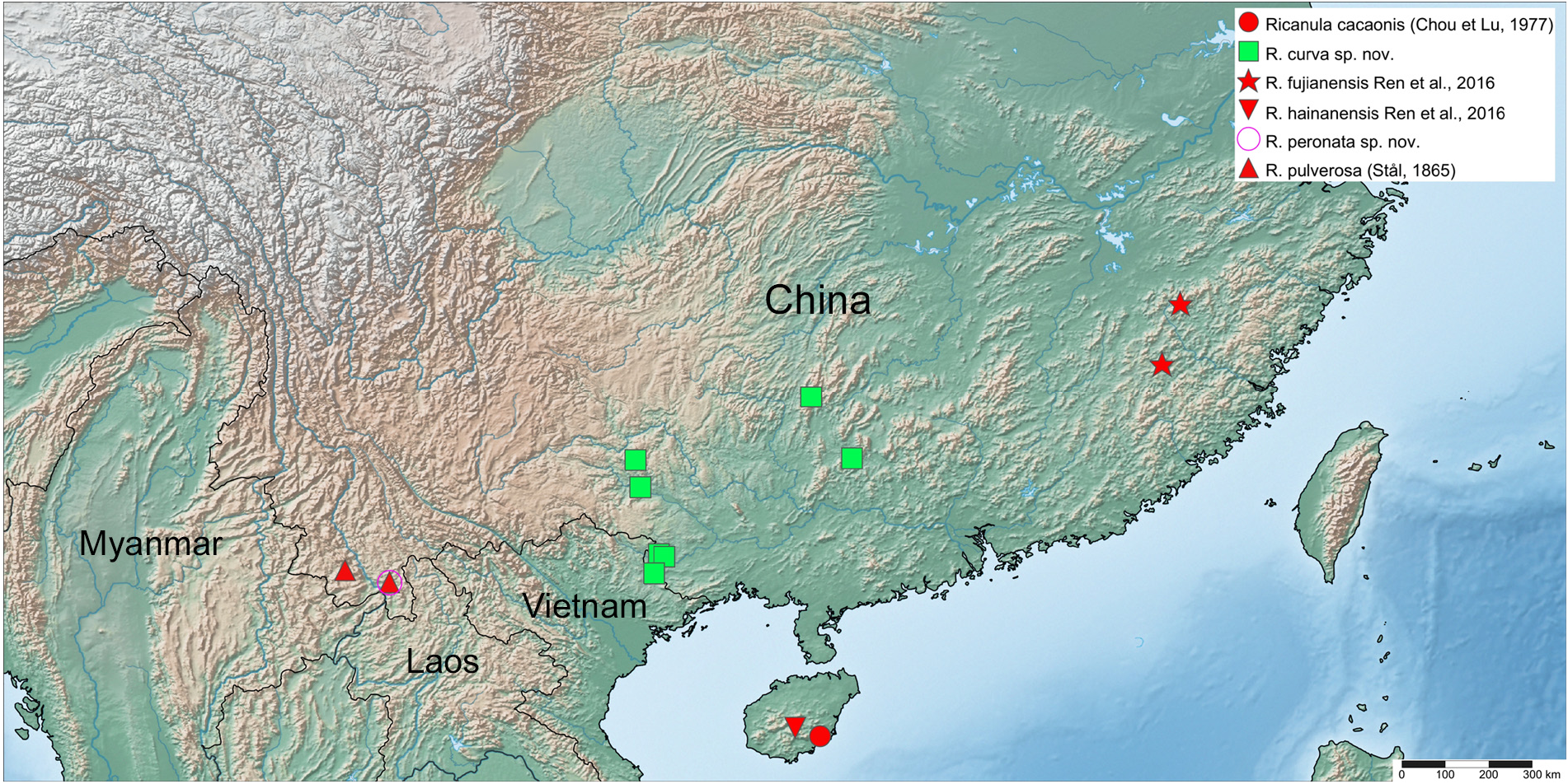
Distribution. China (Province Yunnan).


No known copyright restrictions apply. See Agosti, D., Egloff, W., 2009. Taxonomic information exchange and copyright: the Plazi approach. BMC Research Notes 2009, 2:53 for further explanation.
|
Kingdom |
|
|
Phylum |
|
|
Class |
|
|
Order |
|
|
Family |
|
|
Genus |