
Phaenocora variodentata Meixner, 1915
|
publication ID |
https://doi.org/ 10.11646/zootaxa.3889.3.1 |
|
publication LSID |
lsid:zoobank.org:pub:67896601-F3C6-44F2-A237-78120C8EA5DB |
|
DOI |
https://doi.org/10.5281/zenodo.5660149 |
|
persistent identifier |
https://treatment.plazi.org/id/CF039A58-FFCF-C53D-17C4-0A9CE07FFB3B |
|
treatment provided by |
Plazi |
|
scientific name |
Phaenocora variodentata Meixner, 1915 |
| status |
|
Phaenocora variodentata Meixner, 1915 View in CoL
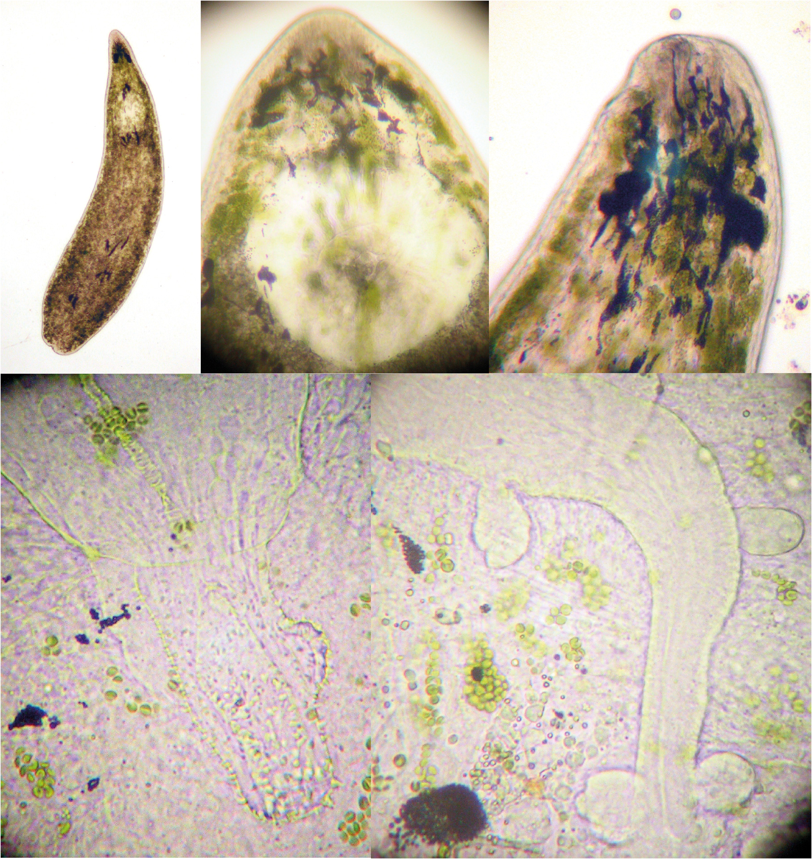
( Figs 8 View FIGURE 8 A–D, 14F1, F2)
Phaenocora variodentata Meixner 1915: 536 View in CoL –541; Beklemischev 1929: 538; Gilbert 1935: 284 –286, 304, 327, 334, 343, 356, 359, 364, 369, tables 1, 2; Beauchamp 1936: 151.
Phaenocora sinensis Wang & Sun 2011: 159 View in CoL –164, Figs 1–9 View FIGURE 1 .
Known distribution: Dürrenstein ( Austria) ( Meixner 1915); Baoan, Shenzhen, Guangdong ( China) (22°38’N; 113°54’E) ( Wang & Sun 2011).
Material examined: The holotype of P. sinensis (a whole mount) ( SMNH Type-8562; formerly SU-C, no. SZ 200903 - I-1) and the paratypes of P. s i n e ns i s (one whole mount) ( SMNH Type-8563; formerly SU-C, no. SZ 200903 - I-2) and (serially-sectioned specimens) ( SMNH nos Type-8564–8565; formerly SU-C, nos SZ 200903 - I- 3; SZ 200903 - I-4]).
Diagnosis: Animals about 1.4–1.8 mm long. Body yellowish grey. Pigmentation absent or present at the anterior part with brown-yellow pigment and five red-brown spots (one elongated, medially situated, and on each side two round ones). Visible eyes absent. Zoochlorellae not mentioned in literature. Male copulatory organ of the duplex-type IIIB. Penis papilla with dorsally three thick, conical spines, but no base plate. At the distal side of the invaginated penis papilla there are long, thin spines of different sizes. Female genital system of the EVELINAE - type.
Descriptive notes: The studied animals are about 1.7 mm long. The body has a bullet-like shape. Visible eyes and zoochlorellae are absent. The testes are somewhat lobular and extend from the caudal end of the pharynx to the caudal end of the body.
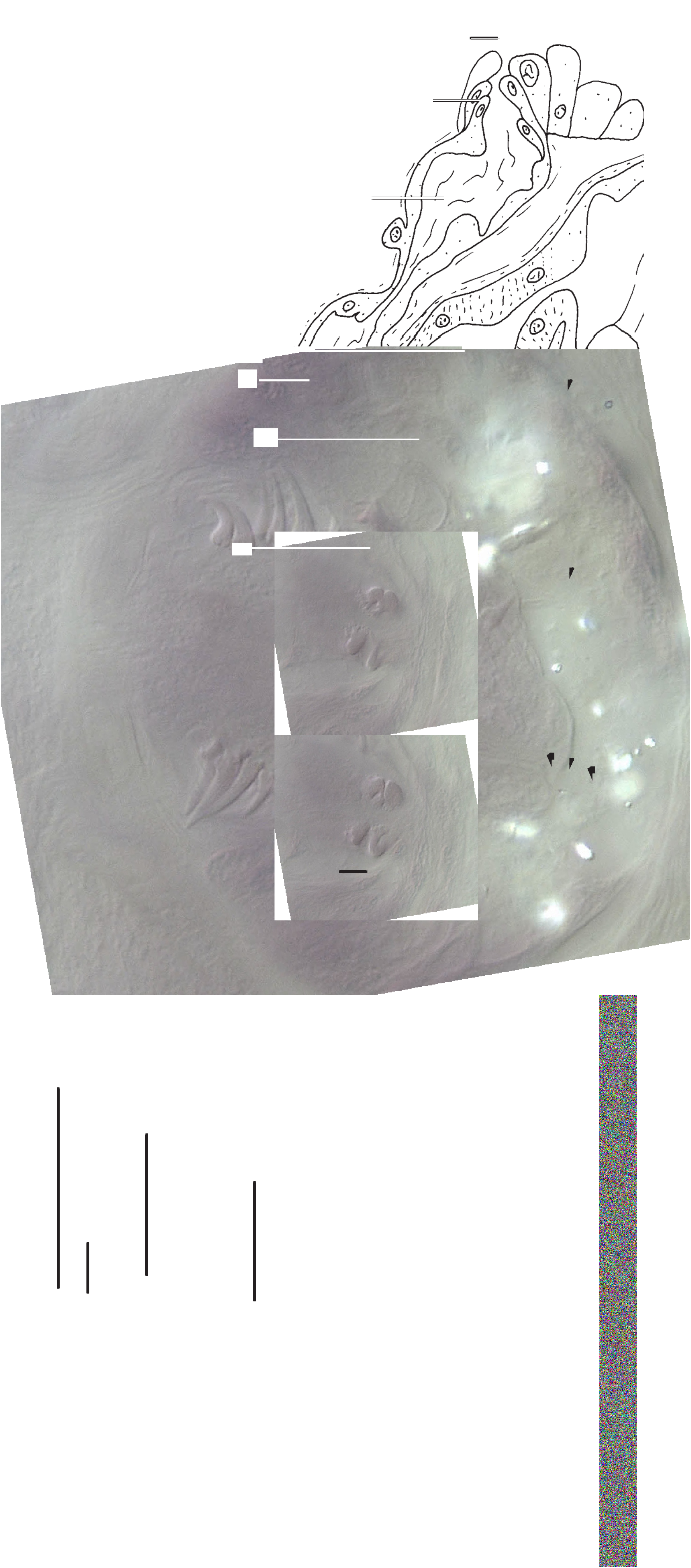
The male copulatory organ ( Figs 8 View FIGURE 8 B–D) is of the duplex-type IIIB. The penis papilla has three huge spines on the dorsal side ( Fig. 8 View FIGURE 8 C3), and at the base it possess long slender spines ( Fig. 8 View FIGURE 8 D: sp1) with those on the opposite side almost with the same appearance although a bit smaller and more curved ( Fig. 8 View FIGURE 8 D: sp2). Additionally, the copulatory organ is lined with several spines ranging from very small ( Fig. 8 View FIGURE 8 D: sp3) to the long slender ones at the base.
The female genital system ( Figs 8 View FIGURE 8 A, 8B) is connected to the gut. A burso-intestinal duct ( Fig. 8 View FIGURE 8 A: dbi) runs towards the intestinal bursa ( Figs 8 View FIGURE 8 A, 8B: bi). This bursa receives the female genital canal ( Figs 8 View FIGURE 8 A, 8B: fgc) more or less in its midway. The female genital canal is wider at its proximal part just before it is attached to the bursa. The oviduct ( Figs 8 View FIGURE 8 A, 8B: od) opens between the female genital canal and the intestinal bursa.
Discussion: Wang & Sun (2011) briefly stated that their material most resembled P. kepneri and P. s u bs a l i n a, without any further explanation. Wang & Sun (2011) further considered P. sinensis as a separate species because the testes of P. subsalina are merely a fourth of its body length and the vitellaria of P. kepneri are arranged in a netlike manner, both observations in contrast to their newly proposed P. sinensis ( Wang & Sun 2011) . As discussed above (see GENERAL MORPHOLOGY section) the only sound basis, at least at this moment, for species identification is the morphology of the reproductive system. The male system of P. sinensis is of the duplex-type IIIB and the female system is of the EVELINAE - type, a combination only found in four other species: P. clavigera , P. highlandensis , P. variodentata and P. t y ph l op s. The specimens now labelled P. sinensis differ clearly from P. highlandensis because the opening of the oviduct is not displaced, and differ from P. clavigera in that a glandular papilla is lacking. P. variodentata and P. sinensis differ from P. typhlops in the detailed morphology of the cirral spines. Although the original description of P. sinensis mentions that the cirrus dorsally bears four spines that are oriented as the wings of a butterfly ( Fig. 8 View FIGURE 8 C1), we found only three spines on the same material, one of which has a split base ( Figs 8 View FIGURE 8 C1–C3: * indicates the two parts of the split spine, arrow indicates the same place on different pictures). This particular cirrus morphology is identical with the one of P. variodentata . As such, the latter species only differs from P. sinensis in the fact that it has five clear spots of dermal pigmentation, which are absent in P. sinensis . Because pigmentation is known to show intraspecific variation (see our notes on P. unipunctata ) we consider P. sinensis a junior synonym of P. variodentata , at least until molecular data become available.
| SMNH |
Saskatchewan Museum of Natural History |
No known copyright restrictions apply. See Agosti, D., Egloff, W., 2009. Taxonomic information exchange and copyright: the Plazi approach. BMC Research Notes 2009, 2:53 for further explanation.
|
Kingdom |
|
|
Phylum |
|
|
Class |
|
|
Order |
|
|
Family |
|
|
Genus |
Phaenocora variodentata Meixner, 1915
| Houben, Albrecht M., Steenkiste, Niels Van & Artois, Tom J. 2014 |
Phaenocora sinensis
| Wang 2011: 159 |
Phaenocora variodentata
| Beauchamp 1936: 151 |
| Gilbert 1935: 284 |
| Beklemischev 1929: 538 |
| Meixner 1915: 536 |