
Pherusa papillata ( Johnson, 1901 )
|
publication ID |
https://doi.org/ 10.11646/zootaxa.3886.1.1 |
|
publication LSID |
lsid:zoobank.org:pub:6ADD860C-D60C-448D-BC11-19EDB74013EE |
|
persistent identifier |
https://treatment.plazi.org/id/7A4987D3-3266-FFB9-FF37-FDEB2C9EFBDF |
|
treatment provided by |
Felipe |
|
scientific name |
Pherusa papillata ( Johnson, 1901 ) |
| status |
|
Pherusa papillata ( Johnson, 1901) View in CoL
Figure 12 View FIGURE 12
Trophonia papillata Johnson, 1901:416 View in CoL , Pl. 12, Figs 111, 123.— Moore 1908:356, 1909a:284.
Stylarioides papillata .— Moore 1923:221–222 (partim).— Hartman 1938:14.
Pherusa papillata View in CoL .— Hartman 1961:120–121, Pl. 24, Figs 1–7 View FIGURE 1 View FIGURE 2 View FIGURE 3 View FIGURE 4 View FIGURE 5 View FIGURE 6 View FIGURE 7 (syn.), 1969:303–304, Figs 1–6 View FIGURE 1 View FIGURE 2 View FIGURE 3 View FIGURE 4 View FIGURE 5 View FIGURE 6 .— Blake 2000:19–20, Fig. 1.8 View FIGURE 1 .
Type material. Northeastern Pacific Ocean. Two syntypes ( MCZ 1900 View Materials ), Port Orchard, Puget Sound , Washington, 2 Jul. 1898, A. Robertson, coll.
Additional material. Northestern Pacific Ocean. Twenty specimens ( USNM 43821 ), 16 complete, San Juan Archipelago, Washington (no further data), M.H. Pettibone, coll. (complete ones 25–61 mm long, 2.5–6.0 mm wide, cephalic cage 5–8 mm long, 70–89 chaetigers; first neurohooks from chaetiger 4 (invariable); gonopodial lobes in chaetigers 5–6; 6–7 notochaetae per bundle, 5 in the smallest one; 10–14 transverse series of dorsal papillae (mean 12) per segment). One specimen ( USNM 1260294 ), SW off mouth of Columbia River (46°08.6' N, 124°13.9' W), Oregon, anchor dredge, 92 m, 21 May 1964, M.S. Alton, coll. (anterior end almost completely everted) GoogleMaps .
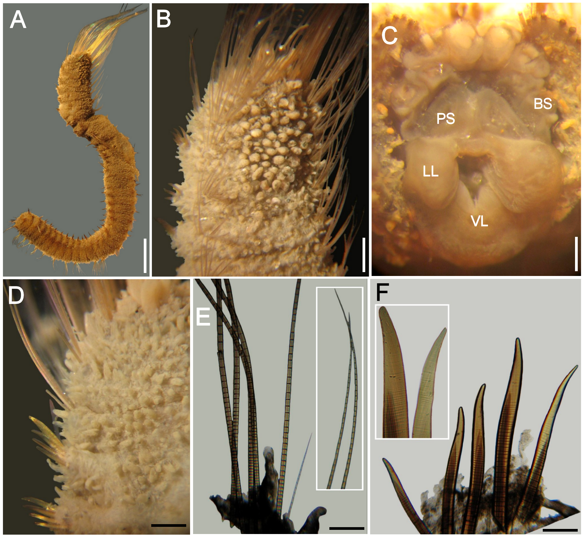
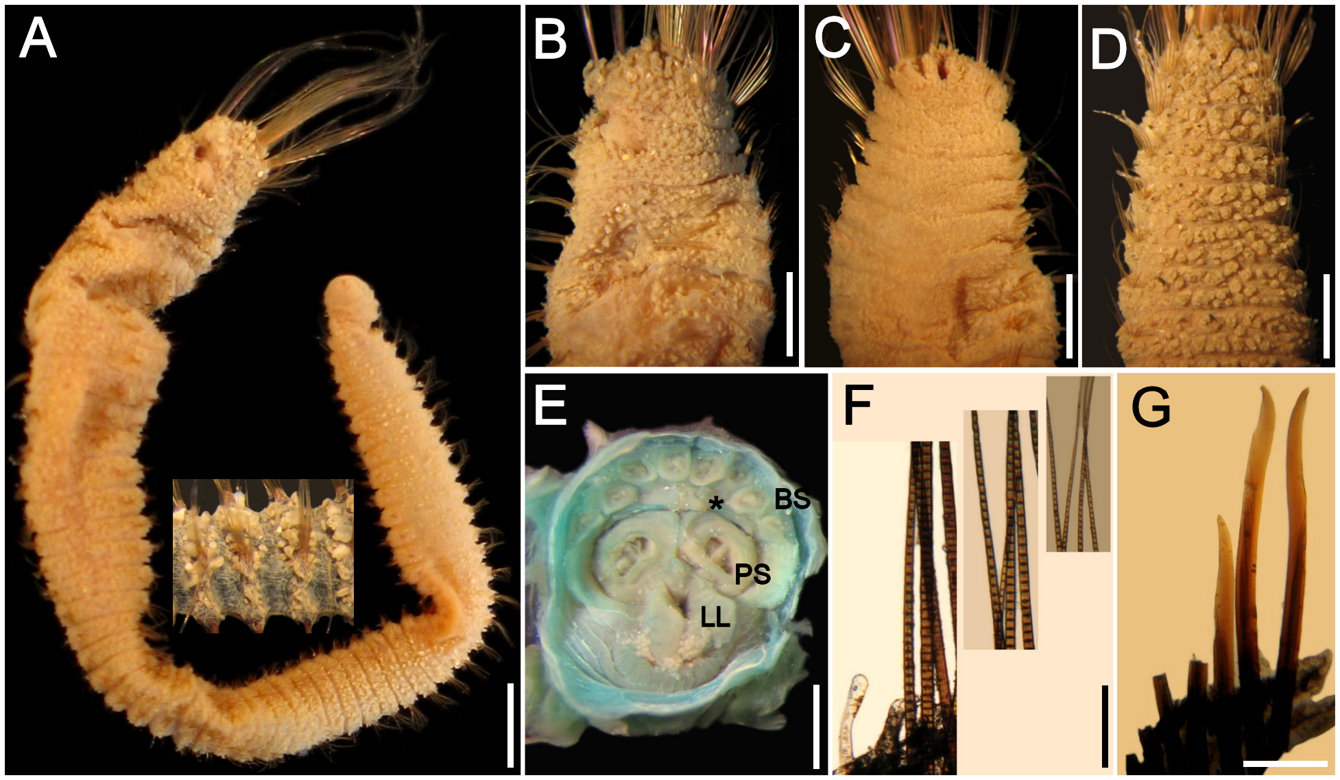
Description. Largest syntype (MCZ 1900) cylindrical, pale, posteriorly tapered ( Fig. 12A View FIGURE 12 ); 74 long, 4 mm wide, cephalic cage 7 mm long, 87 chaetigers. Body papillae with very little sediment, filiform, globose due to adherent sediment, completely covering the segments ( Fig. 12B, C View FIGURE 12 ), about 7 rows per segment on chaetigers 2–3, then up to 12 rows per segment (up to 8 on anterior fragment).
Cephalic hood barely exposed, margin papillated. Prostomium low cone; eyes barely visible ( Fig. 12C View FIGURE 12 ); caruncle reduced. Palps as long as branchiae; palp keels low. Lateral lips well developed; ventral and dorsal lips reduced.
Branchiae cirriform, distorted (observed in non-type specimen), arranged in two rows ( Fig. 12D View FIGURE 12 ), all of the same length and width, resembling palps. Nephridial lobes not seen.
Cephalic cage chaetae as long as 1/12 body length, almost twice as long as body width. Chaetigers 1–3 forming cephalic cage; chaetae arranged in short series. Chaetigers 1–2 with longest chaetae, those in chaetiger 3 about twice as long as following ones, but about half as long as those in previous chaetigers. First notopodia with 12 chaetae, first neuropodia with 10 chaetae, following chaetigers with fewer chaetae.
Anterior dorsal margin of first chaetiger smooth (anterior fragment), or with a short lobe (complete specimen). Chaetigers 1–3 of similar length. Chaetal transition from cephalic cage to body chaetae abrupt; anchylosed, falcate blunt neurohooks start from chaetiger 4. Gonopodial lobes reduced to flat rounded areas, visible on chaetigers 5 and 6 (only in complete syntype).
Parapodia slightly developed; chaetae emerge from low transverse lobes. Parapodia lateral; medial neuropodia ventrolateral. Notopodia and neuropodia without long papillae.
Medial notochaetae arranged in transverse series; all multiarticulated capillaries, articles short basally, medium-sized medially, long distally, 6–8 per fascicle ( Fig. 12F View FIGURE 12 , insets), as long as 1/5 body width. Neurochaetae multiarticulated capillaries in chaetigers 1–3; anchylosed, slightly falcate, reddish-brown neurohooks from chaetiger 4, arranged in transverse series ( Fig. 12E, F View FIGURE 12 inset), generally 4 per fascicle, reduced to 3 in prepygidial chaetigers.
Posterior end tapered into a cone; pygidium with anus dorsoterminal, without anal cirri.
Remarks. Pherusa papillata ( Johnson, 1901) is a member of the group of species without sediment cover on their body papillae. This and the fact that their neurohooks start on chaetiger 4, means that P. papillata resembles P. hobsonae , P. moorei , P. mikacae , and P. rullieri n. sp. However, as both species have neurohooks falcate, and 10–20 transverse series of papillae per segment, P. papillata is most similar to P. mikacae ; these two species differ in the integument pigmentation, relative shape and number of transverse rows of papillae, and number of neurohooks in medial chaetigers. In P. papillata the integument is pale, dorsal papillae are filiform, globose, arranged in 10–12 transverse series per segment, and there are 3–4 neurohooks per bundle, whereas in P. mikacae integument is brownish, dorsal papillae are depressed and arranged in more series (18–20) per segment, and there are more (5) neurohooks per bundle.
Distribution. Northeastern Pacific Ocean, off British Columbia to California, in subtidal shallow depths (up to 92 m). The record by Monro (1933:1058) is questionable.
Pherusa rullieri n. sp.
Figure 13 View FIGURE 13
Stylarioides plumosa View in CoL .— Rullier, 1965:47 (non Müller, 1776).
Type material. Western Africa, Gulf of Guinea. Holotype ( MNHN 1556 View Materials ), Benin , Sta. 1 (06º10' N, 02º10' E), 55 m, greyish muddy sand, 8 Oct. 1963, A. Crosnier, coll. GoogleMaps
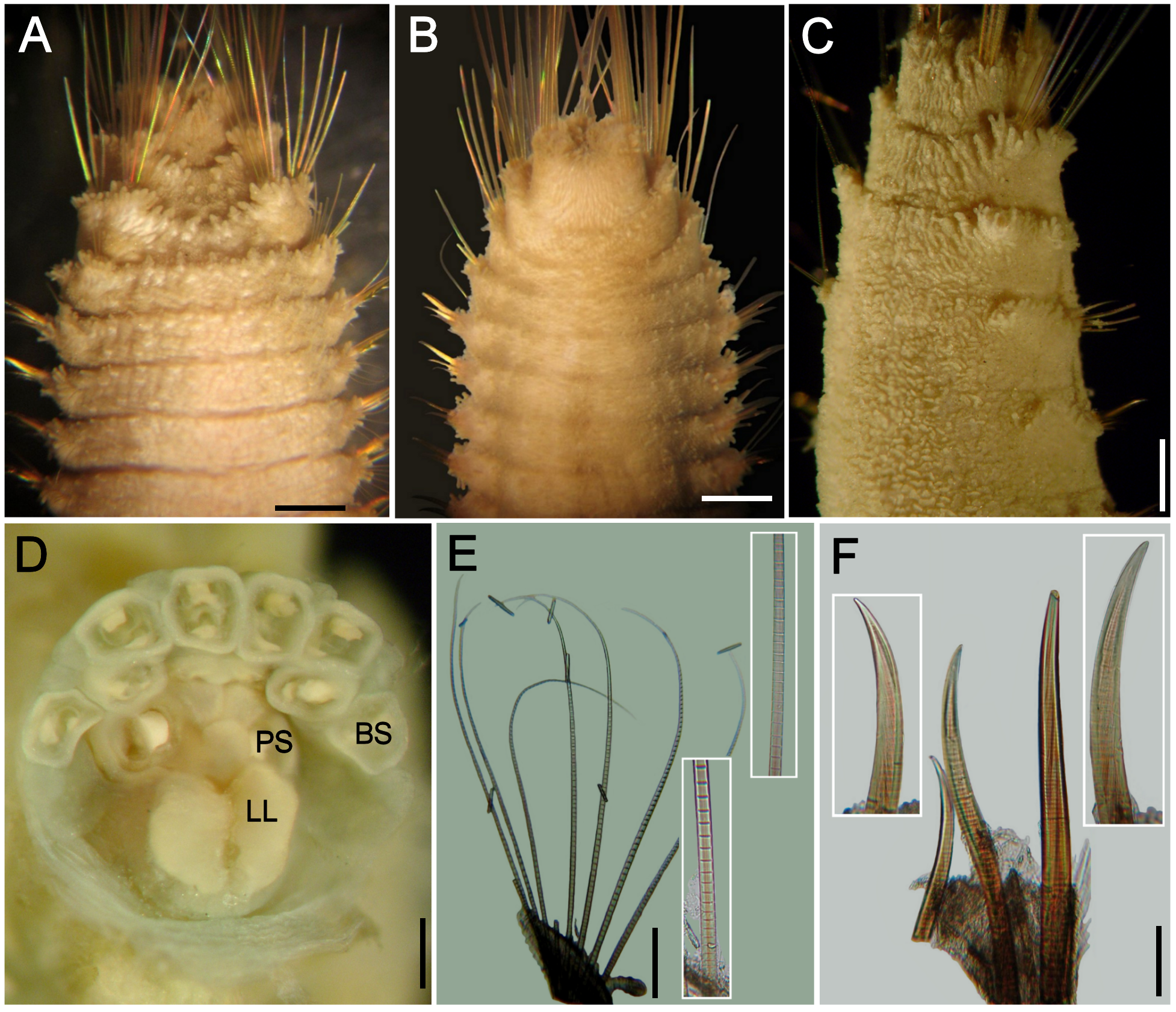
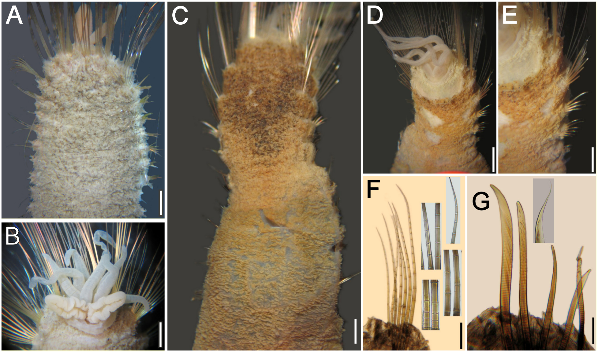
Description. Holotype (MNHN 1556) soft, apparently preserved in ethanol or already rotting when fixed with formalin ( Fig. 13A View FIGURE 13 ); body cylindrical, tapered posteriorly, broken into two parts, some medial segments missing. Anterior fragment 23 mm long, 6 mm wide, cephalic cage 12.5 mm long, 22 chaetigers; posterior fragment 20 mm long, 3 mm wide, 25 chaetigers. Body papillae larger over few anterior chaetigers, smaller throughout the body; most papillae depressed, about 20–26 transverse rows per segment.
Cephalic hood papillose, barely exposed ( Fig. 13B, C View FIGURE 13 ); not dissected to avoid further damage. Cephalic cage chaetae twice as long as body width. Chaetigers 1–2 forming cephalic cage but second chaetiger with chaetae less than half as long as those present in chaetiger 1. Chaetiger 3 with chaetae half as long as those present in chaetiger 2, but twice as long as those present in following chaetigers. Chaetiger 1 with 18 noto- and 10 neurochaetae, arranged in a sinusoid series; chaetiger 2 with 11–12 noto- and 14 neurochaetae, arranged in a curved series.
Anterior dorsal margin of first chaetiger without large papillae, crossed by first notochaetae. Anterior chaetigers (2–4) with larger papillae arranged in 2–3 irregular, transverse rows. Chaetigers 1–3 decreasing progressively in length. Chaetal transition from cephalic cage to body chaetae abrupt; long, slightly curved, aristate neurohooks start in chaetiger 4. Gonopodial lobes not seen.
Parapodia poorly-developed, slightly larger along anterior region, chaetae emerge fom low lobes. Parapodia lateral, medial neuropodia ventrolateral. Notopodia and neuropodia, with 3–4 digitate, large interramal papillae.
Medial notochaetae arranged in transverse or oblique series; all multiarticulated capillaries, articles short basally, long articles medially, slightly decreasing in size towards distal region ( Fig. 13D–F View FIGURE 13 ); 10–12 per fascicle, as long as ¼–1/3 body width Neurochaetae long, multiarticulated capillaries in chaetigers 1–3; anchylosed, falcate, tapered, aristate, light-brown neurohooks from chaetiger 4, arranged in transverse series, mostly 8 per fascicle throughout the body, aristae often broken ( Fig. 13E View FIGURE 13 ).
Posterior end unknown.
Remarks. Pherusa rullieri n. sp. resembles those other species with body papillae without sediment cover and with neurohooks from chaetiger 4: P. hobsonae , P. mikacae , P. moorei , and P. papillata ( Johnson, 1901) . However, P. rullieri is most similar to P. moorei and to P. sibogae n. comb. by having medial and posterior neurohooks slightly curved, aristate. However, P. moorei has a cephalic cage 4 x longer than body width, whereas in P. rullieri and P. sibogae it is only twice as long as body width. These two latter species differ because in P. rullieri body papillae are abundant, without sediment particles whereas in P. sibogae body papillae are scarce and there are sand particles over papillae and tunic, although they do not form tubercles.
Etymology. This species is named after the late Dr François Rullier in recognition of his many publications on polychaetes, and especially because he reported this specimen in one of his contributions. The epithet is a noun in the genitive case.
Type locality. Benin, SE off Cotonou , 55 m depth .
Distribution. Only known from the type locality in the Gulf of Guinea.

No known copyright restrictions apply. See Agosti, D., Egloff, W., 2009. Taxonomic information exchange and copyright: the Plazi approach. BMC Research Notes 2009, 2:53 for further explanation.
|
Kingdom |
|
|
Phylum |
|
|
Class |
|
|
Order |
|
|
Family |
|
|
Genus |
Pherusa papillata ( Johnson, 1901 )
| Salazar-Vallejo, Sergio I. 2014 |
Stylarioides plumosa
| Rullier, F. 1965: 47 |
Pherusa papillata
| Blake, J. A. 2000: 19 |
| Hartman, O. 1961: 120 |
Stylarioides papillata
| Hartman, O. 1938: 14 |
| Moore, J. P. 1923: 221 |
Trophonia papillata
| Moore, J. P. 1909: 284 |
| Moore, J. P. 1908: 356 |
| Johnson, H. P. 1901: 416 |