
Plategeocranus sulcatus ( Karsch, 1884 )
|
publication ID |
https://doi.org/ 10.5281/zenodo.205116 |
|
DOI |
https://doi.org/10.5281/zenodo.5624992 |
|
persistent identifier |
https://treatment.plazi.org/id/03B787EF-FF8D-600F-8CB5-79D1FD6C465F |
|
treatment provided by |
Plazi |
|
scientific name |
Plategeocranus sulcatus ( Karsch, 1884 ) |
| status |
|
Redescription of Plategeocranus sulcatus ( Karsch, 1884)
Adult ( Figs. 1 View FIGURES 1 – 8 –29). Measurements: body length 465–610 µm (n=6), width 280–380 µm (n=4), height 180–270 µm (n=3).
General form and color ( Figs. 1–3 View FIGURES 1 – 8 , 23–26): Body with proterosoma comprising about 1/3 of total length, height slightly less than half length; contour rounded in lateral aspect (Fig. 21; rather flat contour of cuticular remnant in Figs. 3 View FIGURES 1 – 8 , 23 artifactual). Conspicuous system of longitudinal folds on notogaster. Dark-brown with lighter foveolae on notogaster and ventral plate when seen in transmitted light.
Integument ( Figs. 1–5 View FIGURES 1 – 8 , 13, 18 View FIGURES 9 – 20 , 21–27): Foveolate in interlamellar area of prodorsum and on notogaster and ventral plate, reticulate on legs, pedotecta and lateral regions of body. Cerotegument macrogranular (granules up to 2.5 µm) on prodorsum, lateral regions, notogaster and legs; on notogaster granules trace reticulation, forming netlike pattern (Fig. 26).
Prodorsum ( Figs. 1–3 View FIGURES 1 – 8 , 21–24): Prodorsum and notogaster apparently fused, but margins well-delineated by uninterrupted dorsosejugal suture. Rostrum rounded anteriorly, laterally without genal incision or tooth. Prodorsum with system of well-developed interconnected ridges ( Figs. 1 View FIGURES 1 – 8 , 24). Lamella developed as simple costate ridge, pair slightly converging distad, each with small terminal tubercle bearing lamellar seta (le). Tutorium similar in form, with proximal third near and almost parallel to lamella, then directed ventrad to end in small tooth, just dorsal to rostral margin. Additional small lateral carina present dorsal to each acetabulum I. Two long transverse ridges present: anterior ridge connecting distal ends of both lamellae and tutoria; posterior ridge connecting lamellae at their base, then running obliquely posteroventrad to bothridia and on to dorsal corner of large pedotectum I. Pedotectum II present, about half size of I. Rostral (ro) setae set widely apart on small tubercles, ro and le of similar length, 40– 60 (n=4) and 45 µm, respectively, acuminate, slightly barbed, some barbs with granules of cerotegument; interlamellar seta (in; Figs. 7, 8 View FIGURES 1 – 8 ) 24–30 µm (n=2), stiff, barbed, with several wide, untapered branches; exobothridial seta (ex) minute (ca. 5 µm), smooth. Bothridial seta bo ( Fig. 6 View FIGURES 1 – 8 ) 47–50 µm (n=2) curved posterodorsad, with narrow stalk and long, almost drop-shaped, slightly flattened head roughened with minute spicules (sometimes hidden by granules of cerotegument); bothridium large, cup-shaped, with spiral internal ridge. Mutual distances of setal pairs ro–ro, le–le, in–in, and bo–bo 61–65 (n=3), 55, 47–60 (n=2), and 140–150 (n=2) µm, respectively.
Lateral podosomal region ( Figs. 3 View FIGURES 1 – 8 , 21, 23): Foveolar pattern augmented by oblique ridge running from humeral region to between acetabula III and IV and another to acetabulum IV; cuticle without sculpturing dorsal to acetabulum IV. Narrow posterodorsal fold partly covers acetabulum IV. Discidium and circumpedal carina absent.
Gnathosoma ( Figs. 2 View FIGURES 1 – 8 , 17–20 View FIGURES 9 – 20 ): Subcapitulum diarthric; rutellum pantelobasic, with ventral expansion, conspicuous teeth and finger-like mesial projection ( Fig. 18 View FIGURES 9 – 20 ). Palp ( Figs. 19–20 View FIGURES 9 – 20 ) five-segmented, with setal formula 0–2– 1–3–9(1); solenidion ω recumbent, independent of seta acm. Chelicera known only from partial remnant ( Fig. 17 View FIGURES 9 – 20 ): chelate-dentate; seta cha (44 µm) posterior to chb (21 µm); thin light-colored spine (spi) present on antiaxial surface; small fragment of porose area (por) observed posterior to movable digit.
Notogaster ( Figs. 1, 3 View FIGURES 1 – 8 , 21–24): Slightly longer than wide. Surface with distinct topography of folds, expressed as paired apparent grooves and complementary ridges. Anteromedial region with small pair of weak, branched ridges effacing behind level of c -setae. Several deep grooves accentuated by complementary ridges converge in each humeral region ( Figs. 1 View FIGURES 1 – 8 , 22, 24). Medial pair of grooves rather shallow, delineating narrow wedge-shaped central area. Lateral to these run two deeper grooves with complementary ridges: mesial groove almost straight, lateral groove curved parallel to body outline, both converging at humeral region and also posteriorly, near border of notogaster, and leaving short ridge between them. Both medial and lateral ridges well visible even in transmitted light (Fig. 24).With 12 pairs of setae or their alveoli discerned: setae thin, smooth or slightly roughened, strongly curved posteriad. Seta c 1 situated close to branched anterior ridges, c 2 and c 3 close together anterior to, and aligned with, medial longitudinal ridge; la–lm and h 3 –h 2 aligned along lateral ridge; h 1 and setal row p inserted parallel to posterolateral margin of notogaster. Typical five pairs of lyrifissures present, situated as in Fig. 1 View FIGURES 1 – 8 , aligned within lateral groove; ia appears minute in dorsal orientation, but only slightly smaller than others in lateral orientation. Opisthonotal gland opening (gla on Fig. 1 View FIGURES 1 – 8 ) present but very small and difficult to observe.
Ventral region ( Figs. 2, 4, 5 View FIGURES 1 – 8 , 25): Internal epimeral borders 2 and sj distinct, meeting and blending with broad sternal border in midline; epimeral border 3 not evident, so epimeral fields 3 and 4 merged; epimeral border 4 weakly defined, merging with sternal border medially and effacing before approaching acetabulum IV. Apodeme 1 present but poorly seen; apodemes 2 and sj distinct, both of typical form, projecting internally from respective borders; apodemes 3 and 4 not observed. Epimeral formula (n=1) 4–1–3–3 (both alveoli 4c present, but right one not discernible in orientation of figures); setae thin, smooth, straight or slightly curved, about 15–20 µm long (n=2). In transmitted light, absence of foveolae creates impression of weak circumgenital ring that extends around anterior region of anal plates. Distribution of genital setae asymmetrical in single observation, five on left plate, six on right; preserved (anterior) setae thin, filiform. One pair of aggenital (ag), two pairs of anal (an) and three pairs of adanal (ad) setae present; length of ad 2 10 µm. Lyrifissure iad (length as measured on cuticular remnant 15 µm; n=2) located halfway between anal margin and posterolateral border of ventral plate, outside setal row ad.
Legs, general aspects ( Figs. 9–16 View FIGURES 9 – 20 , 21, 22, 27–29): Relatively long (legs I and IV each approximately 0.6 body length), slender, with large apophysis distally on tibiae I and II and at midlength of tarsus I, bearing respective solenidia ( Figs. 9, 10 View FIGURES 9 – 20 , 27–29). Distally, tarsi extremely narrowed, with obvious sclerotization ending well proximal to claws, continued as apparently flexible, hyaline stalk bearing setae (p) and tridactylous apotele (Fig. 27). Terminal soft cuticle without pulvillus. Lateral and empodial claws well-developed, approximately equal in size and form, each with at least two dorsal rows of short spines. Proral (p) setae of tarsi II–IV and all unguinal (u) setae with comb of long, fine cilia along ventral side. Tarsal lyrifissures displaced antiaxially; on legs I and II located under distal projection of tibia and difficult to observe (on leg I, observed only in specimen MGCP Ar 89 where cuticular remnant of tarsus shrank from articulation). Some lateral setae strongly curved and barbed along dorsal side; other setae more or less curved and barbed, some covered with granules of cerotegument. Setal homologies indicated in Table 1 View TABLE 1 .
tures are indicated for the instar in which they were first observed and are present in subsequent instars.1 1Setae in parentheses represent pairs; dash indicates no additions;? indicates poorly visible, but no additional setae were discerned. La, Tn, Ad = larva, tritonymph, adult, respectively.
2Seta e (famulus) was not discerned in the larva but probably is present; among extant oribatid mites it is not known to be absent from tarsus I.
3 Setae and solenidia in this row are present on tritonymphs, but as no protonymphs or deutonymphs were available for study they may have been added in any of the nymphal instars. Among extant oribatid mites, setae are rarely added between larva and protonymph.
Leg I ( Figs. 9–11 View FIGURES 9 – 20 , 27–29). Tarsus: solenidia ω1 and ω 2 set on separate long apophyses; setae (p) appear smooth, probably eupathidial; (a), s, tc', ft', pl', (pv) more robust than (it), tc", pl", l" and v'; famulus e simple, set directly on surface. Tibia: solenidion φ1 long, flagellate, inserted on long, subcylindrical apophysis; φ2 thinner, shorter, set on smaller apophysis; setae l', v' more robust than l "and v". Genu: solenidion σ thin, filiform, curved; seta l' more robust than l", v'. Femur: with dorsal and ventral porose areas, large seta d and smaller, curved and barbed setae (l) and bv". Trochanter: seta v' longer than segment, barbed, strongly curved. Setal formula: 1–4–3(1)–4(2)–19(2).
Leg II ( Figs. 12, 13 View FIGURES 9 – 20 ). Tarsus: bears single solenidion ω, inserted on apophysis much smaller than that of tarsus I; short, blunt, barbed seta ft" lateral to it, on separate tubercle; setae (it), (tc), (a) and s with short barbs, ft', (pv) and l" rough, latter strongly curved. Tibia: φ medium long, inserted on long apophysis; l', v' longer and thicker than l", v". Genu: σ thin, attenuate, geniculate; setae thin, almost smooth; (l) curved, v' almost straight. Femur: with large dorsal and ventral porose areas; thick seta d and smaller curved setae (l) and bv". Trochanter: seta v' longer than segment, barbed, strongly curved. Setal formula: 1–4–3(1)–4(1)–16(1).
Leg III ( Figs. 14, 15 View FIGURES 9 – 20 ). Tarsus: setae (a), s, (it), (tc), (ft), (pv) of similar sizes, some ventral setae ciliate. Tibia: φ short, without apophysis; setae l', (v) present. Genu: short thin σ and small thin seta l', the latter poorly visible. Femur: with lobe-like anteroventral and antiaxial anterolateral projections forming cup-like insertion for genu, and large posterodorsal porose area; large barbed seta d and smaller, curved and barbed l', and ev' present. Trochanter: large and aligned with femur (not noticeably flexed in any specimen; Fig. 21), with proximodorsal porose area; thin smooth v' and small, curved and barbed l' seta. Setal formula: 2–3–2–3(1)–15.
Leg IV ( Figs. 16 View FIGURES 9 – 20 , 21). Tarsus: setae (tc), ft" rough, (a), s, (pv) ciliate. Tibia: long and thick-walled, slightly arched ventrad; φ short, seta v' almost straight, l' and v" curved (the latter lost from drawn leg). Genu with short thin seta d visible on only one specimen. Femur with large posterodorsal porose area and lobe-like projection present as on femur III; setae d and ev' visible on one specimen (MGCP 89), d and paraxial alveolus on other specimen (MGCP 46). Trochanter very large and aligned with femur, with thin seta v' (seen in one specimen, MGCP 89). Setal formula: 1–? –1–3(1)–12.
FIGURES 21–29. Plategeocranus sulcatus ( Karsch, 1884) , adults (samples: 21. MGCP Ar 89. 22. GMUG 33016. 23–25. MGCP Ar 46. 26. MGCP Ar 35, 27–29. SIZK K-8945). 1, 2. Dry amber inclusions in reflected light. 21. Lateral view. 22. Dorsal view. 23–25. Cuticular remnants from the broken inclusion; note scale same as in 21–22. 23. Lateral view, hysterosoma flattened due to displacement of ventral plate which is pressed into notogaster. 24. Oblique dorsal view. 25. Oblique ventral view. 26. Fragment of notogastral cuticle with cerotegument and setal alveolus. 27–29. Leg I right, antiaxial view. 27. Tarsus and distal fragment of tibia. Arrows point to apophyses of solenidia φ1 and ω1. 28. Apophyses of tarsal solenidia, apophysis of ω2 (arrows) visible by transparency. 29. Apophyses of tibial solenidia, apophysis φ2 (arrows) visible by transparency. Scale bars: 21–25 = 100 µm; 26–29 = 10 µm. Figs. 21–25 are combined from 80–95 images each; 27 from 11 images; 28–29 from 3–4 images each.
Immature instars ( Figs. 30–60 View FIGURES 30 – 35 View FIGURES 36 – 46 View FIGURES 47 – 50 View FIGURES 51 – 60 )
Larva ( Figs. 30–35 View FIGURES 30 – 35 , 36–41 View FIGURES 36 – 46 ). Measurements: 210x130 µm.
General form and color: Oval, slightly broadened posteriorly, with proterosoma comprising about half total length; legs about 2/3 of body length. Color light brown in transmitted light.
Integument ( Fig. 30, 32 View FIGURES 30 – 35 , 36–38, 41 View FIGURES 36 – 46 ): Cuticle of hysterosoma strongly plicate; that of prodorsum with few weak folds, epimeral region smooth. Cerotegument macrogranular (granules 2–3 µm) on most of body and legs, best seen on folds of cuticle ( Fig. 38 View FIGURES 36 – 46 ); present on setae ro and bo, not seen in epimeral region; microgranular (granules about 0.5 µm) in dorsosejugal region.
Prodorsum ( Figs. 30, 32 View FIGURES 30 – 35 , 36, 37 View FIGURES 36 – 46 ): With pair of weakly costate lamellae, lacking cusps but each bearing alveolus of seta le at anterior end (setae not observed). Well-defined, narrow transverse fold connects lamellae posteriorly, with broader fold prolonging each end toward respective bothridium. Pair of broad folds run from seta in toward base of leg I. Cuticular folds form pedotectum-like projection over base of leg I and II. Seta ro (10 µm) acuminate, narrow, finely barbed, covered with granules of cerotegument; pair closer together than other prodorsal setae. Seta in (at least 5 µm long) stiff, rather thick, blunt, set with spines ( Fig. 31 View FIGURES 30 – 35 ); left with two branches, right with three; inserted on darker sclerite or tubercle. Seta bo (at least 18 µm long) clavate, with head slightly flattened and spinose. Mutual distances of setal pairs ro–ro, le–le, in–in, and bo–bo 16, 25, 29, and 65 µm, respectively.
Gastronotic region ( Figs. 30, 32 View FIGURES 30 – 35 , 36, 37, 39 View FIGURES 36 – 46 ): With large dark folds forming elevated belt around smoother centrodorsal area; folds approximately transverse anteriorly, chevron-like posteriorly. Seta c 3 slightly roughened, small (ca. 3 µm) but longest of visible setae; minute setae c 1, c 2, and thin, smooth setae da–dm well visible; seta la and alveolus lm seen only on right side; alveolus lp seen (with some doubt) on left side. Discernable in ventral aspect ( Fig. 32 View FIGURES 30 – 35 ): setae h 2 and h 3, pair of oval structures that probably represent cupules ih, and pair of darker-colored round spots that might be opisthonotal gland openings (gla?). Seta h 1 not discerned, either in ventral or dorsal aspect.
Gnathosoma ( Fig. 32 View FIGURES 30 – 35 ): Subcaptiulum diarthric; chelicera chelate-dentate, rutellum atelobasic, with ventral lobe; palps poorly seen.
Coxisternum and paraprocts ( Figs. 32 View FIGURES 30 – 35 , 37, 39 View FIGURES 36 – 46 ): Surface flat, without conspicuous features. Apodemes 2 and sj reach less than half distance to midline; no evidence of porosity or other respiratory elements. Claparède's organ ( Fig. 32 View FIGURES 30 – 35 cla) rather large, details poorly discerned but with posterior part well-defined and more darkly colored than anterior ( Fig. 39 View FIGURES 36 – 46 ). Discernable epimeral setation (I–III) 2–1–2, but seta 1c probably present as scale over cla, in typical fashion. Presumptive genital region with transverse folds. Two pairs of thin, minute pseudanal setae present on paraprocts (segment PS); no third seta or alveolus discerned.
Legs, general aspects ( Figs. 33–35 View FIGURES 30 – 35 , 36, 37, 40, 41 View FIGURES 36 – 46 ): All about as long as gastronotic region, legs III longest; robust; tarsi with long, distal hyaline stalks beyond unguinal setae, terminally supporting proral setal pair (p), pulvillus and strong empodial claw. Setae and solenidia indicated in Table 1 View TABLE 1 ; famulus not discernable in single specimen. Setae (p) on legs II–III and (u) on all legs with ventral comb of long ciliae. Some setae strongly curved, all more or less barbed.
Leg I ( Figs. 33 View FIGURES 30 – 35 , 41 View FIGURES 36 – 46 ): Tarsus: solenidion ω about as long as tarsus, inserted on long apophysis; setae (tc) long, almost smooth; ft', (a) and s almost equal in length, slightly barbed, straight; (pv) slightly and (pl) strongly curved, barbed; seta ft" absent. Famulus not discerned. Tibia: solenidion φ about twice length of ω, inserted on long, narrow apophysis equal to almost 1/3 total length of segment; setal pair (l) strongly curved, barbed; v' small. Genu: almost cylindrical, with one pair of curved setae (l) and attenuate solenidion σ. Femur: with longitudinal ventral ridge and ventral porose area; barbed robust seta d larger than bv". Trochanter: with large dorsal and smaller ventral spine distally, no setae discernable. Setal formula: 0–2–2(1)–3(1)–14(1); if present, famulus not included in tarsal count.
Leg II ( Fig. 34 View FIGURES 30 – 35 ): Tarsus: with wide low projection on which solenidion ω and seta ft" insert; setation as on tarsus I except (pl) absent and ft" present; setae rather long, straight and almost smooth, ft' thickest. Tibia: solenidion φ almost as long as ω, inserted on apophysis of about 1/6 tibial length; barbed, curved seta l' and straighter, smooth v' present. Genu and femur as on leg I; trochanter with one dorsal notch. Setal formula: 0–2–2(1)–2(1)–13(1).
Leg III ( Figs. 35 View FIGURES 30 – 35 , 40 View FIGURES 36 – 46 ): Tarsus: with setae (tc), ft", (a), s long, straight, smooth; (pv) and ft' shorter and curved. Tibia: without apophysis, small solenidion φ and seta v' (see Remark 2) present. Genu: with small solenidion σ and seta l'. Femur: with lateroventral porose area, large rough seta d and smaller, smooth ev'. Trochanter: with strong dorsal spine, no setae discerned. Setal formula: 0–2–1(1)–1(1)–13.
Tritonymph ( Figs. 42–60 View FIGURES 36 – 46 View FIGURES 47 – 50 View FIGURES 51 – 60 ). Measurements: 430x250 µm, measured with scalps.
General form and color ( Figs. 42, 43 View FIGURES 36 – 46 ): About twice longer than wide, prodorsum comprises about 1/3 of total length. With strongly eccentric stack of exuvial scalps which slightly overhangs posteriorly. Legs long, slender; IV longest, slightly longer than body. Light-brown in transmitted light.
Integument ( Figs. 42–45 View FIGURES 36 – 46 , 47–50 View FIGURES 47 – 50 ): Foveolate in epimeral region, foveolate or reticulate on legs, rough with small folds in centrodorsal areas of scalps, almost smooth on prodorsum. Cerotegument macrogranular (granules up to 2.5 µm) on legs, on anterior and central parts of prodorsum, on pedotecta and in gastronotic region; microgranular (granules up to 0.5 µm) on dorsal side of prodorsum anterior to bothridia and in dorsosejugal fold; not discerned in epimeral region.
Prodorsum ( Figs. 42, 43, 45 View FIGURES 36 – 46 , 47, 50 View FIGURES 47 – 50 ): With pair of linear, costate lamellae, lacking anterior tubercle or cusp. Lamellae proportionally closer together than in larva; posterior ends connected by wide ridge that extends as oblique ridge to reach bothridia. Second pair of longitudinal ridges runs lateral to lamellae to form large folds anterior to acetabulum I. Insertions of legs I and II protected by pedotectum-like folds. Seta ro (ca. 35 µm) set on small isolated tubercle, seta le (ca. 13 µm) inserts at end of lamella; both rough, acuminate, more or less covered with cerotegument. Seta in (at least 10 µm long), stiff, with several blunt, spinose branches ( Figs. 45 View FIGURES 36 – 46 , 48 View FIGURES 47 – 50 ). Bothridium cup-shaped, with spiral internal ridge; seta bo (ca. 40 µm) with narrow stalk and drop-like, slightly flattened head, finely spinose and partly covered with cerotegument ( Fig. 45 View FIGURES 36 – 46 ). Alveolus of seta ex visible lateral to bothridium. Mutual distances of setal pairs ro–ro, le–le, in–in, and bo–bo 37, 26, 60, and 100 µm, respectively.
Gastronotic region ( Figs. 42, 43 View FIGURES 36 – 46 , 47, 50 View FIGURES 47 – 50 ): Cuticle with system of large folds, most accentuated in region lateral to exuvial scalps. Three tightly appressed exuvial scalps in rather flat and strongly eccentric stack; each centered well posterior to scalp underneath. Each scalp bears few cerotegument granules anteriorly; transverse anterior fold and pair of folds running posteriorly from humeral region form angle in humeral area of each scalp, especially visible on dry specimens ( Fig. 42 View FIGURES 36 – 46 , right half); larval scalp (SL) plicate throughout. Setation of scalps probably only partly discerned; all visible setae minute. Larval scalp with alveoli of setae c 1, c 2 and da discernable; on ventral side, pair of very thin setae (? on Fig. 50 View FIGURES 47 – 50 ) may represent dp of larval scalp. On protonymphal (SPN) and deutonymphal (SDN) scalps, three pairs of minute setae discernable, those on SDN being about twice length of those on SPN. Visible tritonymphal setae include c 1, c 2 (not less than 6 µm each), curved, slightly rough, c 3 (not shorter than 18 µm), with thin barbs, and apparently smooth p 3 (at least 6 µm long) on ventral side. No cupules or glands discernible.
Ventral region ( Figs. 43 View FIGURES 36 – 46 , 50 View FIGURES 47 – 50 ): Epimeral region much damaged. Alveoli of epimeral setae on cuticular remnants (shown with thin solid lines on Fig. 50 View FIGURES 47 – 50 ), displaced far from respective setae preserved in amber imprint (connected by dotted line; see Sidorchuk & Norton 2011). Setae smooth, thin, curved; 10–15 µm, medial longest); setal formula 3–1–3–3. Genital region with somewhat convex (bulging) triangle of smooth cuticle, well circumscribed anteriorly and separated from epimere IV by pair of oblique, narrow, deep grooves containing striated (apparently softer) cuticle ( Fig. 43 View FIGURES 36 – 46 , arrows). Pair of similar grooves circumscribe genital region posteriorly, interrupted medially by narrow anal region. Genital valves together form triangular shape, flanked by triangular aggenital sclerite. Five pairs of genital setae filiform, all at least 10 µm long, arranged in longitudinal row along medial edge of valve; aggenital seta (ag; at least 11 µm) barbed, inserted close to posterior groove. Two pairs of setae (an 2 ca. 7 µm, an 1 minute) on anal valves (AN). Two adanal (ad, 6–8 µm) setae on left adanal valve well visible, presence of third (ad 1?) uncertain.
Gnathosoma ( Fig. 50 View FIGURES 47 – 50 ): Subcapitulum diarthric, with long smooth setae h, m, a, and two pairs of thin smooth adoral setae visible; rutellum atelobasic, with ventral lobe. Palp and chelicera not observable.
Legs, general aspects ( Figs. 42–44, 46 View FIGURES 36 – 46 , 51–60 View FIGURES 51 – 60 ): Long, tarsi with well-developed ambulacral stalk, pulvillus and strong empodial claw. Complement of setae and solenidia indicated in Table 1 View TABLE 1 . Setae (p) of legs II–IV and all setae (u) with ventral comb of thin cilia, other setae either strongly curved and well-barbed or curved only slightly and almost smooth. Porose areas present on all femora and (uncertainly) on trochanters IV.
Leg I ( Figs. 44 View FIGURES 36 – 46 , 51–53 View FIGURES 51 – 60 ): Tarsus: solenidion ω 1 set on large apophysis, about three times length of ω2, which inserts on smaller apophysis. Famulus distinct, as long as apophysis of ω2, blunt, inserted in large alveolus or pit. Setae (p) smooth; setae (pl) curved, rough; ft' rather short, robust, almost smooth; (pv) slightly curved and barbed, setae (a), s, (it), and (tc) nearly straight and almost smooth; seta ft" absent. Tibia: long, gradually thinner distally; flagellate solenidion φ 1 set on large apophysis, that of short pin-like φ2 smaller. Setae (l) and v' of similar lengths, barbed, (l) strongly curved. Genu: solenidion σ short, curved, inserted on wide elevation. Barbed curved setae l' (large) and l" (very small) present. Femur: with large ventral porose area, reaching almost to proximal stalk of segment; with three setae: d, l' and bv". Trochanter (not shown) cylindrical, difficult to observe. Setal formula (including famulus):?–3–?(1)–3(2)–17(2).
Leg II ( Figs. 54–58 View FIGURES 51 – 60 ): Tarsus: solenidion ω set on small tubercle (ω2 absent). Seta ft" blunt, barbed, positioned lateral to ω on separate tubercle; seta ft' barbed, curved; other setae (( a), s, (pv), (it), (tc )) less curved, almost smooth. Tibia: solenidion φ shorter than ω of tarsus, blunt, set on apophysis. Setae (l) and v' barbed, more or less curved. Genu: solenidion σ curved, shorter than φ, blunt. Seta l" small and strongly curved, l' and v' as on leg I. Femur: as on leg I, but porose area somewhat smaller, not reaching proximal stalk of segment. Trochanter: cylindrical, with seta v' filiform, smooth. Setal formula: 1–3–3(1)–3(1)–15(1).
Leg III ( Fig. 59 View FIGURES 51 – 60 ): Tarsus: setae (a), s, (pv), (it) similar to those on leg II; setae (tc) thin, pair equally long (difference in drawing due to foreshortening), (ft) shorter and more robust; all setae almost smooth. Tibia: with short, blunt, curved solenidon φ set directly on surface; setae l' and v' barbed. Genu: thin, needle-like solenidion σ and single curved seta l' present. Femur: with posteroventral porose area and two barbed setae, ev' and d. Trochanter: large, without spines, similar to that in adult; no porosity or setae discerned. Setal formula:?–2–1(1)–2(1)–15.
Leg IV ( Figs. 46 View FIGURES 36 – 46 , 60 View FIGURES 51 – 60 ): Longest leg, all segments except genu considerably elongated. Tarsus: seta ft' small, straight; setae (a), s, (pv), and (tc) similar to those on tarsus III. Tibia: with tapered solenidion φ, curved barbed seta l' and straighter v'. Genu with smooth, tapered seta d. Femur: very long, with posteroventral porose area and setae ev' and d. Trochanter: very long, with two rounded lobes embracing base of femur; some pores visible on posterior side of ventral surface; no setae observed. Setal formula:?–2–1–2(1)–12.
Designation of neotype for Plategeocranus sulcatus ( Karsch, 1884) . In the original description of Nothrus sulcatus, Karsch (1884) mentioned no type or other individual specimen. Along with his redescription of the species and the proposal of Plategeocranus, Sellnick (1918) noted that Karsch’s specimens were already impossible to locate, and listed seven non-type specimens from Königsberg University's collection (now themselves lost) on which his work was based. In his second work on amber oribatid mites, Sellnick (1931) listed two specimens of P. sulcatus from the Klebs collection and 10 from the Fritsch collection. Eight of the latter are now housed in two museums in Kaliningrad (see Introduction and Materials); as they were certainly viewed and identified by Sellnick, we believe these to be the best choices for designation of neotype and paraneotypes.
Two amber pieces with Plategeocranus sulcatus (one polished piece and one slide-mounted), which we were told came from the Königsberg collection, are now housed in the GMUG (numbers G3.167 and #33016 respectively). Specimen #33016 (Fig. 22) is particularly good, the best specimen we examined in this study. However, their numbers cannot be linked to samples referred to by Sellnick (U35–41) in 1918.
For neotype we designate specimen # 39 in the MWO collection (number 186 in the original Fritsch collection). Of the eight specimens, this shows the most complete set of characters ( Figs. 61–63 View FIGURES 61 – 63 ). Visible on the prodorsum are the pedotecta, the system of lamellar and tutorial ridges, setae ro, le, in, bo and bothridia. The gnathosoma is obscured by a large bubble and the ventral surface is partly obscured by "mist"; however, half of the epimeral region is visible and the anal and genital openings can be vaguely perceived through the mist. The notogaster shows all major folds and ridges, humeral angles and smaller folds in its anterior part. Legs readily show shapes and proportions of segments, claws and some strongly curved setae; large apophyses on tibiae and tarsi I are also visible, though orientation is not favorable. We designate as paraneotypes the remaining seven specimens listed by Sellnick (1931): KMA specimens 197-22 [55?; see Remark 5, below] and 197-54 [54], and MWO specimens 22 [226], 30 [178], 33 [223], 35 [123] and 37 [224].


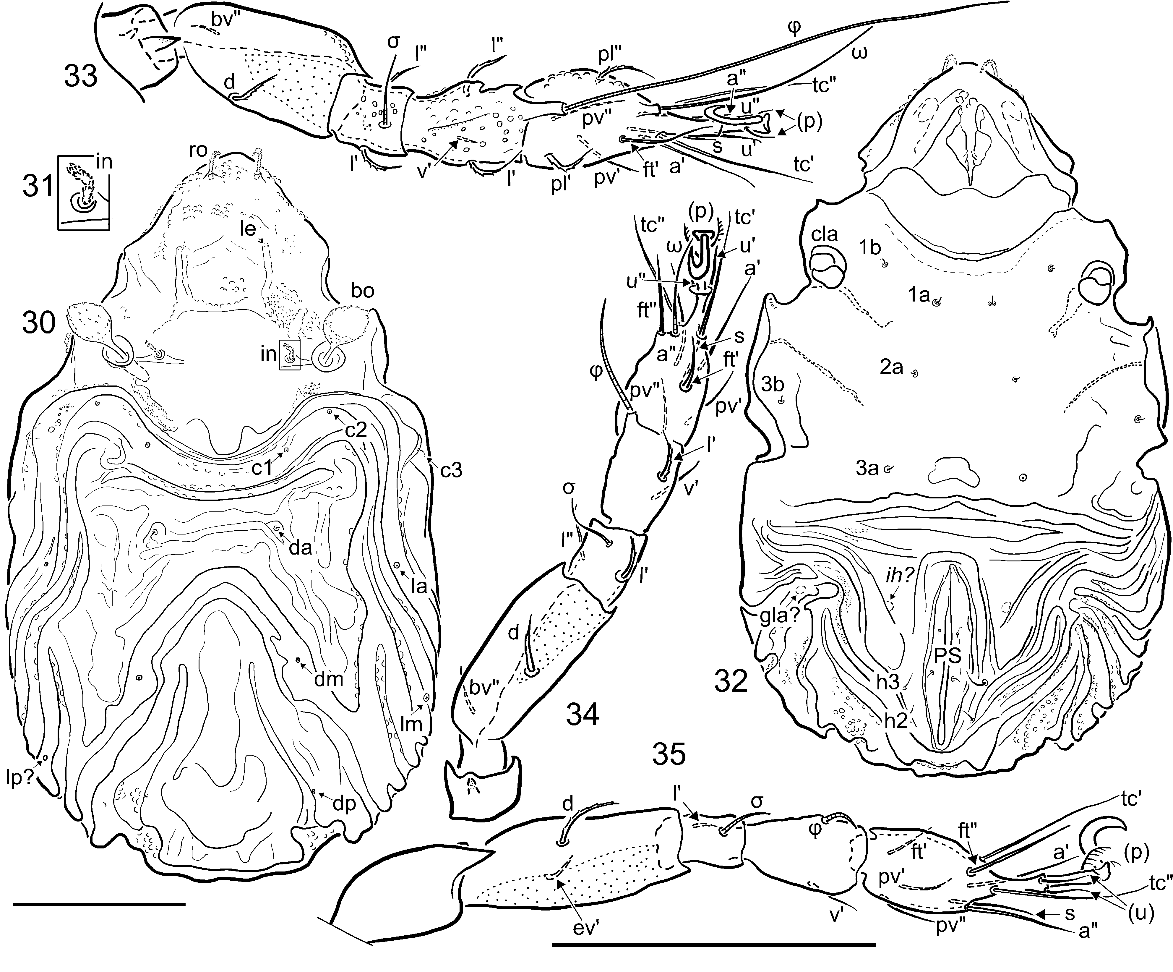

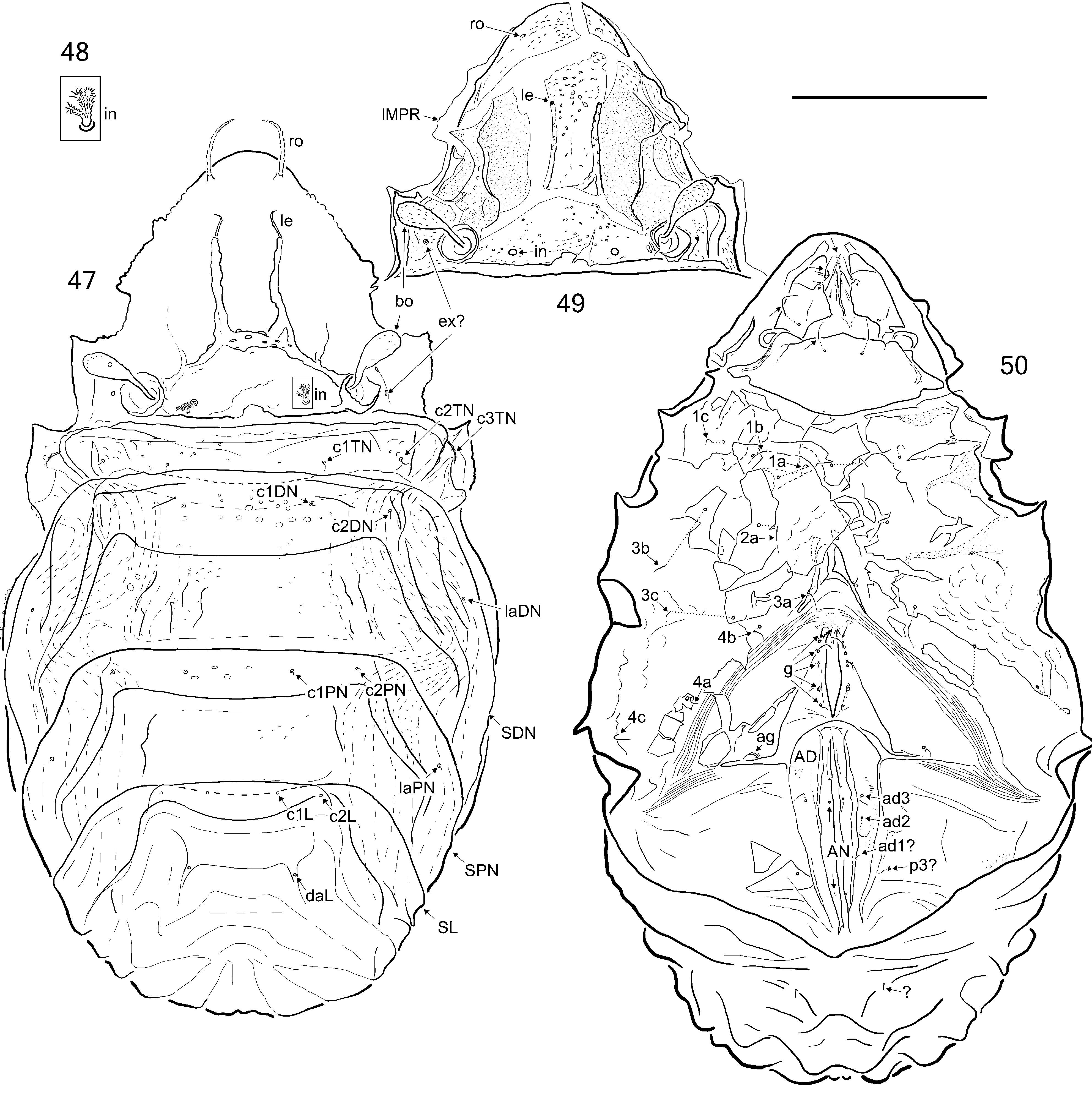


TABLE 1. Ontogeny of setae (Roman letters) and solenidia (Greek letters) in Plategeocranus sulcatus (Karsch, 1884). Struc-
| Trochanter Femur | Genu | Tibia | Tarsus | |
|---|---|---|---|---|
| Leg I | ||||
| La2 Tn3 Ad | - d, bv "? l ' v ' l " | (l), σ? v ' | v ', (l), φ1 φ2 v " | ft ', (pl), (pv), (tc), (a), (u), (p), s, ω1 (it), e, ω2 v ', l " |
| Leg II | ||||
| La | - d, bv " | (l), σ | v ', l ', φ | (ft), (pv), (tc), (a), (u), (p), s, ω |
| Tn3 | v ' l ' | v ' | l " | (it) |
| Ad | - l " | - | v " | l " |
| Leg III La | - d, ev ' | l ', σ | v ', φ | (ft), (pv), (tc), (a), (u), (p), s |
| Tn3 Ad | ? - v ', l ' l ' | - - | l ' v " | (it) - |
| Leg IV Tn3 | ? d, ev ' | d | v ', l ', φ | ft", (pv), (tc), (a), (u), (p), s |
| Ad | -? | - | v " | - |
No known copyright restrictions apply. See Agosti, D., Egloff, W., 2009. Taxonomic information exchange and copyright: the Plazi approach. BMC Research Notes 2009, 2:53 for further explanation.