
Pravistylus odontopygeus, Stiller, 2010
|
publication ID |
https://doi.org/ 10.11646/zootaxa.2468.1.1 |
|
persistent identifier |
https://treatment.plazi.org/id/03EFD356-FFFF-FFF7-6CFF-71708E66D609 |
|
treatment provided by |
Felipe |
|
scientific name |
Pravistylus odontopygeus |
| status |
sp. nov. |
Pravistylus odontopygeus View in CoL sp. n.
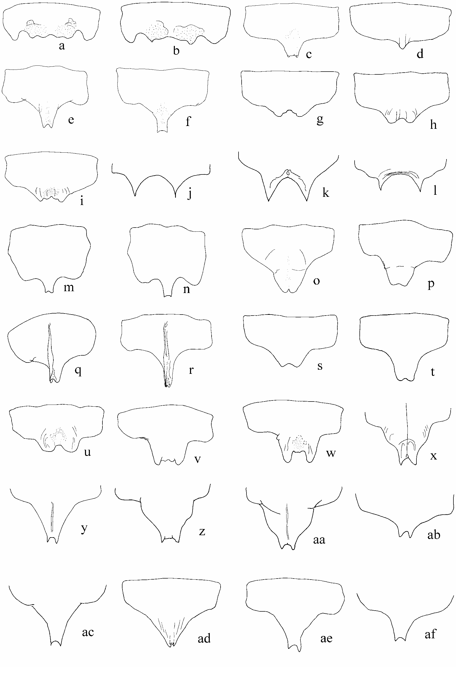
( Figs 1 y View FIGURE 1 ; 2 x View FIGURE 2 ; 3 View FIGURE 3 ah; 4 af; 5 x; 6 ad; 7 bl; 8 at)
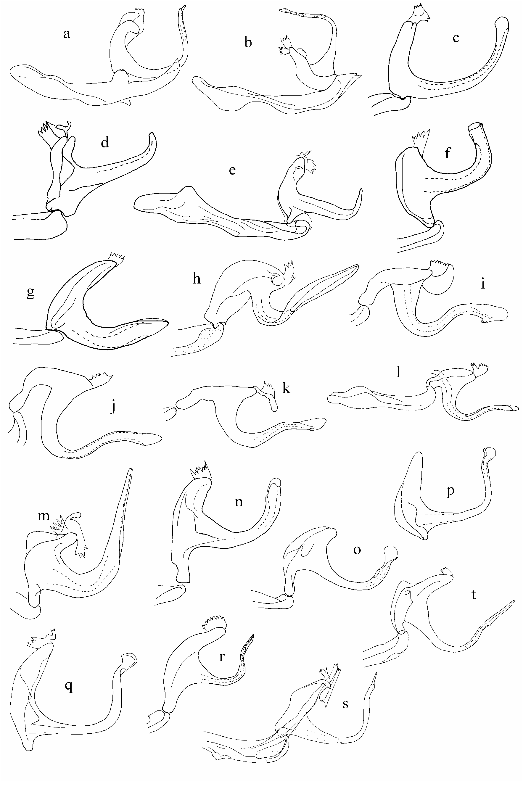
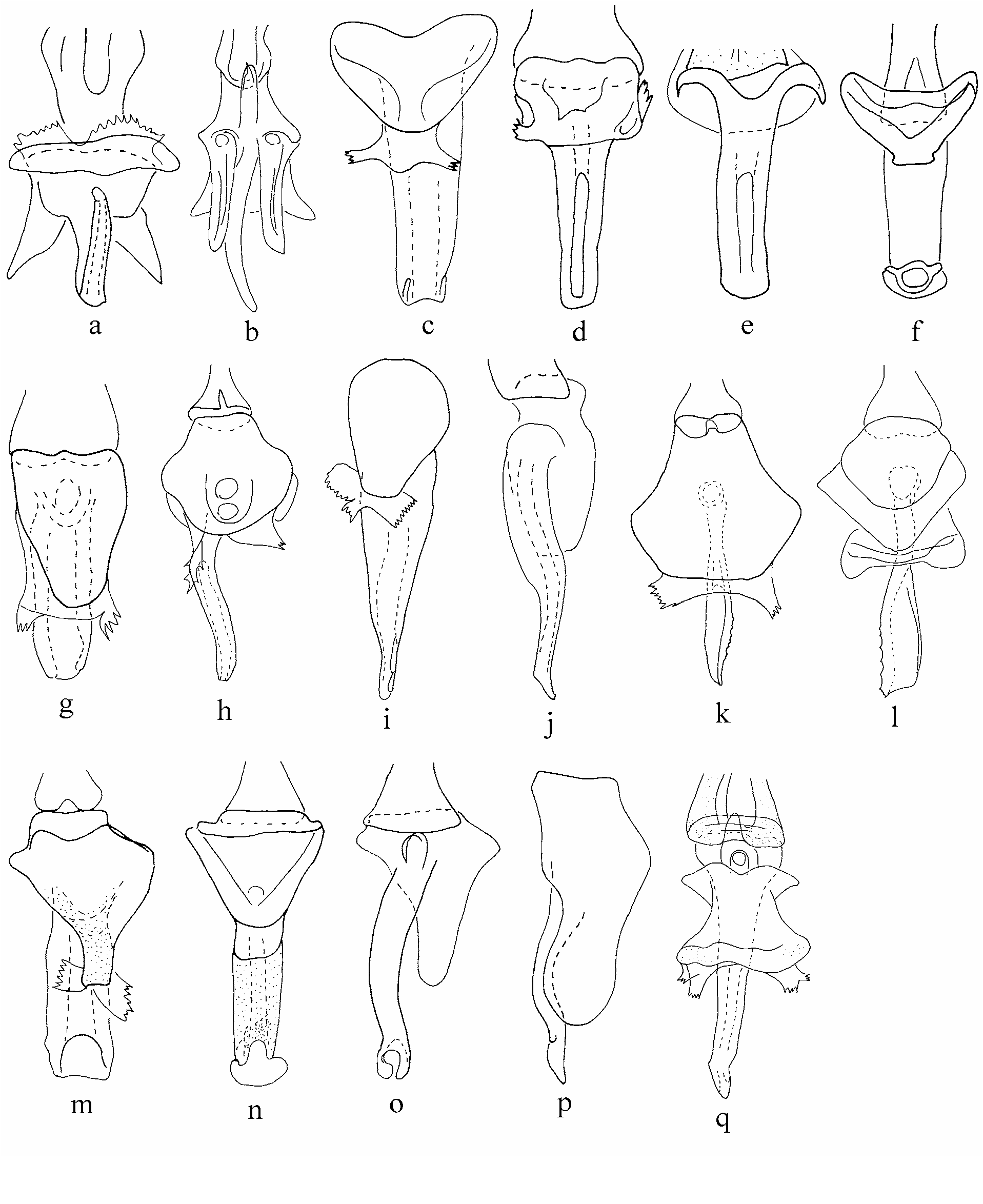
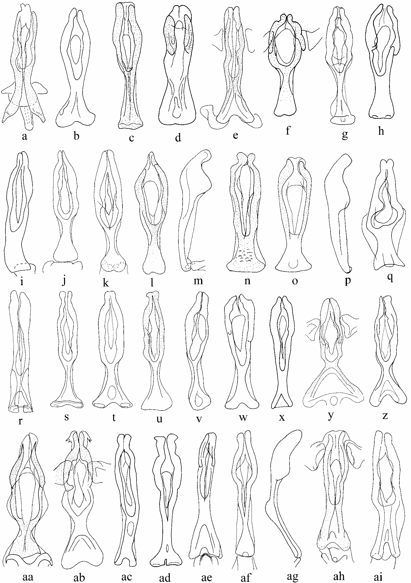
Diagnosis. Plate apex rectangular, apex uniformly merged into lateral margin, both plate apices usually of equal length ( Fig. 2 x View FIGURE 2 ). Pygofer lobe ventrobasally with short, sclerotized tooth, arising from medial surface ( Fig. 1 y View FIGURE 1 ). Aedeagus, in lateral view, with shaft narrow, uniformly C-shaped ( Fig. 3 View FIGURE 3 ah). Female sternite 7 as in Fig. 7 View FIGURE 7 bl.
Etymology. Greek, for the short tooth (odontos) on the pygofer (pyge) lobe.
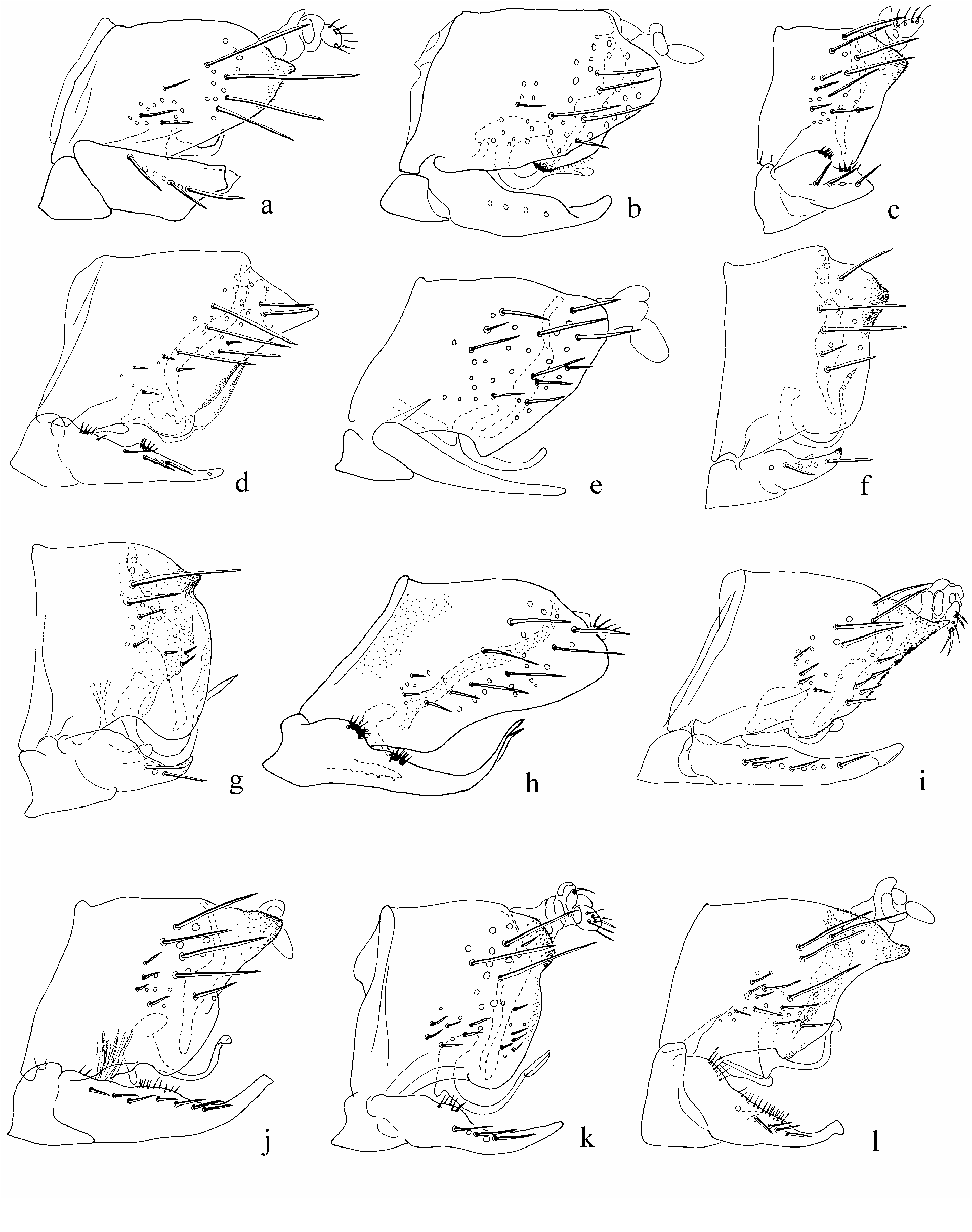
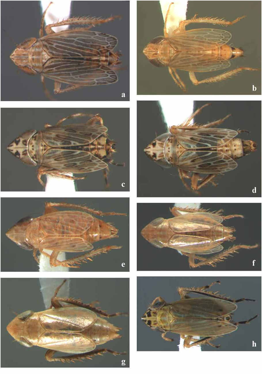
Male and female. Ochraceous, sometimes with paired fuscous markings on vertex and inner anteapical cell ( Fig. 8 View FIGURE 8 at).
Male. Dimensions. (n = 5) Length: apex of vertex to apex of tegmina 2.5 mm; apex of vertex to apex of abdomen 2.5–2.6 mm; vertex medially 0.4 mm; vertex next to eye 0.3 mm; pronotum medially 0.3 mm. Width: head 0.8–0.9 mm; pronotum 0.8 mm. Ocellar diameter 28.0 µm; ocellocular distance 24.5–37.1 µm.
Genital capsule. Pygofer, in lateral view, with ventroposterior margin broadly rounded ( Fig. 1 y View FIGURE 1 ), ventral part with membranous region invaded by long, narrow, sclerotized process ( Fig. 1 y View FIGURE 1 ). Pygofer lobe acutely triangular, sclerotized, with denticulate microsculpture; ventroposterior margin with short, sclerotized, ventral tooth ( Fig. 1 y View FIGURE 1 ); base of lobe about one third as wide as width of pygofer. Plate apex truncate, rectangular, apices usually of similar length; lateral margins straight, merging uniformly with apex; 7–10 macrosetae, usually uniseriate ( Fig. 2 x View FIGURE 2 ); plate 1.5–1.7 times as long as wide. Aedeagal shaft, in lateral view, arising ventrally from atrium; preatrium reduced; entire shaft uniformly C-shaped ( Fig. 3 View FIGURE 3 ah); dorsal apodeme elongate, curved towards apex of shaft; gonopore elongate, lateroventral ( Fig. 4 View FIGURE 4 af). Style distal part far from anterior medial lobe; apex of apophysis acute; apophysis acutely angled to preapical lobe, preapical lobe acute; apophysis with 3–4 ventral teeth ( Fig. 5 x View FIGURE 5 ). Connective, in dorsal view, with stem apex triangular, wider than stem base ( Fig. 6 View FIGURE 6 ad).
Female. Dimensions. (n = 3) Length: apex of vertex to apex of tegmina 2.6–2.8 mm; apex of vertex to apex of abdomen 3.0– 3.1 mm; vertex medially 0.5 mm; vertex next to eye 0.3 mm; pronotum medially 0.3 mm. Width: head 0.9 mm; pronotum 0.8 mm. Ocellar diameter 26.3–34.4 µm; ocellocular distance 24.6–40.7 µm.
Genitalia. Sternite 7 with base rectangular; hind margin ligula wide, triangular; apex deeply notched ( Fig. 7 View FIGURE 7 bl).
Material examined. Holotype male. South Africa, KwaZulu-Natal. Bushman’s Neck Nature Reserve , 29°53ʹS, 29°11ʹE, 6.iv.1994, M. Stiller, sweeping grass ( SANC) GoogleMaps . Paratypes. 3♂, 14♀. South Africa, Kwa- Zulu-Natal . 1♂, 3♀, same data as holotype GoogleMaps ; Lesotho. 2♂, 11♀, Sehlabathebe Nature Reserve , 29°53ʹS, 29°04ʹE, 1.iv.1994, M. Stiller, sweeping, grass ( BMNH, SANC) GoogleMaps .
Remarks. A combination of characters distinguish P. odontopygeus from a number of similar species. The tooth on the ventral margin of the pygofer lobe ( Fig. 1 y View FIGURE 1 ), the uniformly C-shaped aedeagal shaft and dorsal apodeme ( Fig. 3 View FIGURE 3 ah), and the shape of the female sternite 7 ( Fig. 7 View FIGURE 7 bl) are distinguishing features of P. odontopygeus . Species with a similar shape of the plate are P. caenophallus ( Fig. 2 y View FIGURE 2 ) and P. trunculidiscus sp. n. ( Fig. 2 j View FIGURE 2 ). The modified aedeagus ( Fig. 3 View FIGURE 3 ak) and the absence of the tooth on the pygofer lobe ( Fig. 1 d View FIGURE 1 ) of P. odontopygeus distinguish it from P. caenophallus . Of use in distinguishing P. caenophallus and P. odontopygeus is the position and number of macrosetae on the plate. In P. caenophallus the macrosetae are grouped subapically or somewhat medially ( Fig. 2 y View FIGURE 2 ), whereas in P. odontopygeus the macrosetae extend across most of the lateral margin ( Fig. 2 x View FIGURE 2 ). In P. trunculidiscus the style has the distal part close to the anterior medial arm ( Fig. 5 j View FIGURE 5 ) and the aedeagal shaft is U-shaped ( Fig. 3 z View FIGURE 3 ), but has a similar arrangement of macrosetae, although the apex of the plate is abruptly merged with the lateral margin ( Fig. 2 j View FIGURE 2 ). Specimens of P. odontopygeus and P. indistinctidiscus both occur at Bushman’s Neck and Sehlabathebe. Differentiation between these two species is based on the plate shape, ratio of plate length to width and the shape of the pygofer in lateral view. In P. indistinctidiscus the plate ( Figs 2 View FIGURE 2 ag & ah) is 1.0–1.4 times as long as wide and the pygofer is as in Fig. 1 m View FIGURE 1 . In P. odontopygeus the plate ( Fig. 2 x View FIGURE 2 ) is 1.5–1.7 times as long as wide and the pygofer is as in Fig. 1 y View FIGURE 1 . However the following structures of P. indistinctidiscus and P. odontopygeus share some similarities: style ( Figs 5 q & x View FIGURE 5 , respectively), aedeagus ( Figs 3 View FIGURE 3 af & ah, respectively) and partially sternite 7 of the female ( Figs 7 x View FIGURE 7 –aa & bl, respectively).


| SANC |
Agricultural Research Council-Plant Protection Research Institute |
No known copyright restrictions apply. See Agosti, D., Egloff, W., 2009. Taxonomic information exchange and copyright: the Plazi approach. BMC Research Notes 2009, 2:53 for further explanation.
|
Kingdom |
|
|
Phylum |
|
|
Class |
|
|
Order |
|
|
Family |
|
|
Genus |