
Dasydytes (P.) lamellatus Kisielewski, 1991
|
publication ID |
https://doi.org/ 10.11646/zootaxa.5209.1.3 |
|
publication LSID |
lsid:zoobank.org:pub:E5FADE56-6166-4329-9CE9-625315DB7303 |
|
DOI |
https://doi.org/10.5281/zenodo.7330329 |
|
persistent identifier |
https://treatment.plazi.org/id/0385C448-1D09-FFED-56F4-FC66566EE6B2 |
|
treatment provided by |
Plazi |
|
scientific name |
Dasydytes (P.) lamellatus Kisielewski, 1991 |
| status |
|
Dasydytes (P.) lamellatus Kisielewski, 1991 View in CoL
( Figs. 1–4 View FIGURE 1 View FIGURE 2 View FIGURE 3 View FIGURE 4 ; Table 1 View TABLE 1 )
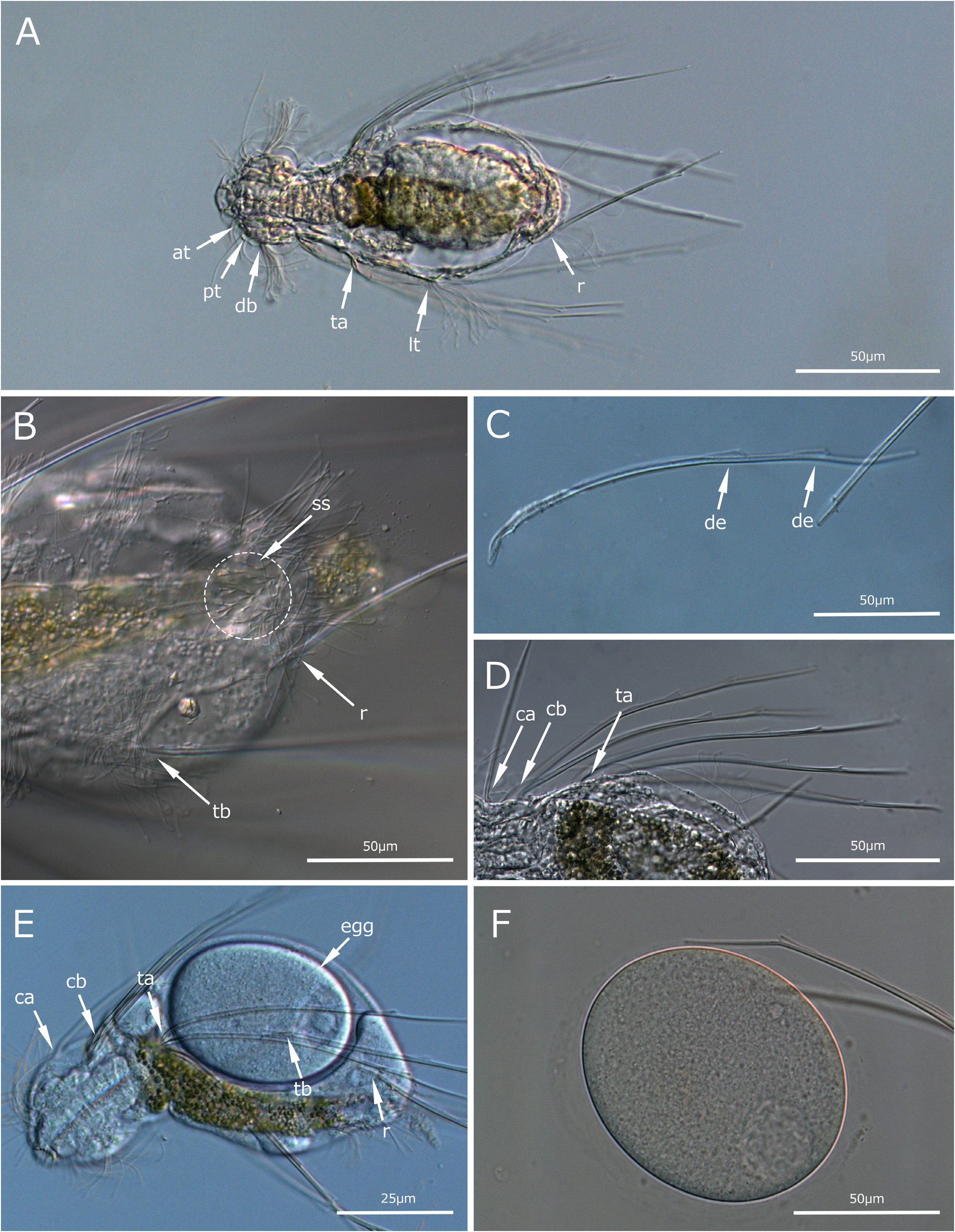
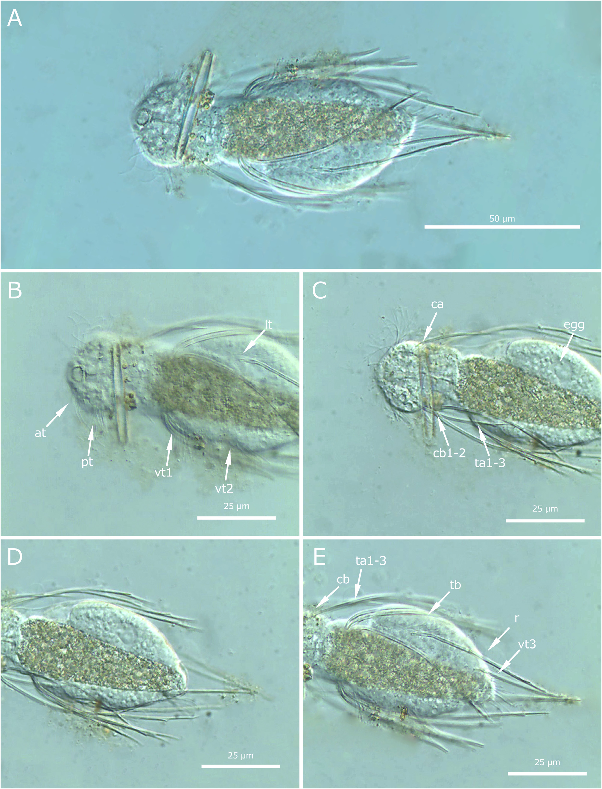
Measurements based on mature specimens from southeastern location: Dasydytes (Prodasydytes) with an elongatedoval body, 200–270 μm in total length, 133–200 μm (spines excluded). Distinct concave head with well-developed lateral lobes (70 μm wide). Distinct neck (37–74 μm wide) considerably narrower than the head and trunk (figs. 1, 2, 3, 4). An oval trunk with a 120 μm maximum width, and a protuberant caudal end (figs. 1A, B, 3A). Cephalion small and compact (fig. 2D), with small lateral pleura and small hypostomion, joined around the ventral portion of the mouth ring (fig. 3D). Subterminal mouth ring (17 μm in diameter) connected to the 46–67 μm long pharynx (fig. 2D).
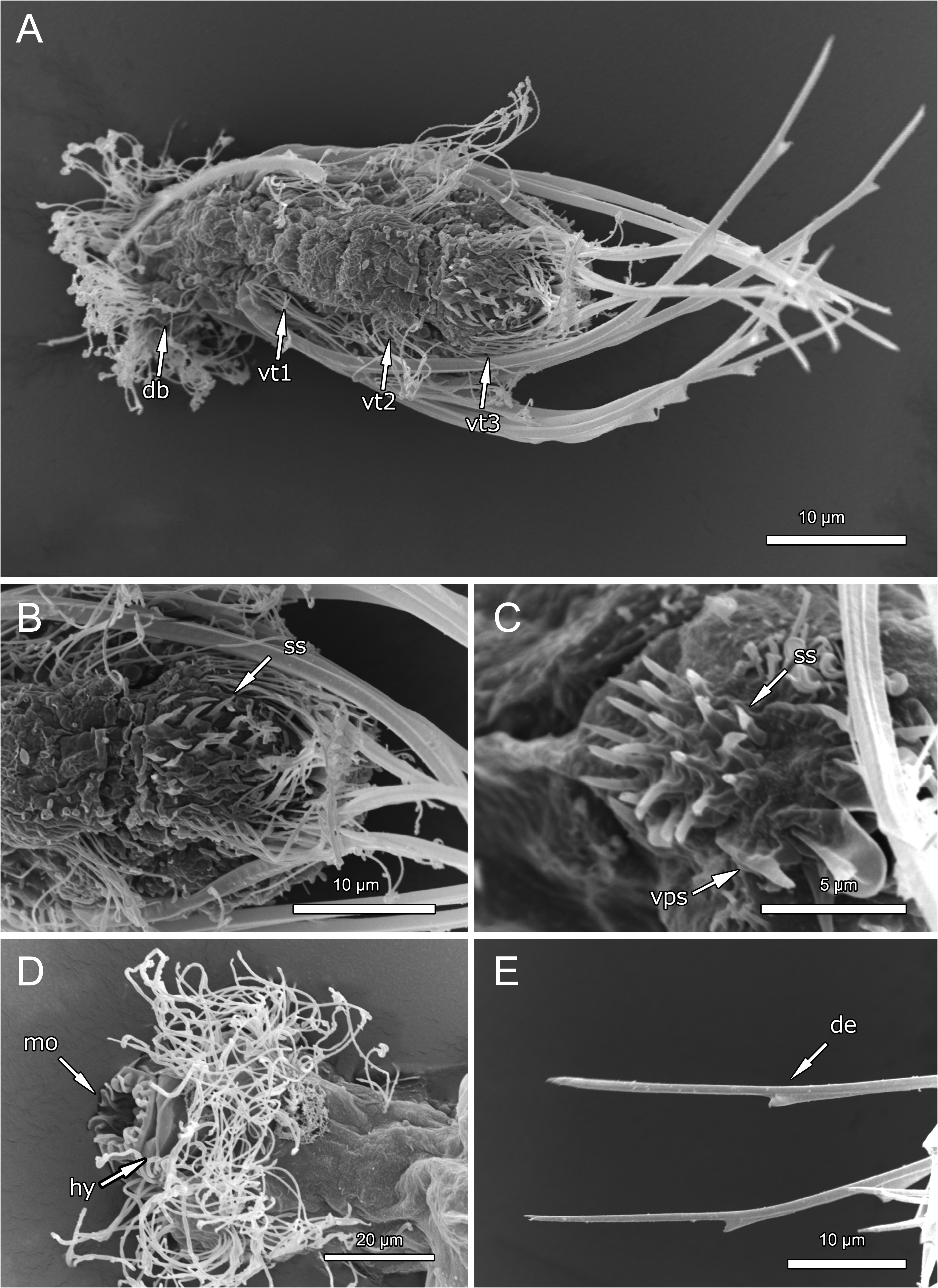
Cephalic ciliature consists of lateral tuft adjacent to the mouth (U6), mediolateral tuft (U10), and posterior pair of conspicuous transverse bands on the lateral and dorsal head (U17) (fig. 2E). A pair of dorsal sensory bristles inserted posteriorly adjacent to the mouth (figs. 1A, 2A, D). Locomotor ciliation on the trunk consists of four-paired tufts at the anterior ventral trunk (U51), middle lateral trunk (U68), posterior ventral trunk (U87) and ventrally adjacent to caudal protuberance (U92) (figs. 2B, C, 3A). Posterior pair of dorsal sensory bristles inserts on the dorsal anterior part of caudal protuberance (30 μm long) (fig. 1B).
Dorsal cephalic spine (ca) (U16) (100 μm long) and two-paired ventral cephalic spines (cb1–2) (U25) (200 μm and 198 μm long, respectively) (figs. 2C, D, 4B, F). Trunk anterior half with three pairs of ventral, double-barbed curved spines inserted directly to the cuticle without scales (figs. 1D, 2D, E). (fig. 1C, D). First group (ta1–3) inserts ventrolateral on the base of the neck at U43 (figs. 1D, 2D). Second ventral group (tb) with single spine (184 μm) at U69 (figs. 2D, 4B). Posterior ventral spine (r) is inserted adjacent to caudal protuberance (135 μm) at U93 (figs. 2D, 4D). Caudal protuberance with 20 short-spined oval ventral scales (ss, fig. 3A–C) marking a medial circle at U90, between posteriormost ventral ciliary tuft (figs. 1B, 2B, 3A–C, B, C, 4A). Additionally, two pairs of keeled, arrowhead-shaped scales on the caudal end of the protuberance at U92 and U93 (fig. 3C), anterior pair inserted lateral to the circle of short-spined scales and medial to posterior ciliary tuft (U92), and posterior pair medial to rear long spine (r) (U93) (fig. 3A–C).
Some specimens were found carrying a single egg taking up ⅓ of the animal’s total body volume, with ovalshape (120 μm long and 100 μm wide) and completely smooth surface without ornamentations (figs. 1E, F, 4C, F).
Remarks: The specimens found in the Atlantic forest (São Paulo state) and Northeastern Cerrado (Piauí state) share many morphological similarities with those described by Kisielewski (1991) from the Amazon forest (Pará state). All specimens exhibit a long dorso-lateral cephalic spine (ca) and two paired ventro-lateral spines at neck (cb), each spine provided with two lateral denticles, while the proximal one has a membrane-like lamella. The number of trunk spines are identical, with paired lateral groups of spines on the trunk (ta–tb) and a pair of posterior rear spines (r). All representatives share the anterior ciliary tufts at the neck base (U51), the characteristic lateral ciliary tuft at the middle trunk (U68), and posterior ventral tuft (U87) and the rearmost being the largest, located posterior medial to the rear spine (r) (U92). However, the trunk ciliature of Atlantic specimens exhibits four-paired ciliary tufts, in contrast to Pará and Piauí ones that have an additional ventral anteriormost tuft at the anterior neck (U30), adding up a five-paired ciliature. The Piauí specimens share much closer dimensions with Pará ones (99 µm and 112 µm in total length, respectively), being smaller than São Paulo specimens (160 µm, see Table 1 View TABLE 1 ). With an elongated-oval body with 99 μm long (spine excluded) and 131 μm (spined included), distinct head (30 μm wide), narrower neck (23 μm wide), oval body (41 μm wide), terminal mouth diameter 6 μm, pharynx 23 μm long. Egg dimension: 41 μm length, 16 μm width. Cephalion and hypostomion not seen.

No known copyright restrictions apply. See Agosti, D., Egloff, W., 2009. Taxonomic information exchange and copyright: the Plazi approach. BMC Research Notes 2009, 2:53 for further explanation.