
Protaustrosimulium pilfreyi ( Davies & Györkös 1988 )
|
publication ID |
https://doi.org/ 10.11646/zootaxa.4521.3.1 |
|
publication LSID |
lsid:zoobank.org:pub:F5736D61-B519-4F13-93FC-FCCAEC9505B4 |
|
DOI |
https://doi.org/10.5281/zenodo.5959753 |
|
persistent identifier |
https://treatment.plazi.org/id/60352B2A-FFAF-FFD0-FF5C-B009FEC812D9 |
|
treatment provided by |
Plazi |
|
scientific name |
Protaustrosimulium pilfreyi ( Davies & Györkös 1988 ) |
| status |
|
Protaustrosimulium pilfreyi ( Davies & Györkös 1988) View in CoL . New combination
( Figs. 1–21 View FIGURES 1–6 View FIGURES 7, 8 View FIGURES 9–13 View FIGURES 14–19 View FIGURES 20, 21 )
Cnephia pilfreyi Davies & Györkös 1988: 107 View in CoL .
pilfreyi View in CoL . Crosskey, 1989: 222. Unplaced species of Prosimuliini View in CoL .
Paracnephia pilfreyi . Crosskey & Howard, 1997:18. New combination. Bugledich, 1999: 328.
" Cnephia " pilfreyi . Moulton, 2000: 98. Moulton, 2003: 47.
Paracnephia pilfreyi . Adler & Crosskey, 2008: 26. Transferred to Simuliini .
Redescription (based in part on original description by Davies & Györkös 1 988: 106).
Adult female. Body: total length 2.5-2.7 mm. Head ( Fig. 1 View FIGURES 1–6 ): medium orange brown with silvery concolourous adpressed hair; width 0.80 mm; depth 0.70 mm; 0.75× width of thorax; frons sub-parallel, broadening markedly dorsally; frons:head ratio 1.0:6.1, frontal angle 70°. Eye: interocular distance ca. 0.13 mm. Antenna: evenly medium orange brown, tapering distally, total length ca. 5.6 mm; scape, pedicel and basal flagellomere 1.5× longer than broad, apical flagellomere IX, twice as long as others. Mouthparts: maxillary palpus, pale brown, apical palpomere V elongated; proportional lengths of III–V palpomeres 1.0:1.0:1.5; sensory organ moderately elongated, 0.33× length of palpomere III, opening large, round, 0.6× vesicle width; mandible with ca. 55 poorly developed inner teeth, smaller proximally, 18 outer teeth; lacinia with 16 and ca. 24 teeth on inner and outer edge respectively; cibarium details not known. Thorax: length not known; scutum medium even brown with three darker vittae; scutellum light brown medially, scutellar depression with long brown hairs; postnotum medium brown, bare; anepisternal (plural) membrane without hairs; katepisternum longer than deep, sulcus distinct. Wing: length 2.6 mm, width 1.3 mm; a:b ratio 1:3; disposition of apices of R 1 and Rs before joining C not recorded; short spinules mixed with setae on distal vein C, but not R, basal section of R haired, R s simple, S c haired ventrally, vein CuA slightly curved, A 1 ending near wing margin; basal (bm) cell present. Haltere: very pale brown, becoming whitish distally in some specimens. Legs: Fore leg and mid leg: coxa and trochanter medium brown (except anterior trochanter surface); femur and tibia light brown darkened slightly at ends and along anterior tibial margin, tarsus medium brown; fore basitarsus narrow, cylindrical, width:length ratio 1:9. Hind leg ( Fig. 2 View FIGURES 1–6 ): and trochanter medium brown, rest of leg light brown except basal tip and distal end of femur, basal and distal quarter, anterior edge of tibia, basal and distal quarter of basitarsus, distal half of second tarsal segment and rest of tarsus dark brown, with fine adpressed concolourous hair except long medium-brown erect hairs on anterior tibial edge; hind basitarsus wider than fore basitarsus, but sides subparallel, width:length ratio 1.0:5.2; calcipala ( Fig. 3 View FIGURES 1–6 ): 1/2 width of basitarsus apex and as wide as long; pedisulcus as wrinkled cuticle; tarsomere II 4 × longer than apical width; claw finely tapered with minute basal tooth on inner surface, heel not markedly expressed ( Fig. 4 View FIGURES 1–6 ). Small intersegmental sclerites between basitarsus and next two tarsal segments, well expressed. Abdomen: basal scale (tergite I) pale brown except medium brown edge, covered with fine concolourous hair but with long posterodorsal erect hairs. Dorsum of segment II with large, medium-brown tergite (0.75× abdominal width), segments III & IV with tergites half abdominal width (tergite III medium brown); segments IV–VIII uniformly pale brown; hairs shorter, lighter and finer on tergites II–IV than for rest of dorsum; ventral surface without sternites. Genitalia ( Fig. 5 View FIGURES 1–6 ): hypogynial valves bluntly triangular with medial edges slightly convergent distally; genital fork anterior arm with anterior third slightly expanded; posterolateral arms plus plate subequal in length to anterior arm. Spermatheca elliptical (length:width ratio 1.4:1.0), darkly pigmented and smooth; presence of internal spines (acanthae) unknown; spermathecal duct with pigment extended for short distance. Cercus ( Fig. 6 View FIGURES 1–6 ) as wide as long, smoothly rounded, anal lobe small with minute central depression.
Male (based on original description). Body: colour not given. Head: width 0.85 mm. Eyes: upper ommatidia diameter 0.034 mm, ca. 16 across and 20 down. Clypeus: medium brown; vestiture of long concolourous hairs. Antenna: total length not given; pedicel swollen, 1.3× wider than first flagellomere. Mouthparts: maxillary palpus, palpomere V 1.4× longer than palpomere IV, sensory vesicle small, spherical, occupying 0.2× length of palpomere III, opening 0.33× vesicle width. Thorax: width 0.7 mm, medium brown with three dark brown vittae; scutellum light brown, anepisternal membrane without hairs, katepisternum browner, sulcus distinct. Wing: length 2.3 mm, width 1.3 mm; short spinules mixed with hairs on vein C, but not R 1 with; basal section of R haired, R s simple, S c with ventral hair; CuA and A 1 curved; basal cell absent. Haltere: stem mainly translucent to yellowish white opaque, knob yellowish white. Legs: pale brown except medium brown at both ends of femur and tibia, and at basal end also on anterior margin of hind basitarsus. Fore basitarsus narrow, cylindrical, width:length ratio 1:8; hind basitarsus somewhat flattened, width:length ratio 1:5; calcipala pronounced. Abdomen: basal scale medium greyish brown with margin and long bordering hairs pale yellowish; distal edges of tergites II–V medium greyish brown, narrowing on posterior segments with tergite VI only half width of tergum; sides of abdomen medium to dark brown becoming paler on sterna. Genitalia: Lost, but observation prior to that indicated the gonocoxa and gonostylus were subequal in length; gonocoxa a truncate cone slightly narrower distally; gonostylus curved medially with 4 or 5 short spines distally on inner margin.
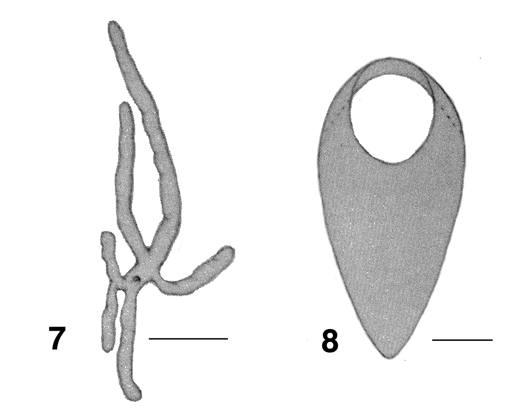
Pupa. (based on original description) Body: length ca. 4.0 mm. Head: facial and epicranial setae present, the latter closely applied beside antennal sheath. Thorax: no details given for cuticle; scutum with three dorsocentral setae. Gill: six filaments (original description, Fig. 7 View FIGURES 7, 8 ), eight (Grampians, Fig. 19 View FIGURES 14–19 ), translucent, thin-walled, inflated; original with three main trunks, anterior one extended horizontally beyond head, with distal upturn, posterior one usually extended horizontally to abdominal segment VII, ventral pair of filaments on short base, two other filaments arising individually near base; Grampians (based on pharate specimen) with short basal trunk, giving rise to single thick filament, three short petioles giving rise each to two filaments, with single filament arising directly from base, no distinct annulations, surface with normal array of trabeculae. Abdomen: armature, tergites I, II, with posterior row of simple hairs, tergite V with one posterior hair, tergites III, IV with posterior row of 4 abruptly tapered hooks, tergites I–IV also with single hair laterally, tergites VI–VIII with fine spine-combs, tergites IX with four grapnel hooks, terminal spines straight, conical, pointing anterodorsally, plus two dorsal hairs; sternites IV–VI with one small medial spine and two lateral finer hairs on sternite V, sternites VI & VII with median membranous area; pleurites absent.
Cocoon (specimens damaged). Apparently shoe-shaped, not completely covering pupa, finely woven without anterior rim, connected ventrally at collar ( Fig. 8 View FIGURES 7, 8 ).
Larva (based on original description and new material). Body ( Fig. 9 View FIGURES 9–13 ): total length 5.6–7.0 mm, colour pale without segmental markings. Head ( Fig. 10 View FIGURES 9–13 ): overall colour moderate brown, darker medially, head spot pattern lightly positive; length 0.75 mm, maximum width 0.53 mm; distance between antennal bases 0.48 mm; head widest at stemmata, lateral margins convex, more so posteriorly; cervical sclerites weakly sclerotized, but distinct and not connected to postocciput. Antenna ( Fig. 11 View FIGURES 9–13 ): extended well beyond labral fan stem; total length 0.65 mm, including elongated apical sensillum, basal and medial articles concolourous medium brown; medial article markedly shorter than basal article, distal article markedly elongated, clear, proportional lengths of basal, medial, and apical articles 1.0:0.25:2.5. Labral fan: stem broad, not markedly pigmented, light brown in early last instar larvae, ca. 50 (original description) and 70 (Grampians material) fine pale rays, length 0.9 mm, mid-ray width 0.007 mm; microtrichial pattern of one longer with five shorter between. Maxilla ( Fig. 12 View FIGURES 9–13 ): not markedly pigmented; palpus elongated and curved, 2.8× as long as basal width; hair tuft at base of palp poorly developed. Mandible ( Figs. 13 View FIGURES 9–13 , 14 View FIGURES 14–19 ): not markedly pigmented; brushes finely expressed; outer teeth ca. half length of larger apical tooth; subapical teeth poorly expressed; ca. 10 fine spinous teeth, two sensilla with marked bases, serration barely evident; blade region long and slightly concave. Postgenal cleft ( Fig. 15 View FIGURES 14–19 ): essentially absent, minute, slot-shaped; posteroventral muscles spots obvious; ratio of hypostoma: genal bridge: postgenal cleft 1.0:6.0:3.0. Hypostoma ( Fig. 16 View FIGURES 14–19 ): domelike; tooth 0 (median) and 4 (lateral), subequal in length, teeth 1–3 small and largely obscured by edge of hypostoma, teeth 5 & 6 small, tooth 7 occasionally present; lateral serrations absent; hypostoma sloped smoothly laterally to edge of genae; 6–8 markedly fine hypostomal setae on each side. Thorax ( Fig. 17 View FIGURES 14–19 ): pale; the immature pharate pupal gill shows poorly, filaments indistinct. Prothoracic proleg: not markedly developed, lateral sclerite narrowed ( Figs. 17, 18 View FIGURES 14–19 ). Abdomen: markedly pale anteriorly, light orange posteriorly, narrow anteriorly with segmental expansions; markedly expanded laterally at segment VI, smoothly tapered posteriorly, amphora-shaped laterally. Ventral tubercles: well expressed. Rectal papillae: three simple lobes, well developed. Anal sclerite ( Figs. 20, 21 View FIGURES 20, 21 ): anterior and posteroventral arms subequal in length, former with basal flange, median region not markedly developed, interarm struts absent; accessory sclerites well developed, extended slightly as semicircular sclerite (Grampians), apparently not so in typical material, clear basal cuticular sclerite underlying circlet of hooks visible, fully sclerotized semicircular sclerite absent. Posterior circlet: numbers of hooks ca. 85 rows of hooks, 14 per row (total ca. 1,200).
Etymology. Named by Davies & Györkös (1988: 111) for Ron Pilfrey, who collected the original material.
Types. Holotype. Davies & Györkös (1988: 107) designated a reared female in ethanol as the holotype, with exuviae and parts mounted on slides. However, as noted previously, this material was never deposited in the Australian National Insect Collection (ANIC). Of the material recovered by Craig (2011) and now deposited in ANIC, just three legs and the head minus the labellum remain; all now in glycerine in a pinned microvial. Label data: [HOLO/ TYPE] [ Paracnephia / pilfreyi ] [ AUSTRALIA, ACT/ Tidbinbilla Rd/ S35.4200° E148.9400° / 15-ix- 1964 / Coll. R. Pilfrey].
Paratypes. Little was recovered of the paratype adults, pupae and larvae originally designated by Davies & Györkös (1988: 107). The following material is stored in glycerine in pinned microvials, as follows: a pupal gill histoblast, a larval mandible plus hypostoma, a single complete immature larva, and a single mature last instar larva. Other material is mounted on slides: two male pupal exuviae, two female pupal exuviae, and a single last instar larval anal sclerite. Label data: as above, but with [Paratype]; all in ANIC .
Additional material. Early last instar larvae of Prot. pilfreyi . [ AUSTRALIA VIC./ Grampians National Park/ Small tributary of Glenelg/ River, ex. Glenelg River. Rd./ 26 September 1996 / Coll. J.K. Moulton], (ca. S37.2500° E142.4200°, elev. 226m.). (ETOH: UASM# 370857), (Slide: UASM# 370929) and personal collection of JKM.
The whereabouts of immature larvae collected by R. Pilfrey from an intermittent stream draining into Paddys River, ACT ( Davies & Györkös, 1988: 107) is unknown.
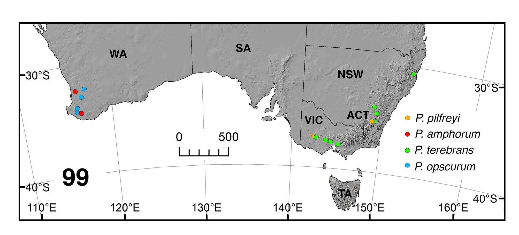
Distribution ( Fig. 99 View FIGURE 99 ). Australian Capital Territory. Davies & Györkös (1988: 107) listed four localities in the Tidbinbilla Road region, south of Canberra; namely a trickle above Tidbinbilla Road draining into Paddys River (ca. S35.4200° E148.9400°, elev. 670m., 15-ix-1964); Gibraltar Creek (S35.4500° E148.9785°, elev. 667m., 10-ix- 1964); Oakey Creek (S35.4145° E148.9456°, elev. 650m., 1-x-1966) and a temporary stream draining into Paddys River (28-vii-1964, 21-ix-1966, 5-x-1966). Victoria. Grampians National Park, Glenelg River Road, small tributary of Glenelg River, 26 September 1996. Coll. J.K. Moulton (ca. S37.2500° E142.4200°, elev. 226m.).
Bionomics. Little is known about the biology of Protaustrosimulium pilfreyi . Attempts were made in 2011 and 2014 to recollect material from Gibraltar Creek (Fig. 97) and the creek at Oakey Farm (Fig. 96); however, no specimens of Prot. pilfreyi were recovered—even though the season corresponded with Pilfrey’s original collections in 1964 and 1966. Oakey Creek yielded Austrosimulium (Novaustrosimulium) furiosum (Skuse) , A. (A.) crassipes (Tonnoir), and Simulium (Nevermannia) ornatipes (Skuse) , while Gibraltar Creek yielded A. (N.) victoriae (Roubaud) and " Paracnephia " orientalis (Mackerras & Mackerras). This latter creek is larger and less ephemeral than the originally described localities (e.g., " trickles crossing Paddy's Road ") and the Oakey Farm creek (cf. Figs. 96 & 97). Davies & Györkös (1988) noted that D.G. Bedo attempted to find more pilfreyi material in 1985 and 1986, but he too was unsuccessful. Dates given in Davies & Györkös (loc. cit.: 107) indicate that Prot. pilfreyi is a late Austral winter/early spring species, as are many of the Gondwanan Australian species. This agrees well with the seasonality of the Prot. pilfreyi population from Grampians National Park.
Tidbinbilla Road is now a fully paved thoroughfare and any water crossing the road flows through a culvert. Agricultural impacts on the surrounding land appear considerable. Similarly, climate change for the Australian Capital Territory (ACT) has seen maximum temperatures increasing since the 1950's, with a concomitant decrease in spring rains. Indeed, the Oakey Farm creek when visited in early October, 2014, was dry. There were serious droughts in the early 2000's and major fires in the Tidbinbilla region during January, 2003. In short, it is possible that Prot. pilfreyi no longer exists in the Tidbinbilla area. The only other known locality for this species is Grampians National Park, Victoria; however, exact coordinates are unknown. Given the general warming trend in the region, perhaps collection efforts should be made earlier in the year.
Remarks. The latitude and longitude (35° 26' S 142° 56' E) given for the type locality of Prot. pilfreyi , by Davies & Györkös (1988: 107) is incorrect; the degrees longitude should be 148° and such corrected coordinates (35° 26' S 148° 56' E) place this near the Tidbinbilla Nature Reserve (a National Park since 1962). The spelling "Oakley Ck" for one locality is incorrect and should be "Oakey". The property on which the original collections were made still existed at the time of writing and is named "Oakey Farm".
While there is good concordance between the Australian Capital Territory ( Davies & Györkös, 1988) and the Grampians larval material described here, differences in pupal gill filament, labral fan ray numbers and accessory sclerites of the larvae are suggestive of a species complex. Indeed, the Grampians larvae are more similar to those of the Western Australian species Prot. amphorum than they are to typical larvae. More material of Prot. pilfreyi is clearly needed.


| ANIC |
Australian National Insect Collection |
No known copyright restrictions apply. See Agosti, D., Egloff, W., 2009. Taxonomic information exchange and copyright: the Plazi approach. BMC Research Notes 2009, 2:53 for further explanation.
|
Kingdom |
|
|
Phylum |
|
|
Class |
|
|
Order |
|
|
Family |
|
|
Genus |
Protaustrosimulium pilfreyi ( Davies & Györkös 1988 )
| Currie, Douglas C., Craig, Douglas A. & Moulton, John K. 2018 |
Paracnephia pilfreyi
| Adler, P. H. & Crosskey, R. W. 2008: 26 |
Paracnephia pilfreyi
| Bugledich, E. - M. A. 1999: 328 |
| Crosskey, R. W. & Howard, T. M. 1997: 18 |
pilfreyi
| Crosskey, R. W. 1989: 222 |
Cnephia pilfreyi Davies & Györkös 1988 : 107
| Davies, D. M. & Gyorkos, H. 1988: 107 |