
Pseudechiniscus santomensis, Fontoura, Paulo, Pilato, Giovanni & Lisi, Oscar, 2010
|
publication ID |
https://doi.org/ 10.5281/zenodo.197214 |
|
DOI |
https://doi.org/10.5281/zenodo.5617902 |
|
persistent identifier |
https://treatment.plazi.org/id/DD6C480A-FFE7-8020-EBC1-DD73FDC4F9FD |
|
treatment provided by |
Plazi |
|
scientific name |
Pseudechiniscus santomensis |
| status |
sp. nov. |
Pseudechiniscus santomensis View in CoL sp. nov.
( Figs. 1 View FIGURE 1 A–D; 2A)
Material examined: 32 specimens, one of them is a two clawed larva.
Type locality: Cascata de São Nicolau (0°16΄ 38ʺN; 6°38΄ 46ʺE), São Tomé Island, Democratic Republic of São Tomé and Príncipe.
Type repository: The holotype (slide No. 5401) and one paratype (slide No. 5402) are deposited in the collection of Binda & Pilato, (Museum of the Department of Animal Biology “Marcello La Greca”, University of Catania, Italy). The other paratypes including the larva (slides CII59 - CII89) are deposited in the collection of P. Fontoura at the Department of Biology, Faculty of Sciences, University of Porto, Portugal.
Specific diagnosis: Pseudechiniscus with tiny projections on the posterior margin of the undivided pseudosegmental plate. Those projections, generally triangular in shape, are sometimes reduced into two small teeth or assume the shape of a wide protruding flap poorly developed with a central indentation; median plate 1 with a transverse fold; median plate 2 divided; median plate 3 undivided; terminal plate not faceted but with two indentations. One intersegmental platelet present on each side of the median plates 1 and 2. Ornamentation of the dorsal plates comprised of dots joined by very delicate striae. Ventral dots unjoined by striae and forming a reticular patched design. Cephalic appendages and very short filaments A, present; all other lateral, dorsal filaments, or projections, absent. Internal claws with a thin and straight spur oriented towards the claw base. No dorsal leg spines. Short papilla on hind legs present; dentate collar, absent.
Description of the holotype: Female; body length 168 µm; colour reddish. Eye spots not observed either in slide mounted specimens or in live specimens. Head plate with a W-shaped fold ( Fig. 1 View FIGURE 1 A, arrow a). Scapular plate divided into an anterior and a posterior portion by a transverse fold ( Fig. 1 View FIGURE 1 A, arrow b); lateral longitudinal folds absent. Median plate 1 divided by a transverse fold into an anterior and posterior portion with clearly different sized dots ( Fig. 1 View FIGURE 1 A, arrow c); median plate 2 also clearly divided but dots similar in size in anterior and posterior portion ( Fig 1 View FIGURE 1 B, arrow a); median plate 3 undivided ( Fig. 1 View FIGURE 1 C, arrow a). An intersegmental platelet on each side of median plates 1 and 2 ( Fig. 1 View FIGURE 1 B, arrows b, c). Median and lateral longitudinal folds absent in all the median plates and in the pseudosegmental plate. Two small triangular projections, backward oriented, extending about 4.2 µm from the posterior margin of the pseudosegmental plate ( Fig. 1 View FIGURE 1 B, arrows d, e). Terminal plate unfaceted with two indentations.
Holotype (largest) Smallest Mean values and range
All the dorsal plates have a dense and regular ornamentation comprised of dots joined by very delicate striae, which are difficult to see ( Fig. 2 View FIGURE 2 A, arrow). Dots are slightly larger on the terminal and pseudosegmental plates (with respectively c. 50 and 56 dots per 100 µm2), dorsal portion of paired plates and posterior portion of the scapular plate. Smallest dots are present on the neck plate, anterior portion of median plate 1, lateral portions of paired plates and on the intersegmental lateral platelets. Leg plates also sculptured but striae joining the dots are not visible ( Fig. 1 View FIGURE 1 A).
Ventral ornamentation comprised of a very fine granulation (without striae joining the dots) forming transverse patches and a reticular design ( Fig. 1 View FIGURE 1 D). A wide patch is present in the head region, two elliptical meshes follow; after the first pair of legs alternate transverse patches of dots and transverse bands of mesh are present; in some bands all the meshes are elongate in shape, in other bands two large roughly triangular meshes are also present.
Length of external and internal buccal cirri 12.8 µm and 9.4 µm respectively; cephalic papilla 4.8 µm long ( Fig. 1 View FIGURE 1 A, arrow d).
Cirri A very short, 18.7 µm long; adjacent clava 4.8 µm long ( Fig. 1 View FIGURE 1 A, arrow e). Apart from the triangular projections of the pseudosegmental plate, all other lateral or dorsal cirri, or any other kind of projection are absent.
No spines on legs I. Hind legs without dentate collar but with a small papilla 2.2 µm long ( Fig. 1 View FIGURE 1 B, arrow f). Length of the external and internal claws of the legs I, 9.0 µm and 8.0 µm respectively; on the legs II and III, external 8.0 µm and internal 8.4 µm; external and internal claws of legs IV, 8.5 µm and 9.2 µm long respectively. A very thin and straight spur oriented towards the claw base is present on internal claws ( Fig. 1 View FIGURE 1 D, arrows).
Eggs unknown.
Remarks: The quantitative and qualitative characters of the paratypes are similar to those of the holotype ( Tab. 1 View TABLE 1 ), but a degree of variability concerning the shape of the posterior margin of the pseudosegmental plate must be stressed. Most specimens (64 %) have two triangular projections, as in the holotype ( Fig. 1 View FIGURE 1 B), which can be reduced to two very small teeth; other specimens (32%) have the posterior margin slightly projected forming only one wide protruding flap, sometimes with a very soft median indentation assuming the shape of two rounded lobes ( Fig. 1 View FIGURE 1 C), Only one paratype (4%) has the posterior margin of the pseudosegmental plate without any kind of projection.
Etymology: The name santomensis refers to the locus typicus: São Tomé Island; santomensis = inhabiting São Tomé.
Differential diagnosis: According to Kristensen (1987), two main groups of species can be distinguished within the genus Pseudechinicus Thulin, 1911: the ‘ victor group’ composed by species with trunk cirri and the ‘ suillus /conifer group’ with species characterised by the absence of trunk cirri. Pseudechiniscus santomensis sp. nov. clearly belongs to the latter.
Within the ‘ suillus /conifer group’, six species have the following five important characters in common with P. santomensis sp. nov: absence of lateral papillae, presence of projections at the posterior margin of the pseudosegmental plate, scapular plate with a transverse fold, terminal plate not faceted, and cuticular ornamentation comprised of dots joined by striae. Those taxa are: P. b a r t k e i Węglarska, 1962; P. quadrilobatus Iharos, 1969c ; P. spinerectus Pilato, Binda, Napolitano & Moncada, 2001 ; P. gullii Pilato & Lisi 2006 , P. pilatoi Li, 2007 and P. yunnanensis Wang, 2009 . In addition, we must stress a problem regarding P. novaezeelandiae ( Richters, 1908a) . The nominal subspecies of this species has lateral papillae, but in the subspecies aspinosus, described by Iharos (1963), these are lacking. In the description of P. novaezeelandiae , and all the subspecies, striae joining the cuticular dots were not described but we noted that they are present, even if very faint, in a specimen from New Zealand (see Pilato et al. 2005). Unfortunately we were not able to examine the subspecies aspinosus and therefore we may only hypothesize that it has cuticular ornamentation similar to that of the nominal subspecies. If the hypothesis is true, P. novaezeelandiae aspinosus would also have the five characters indicated above, and therefore we have included a comparison of P. santomensis sp. nov. to this subspecies. Pseudechiniscus santomensis sp. nov. differs from P. bartkei , P. quadrilobatus , P. gullii , P. pilatoi and P. yunnanensis in having the median plate 2 divided; more dense cuticular granulation, and less visible striae joining the dots ( Fig. 2 View FIGURE 2 ). In addition, it does not have the raised margins of the cuticular plates, which are present in P. gullii , P. quadrilobatus and P. p i l a t o i; the median plate 1 is divided by a transverse fold, unlike P. bartkei , P. quadrilobatus , P. pilatoi and P. yunnanensis ; spurs are present on the internal claws, absent in P. gullii , P. quadrilobatus , P. pilatoi and P. yunnanensis ; and the new species has a different ventral ornamentation to that seen on P. bartkei , P. gullii , P. quadrilobatu s and P. yunnanensis (in the description of P. pilatoi no information was supplied regarding the ventral cuticular ornamentation). Pseudechiniscus santomensis can also be distinguished from P. bartkei in having the pseudosegmental plate undivided and only one intersegmental platelet lateral to the median plates 1 and 2; from P. gullii as the pseudosegmental plate is undivided and with two marginal lobes instead of only one, and the absence of a dip between scapular plate and median plate 1; from P. quadrilobatus as the pseudosegmental plate has two marginal projections instead of only one; from P. pilatoi as the pseudosegmantal plate is not divided and has two marginal projections instead of only one, plus the terminal plate has two indentations (n.b. the description of P. pilatoi does not note presence or absence of intersegmental lateral platelets, which may also be a distinguishing character); and from P. yunnanensis as the terminal plate has two indentations and lateral platelets are present.
P. spinerectus is the species most similar to Pseudechinscus santonensis sp. nov., but the new species differs in having the pseudosegmental plate undivided; the marginal projections of this plate less developed and never erected; lateral longitudinal folds absent in the scapular and paired plates; only one intersegmental platelet lateral to the median plates 1 and 2; striae joining the plate dots slightly less visible ( Fig. 2 View FIGURE 2 A and E); dots on the legs not joined by striae, and some differences in the detail of the ventral cuticular ornamentation. In addition, P. santomensis sp. nov. differs from P. spinerectus in having shorter buccal cirri (external and internal cirri 12.8 µm and 9.4 µm respectively (holotype) and 14.5 µm and 12.1 µm long respectively ( P. spinerectus specimen 135 µm long)); in having shorter cirrus A (<20 µm long in P. santomensis sp. nov. and>32 µm in P. spinerectus ); shorter papilla of hind legs (c. 2.2 µm in the new species and c. 3.0 in P. spinerectus ).
The new species differs from P. navaezeelandiae aspinosus in having the pseudosegmental plate undivided; intersegmental lateral platelets more clearly defined; striae joining the cuticular dots more visible (if ssp aspinosus follows P. navaezeelandiae form of striae); completely different ventral cuticular ornamentation and internal claws with spurs.
It is interesting to note that some authors (e.g. Dastych, 1984; Kathman & Dastych, 1990; McInnes, 1995; Miller et al. 2001) ascribed to specimens of Pseudechiniscus suillus the ventral cuticle with small dots forming a reticular design and, with the exception of Miller et al. (2001), also a dorsal sculpture comprised of dots joined by very delicate striae. However, they also stated that this kind of sculpture seems to be characteristic of localised populations. This kind of cuticle sculpture is not usually observed in populations of P. suillus , and is not mentioned in descriptions made by taxonomists of high reputation (e.g. Dastych, 1984; Maucci, 1986) with light microscope (phase contrast) or scanning electron microscope ( Schuster et al., 1975). According to McInnes (1995) with whom we agree, those specimens do not belong to P. suillus but to different species. For this reason we think superfluous to compare P. santomensis sp. nov. with populations belonging to undetermined species.

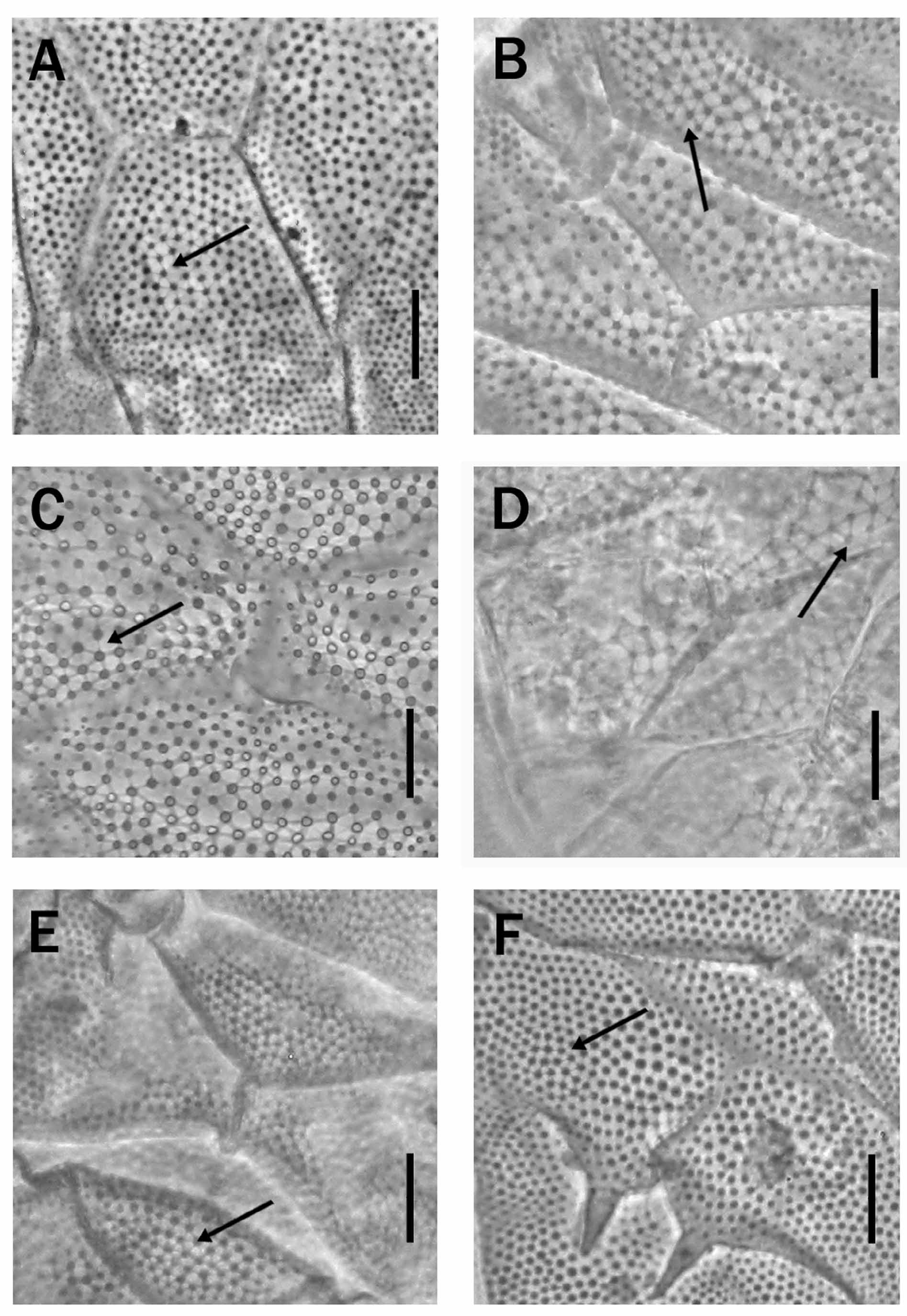
TABLE 1. Measurements (in µm) of the largest (holotype) and the smallest specimens, and mean values (mean; standard deviation; number of specimens (n); minimum and maximum) of P. santomensis sp. nov. (% bo = percentage of the given structure to the body length; % sc = percentage of the given structure to the scapular plate length).
| Character µm | %bo | %sc | µm | %bo | %sc | µm | %bo | %sc |
|---|---|---|---|---|---|---|---|---|
| Body length 168 | - | - | 119 | - | - | 143.6±15.2(21) 119–168 | - | - |
| Scapular plate length 26.2 | 15.6 | - | - | - | - | 23.0±2.2(15) 18.1–26.2 | 15.9±1.6(13) 13.8–18.6 | - |
| Internal buccal 9.4 cirrus | 5.6 | 35.9 | 6.1 | 5.1 | - | 7.8±0.9(22) 6.1–9.8 | 5.4±0.1(21) 4.6–6.7 | 35.1±3.8(14) 28.9–44.7 |
| External buccal 12.8 cirrus | 7.6 | 48.9 | 7.2 | 6.1 | - | 11.1±1.6(22) 7.2–14.4 | 7.8±1.0(21) 5.7–9.6 | 50.0±5.9(14) 39.9–55.7 |
| Cephalic papilla 4.8 | 2.9 | 18.3 | 3.7 | 3.1 | - | 4.3±0.45(22) 3.5–5.4 | 3.0±0.3(20) 2.4–3.6 | 19.0±1.6(14) 15.6–22.7 |
| Clava 4.8 | 2.9 | 18.3 | 3.3 | 2.8 | - | 4.1±0.6(22) 2.7–5.2 | 2.8±0.4(20) 2.3–3.5 | 18.2±3.5(15) 12.3–24.3 |
| Cirrus A 18.7 | 11.1 | 71.4 | 10.5 | 8.8 | - | 17.4±2.3(21) 10.5–21.8 | 12.0±1.4(19) 8.8–14.2 | 77.9±7.7(14) 67.6–96.8 |
| Pseudosegmental 4.2 plate projections | 2.5 | 16.0 | 1.0 | 0.8 | - | 3.6±1.2(20) 1.4–6.3 | 2.5±0.8(18) 1.1–4.1 | 16.8±6.7(15) 6.1–29.4 |
| Internal claws I 9.0 | 5.4 | 34.4 | - | - | - | 8.0±0.7(14) 6.8–9.0 | 5.3±0.2(13) 4.9–5.6 | 34.4±1.9(9) 30.8–37.9 |
| External claws I 8.0 | 4.8 | 30.5 | 5.5 | 4.6 | - | 7.1±0.8(15) 5.5–8.2 | 5.0±0.6(15) 3.8–6.3 | 32.3±5.5(9) 23.7–43.1 |
| Internal claws II 8.4 | 5.0 | 32.1 | - | - | - | 7.8±0.4(13) 7.2–8.4 | 5.3±0.3(12) 5.0–6.2 | 34.0±3.4(11) 30.4–42.5 |
| External claws II 8.0 | 4.8 | 30.5 | - | - | - | 7.2±0.7(14) 5.4–8.1 | 4.9±0.4(13) 4.3–5.9 | 32.5±3.8(10) 27.3–40.3 |
| Internal claws III - | - | - | - | - | - | 7.4±0.5(13) 6.3–8.0 | 5.1±0.3(11) 4.7–5.6 | 32.5±2.7(9) 28.5–37.0 |
| External claws III 8.0 | 4.8 | 30.5 | - | - | - | 7.0±0.6(15) 6.1–7.9 | 4.7±0.4(13) 4.2–5.6 | 31.4±3.5(11) 25.7–38.7 |
| Internal claws IV 9.2 | 5.5 | 35.1 | 6.7 | 5.6 | - | 8.0±0.7(11) 6.8–9.0 | 5.6±0.4(10) 4.7–6.0 | 37.6±3.8(7) 34.6–45.3 |
| External claws IV 8.5 | 5.1 | 32.4 | 6.0 | 5.0 | - | 7.4±0.7(13) 6.7–9.2 | 5.2±0.4(11) 5.1–6.6 | 34.1±3.7(8) 29.8–41.4 |
| Papilla on legs IV 2.2 | 1.3 | 8.4 | 1.8 | 1.5 | - | 2.1±0.2(22) 1.6–2.3 | 1.4±0.2(20) 1.2–1.8 | 9.1±0.7(15) 7.9–10.5 |
No known copyright restrictions apply. See Agosti, D., Egloff, W., 2009. Taxonomic information exchange and copyright: the Plazi approach. BMC Research Notes 2009, 2:53 for further explanation.