
Pseudohexapodibius degenerans ( Biserov, 1990 )
|
publication ID |
https://doi.org/ 10.1093/zoolinnean/zlad129 |
|
DOI |
https://doi.org/10.5281/zenodo.10616540 |
|
persistent identifier |
https://treatment.plazi.org/id/03E04041-6E1E-FFA0-FF57-2C0C6E77F90E |
|
treatment provided by |
Plazi |
|
scientific name |
Pseudohexapodibius degenerans ( Biserov, 1990 ) |
| status |
|
Pseudohexapodibius degenerans ( Biserov, 1990) View in CoL
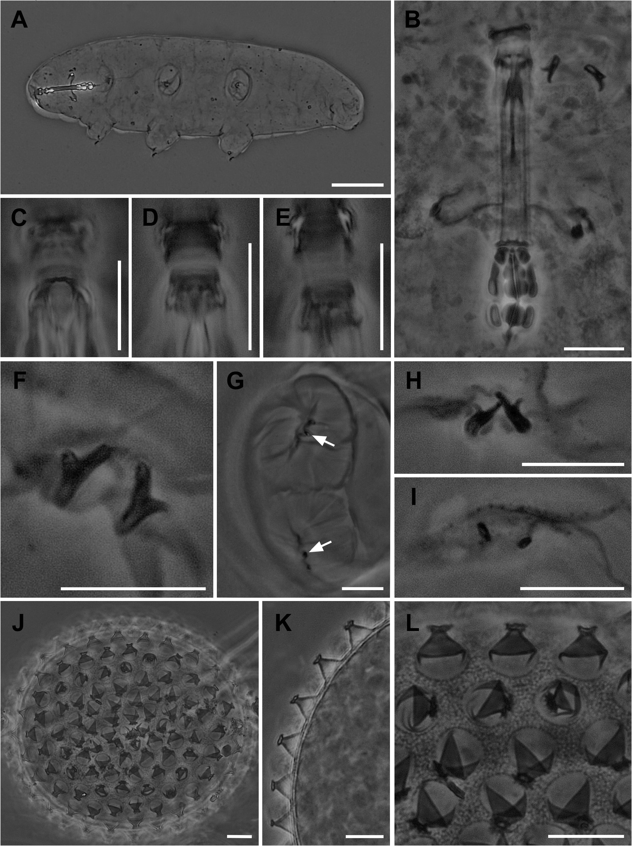
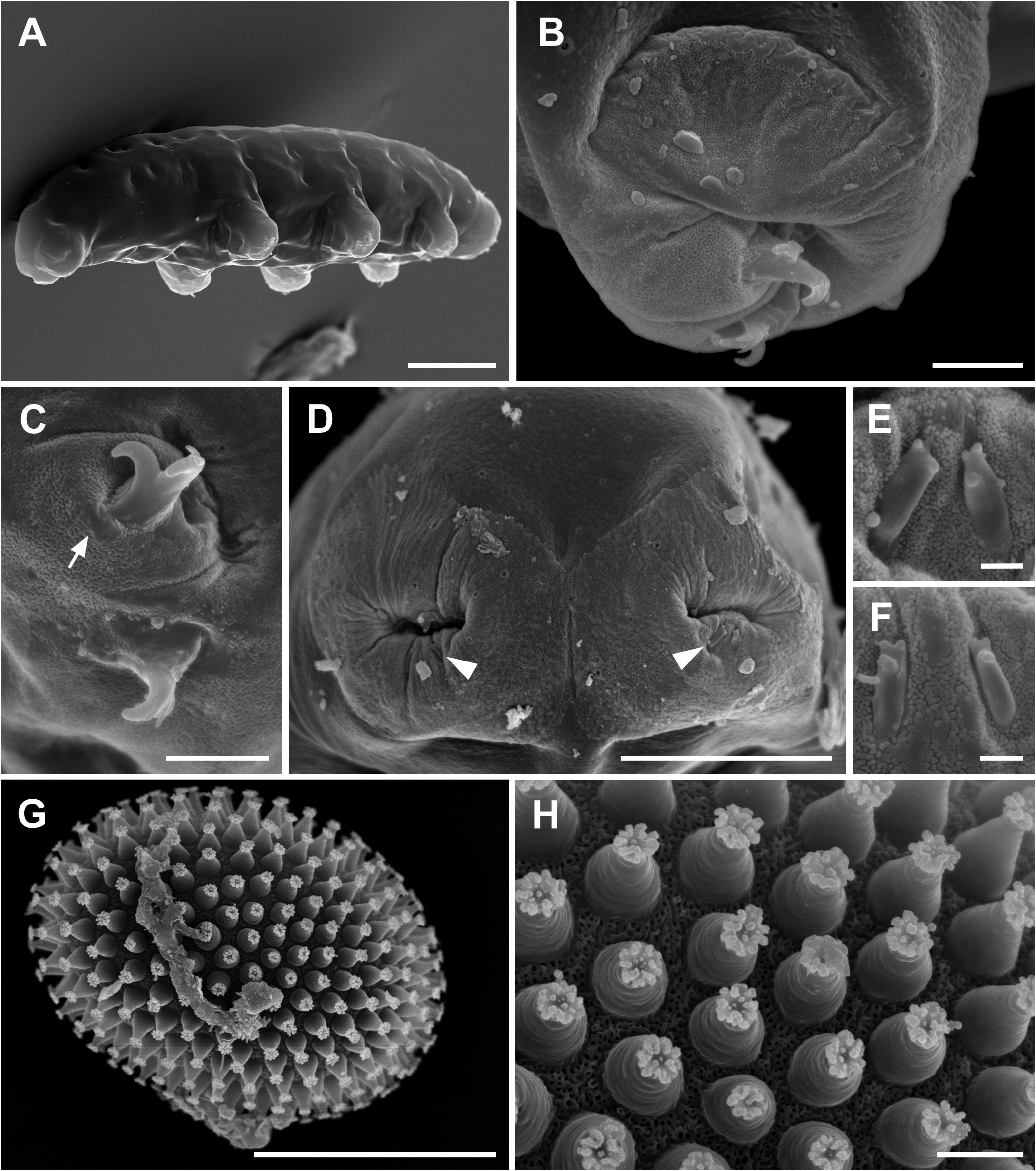
( Figs 8 View Figure 8 , 9 View Figure 9 ; measurements and statistics are in Tables 2 View Table 2 and 3 View Table 3 ; Supporting Information, Table S2 View Table 2 ).
Locality:
54°44ʹ54.82″N, 17°26ʹ15.31″E; 8 m a.s.l., Poland, Pomerania, Słowiński National Park,
Łącka Dune
, clump of grass [(
Corynephorus canescens
(L.) P.Beauv.)] from sand, September 2019, coll. M. Roszkowska and Ł. Kaczmarek.
View Table
View Table
View Figure
View Figure
Material examined: One hundred and fissy-six adults and 22 eggs (slide numbers
SPN1
–
SPN17
).
Type depositories: All specimens and eggs were deposited at the Department of Animal Taxonomy and Ecology, Institute of Environmental Biology, Adam Mickiewicz University in Poznań, Uniwersytetu Poznańskiego 6, 61–614 Poznań, Poland.
DescriptionofthePolishpopulation: Bodytransparentasserfixation in Hoyer’s medium; eyes present in 65% of fixed and measured specimens. Very small, scaưered pores (~0.5 µm in diameter, visible only with SEM; Fig. 9D View Figure 9 ) in the dorsolateral cuticle of body and legs. Very small single granules (visible only with SEM), distributed almost regularly, present on the entire cuticle ( Fig. 9D View Figure 9 ). Legs of the first pair clearly smaller than those of the second and third pairs. The area of the leg cuticle surrounding the claws with a swelling (forming a furbelow-like structure; Figs 8G View Figure 8 , 9B, D View Figure 9 ) covered with spheroidal microdigitations clearly visible only in SEM ( Fig. 9C, E, F View Figure 9 ).
Buccopharyngeal apparatus of the Macrobiotus type, anteroventral mouth with 10 small peribuccal lamellae. Oral cavity armature of the maculatus type, with only third band of teeth (visible with LM). This band of teeth is formed by a system of one continuous dorsal tooth (ridge) and two or three ventral transverse granular teeth (ridges) ( Fig. 8C–E View Figure 8 ). In the pharynx ( Fig. 8B View Figure 8 ): large and triangular pharyngeal apophyses overlapping the first macroplacoid; two rod-shaped macroplacoids, length sequence 2 <1 (in lateral view), and evident triangular microplacoid. First macroplacoid with central constriction. Second macroplacoid without constriction.
Claws I –III of Xerobiotus type, small and compact, with the common tract similar in length to the main branch; main branch with accessory points; lunules absent ( Fig. 8F View Figure 8 ); small cuticular plates at the base of the claws present (visible with SEM; Fig. 9C View Figure 9 ). Claws IV extremely reduced (granular shaped with LM) to very short primary branches without accessory points (sometimes extremally small accessory points and secondary branch also present; visible with SEM), without lunules or thickening under the claws ( Figs 8G–I View Figure 8 , 9D–F View Figure 9 ).
Eggs spherical, white, ornamented and laid freely ( Figs 8J View Figure 8 , 9G View Figure 9 ). The surface between processes of the hufelandi type, i.e. covered with a reticulum formed by a mesh of small densely distributed pores, uniform in size and evenly distributed (however, the mesh seems to be wrinkled with SEM) ( Figs 8K, L View Figure 8 , 9H View Figure 9 ). Egg processes of hufelandi type, with a straight trunk and a relatively small and concave terminal disc. The terminal disc is greatly indented on the disc margin, forming evident but irregular teeth covered with microgranules (visible only with SEM; Fig. 9H View Figure 9 ).
Reproduction: Reproductive mode unknown.
Molecular characterization: One haplotype for cox1, two haplotypes for ITS2 (p-distance 0.5%–5.9%), three haplotypes for 18S, and three haplotypes for 28S genes (GenBank accession numbers in Supporting Information, Table S1 View Table 1 ; p-distances in Supporting Information, Table S8). Sequences of P. degenerans were more similar to X. reductus and X. naginae for cox1 (with p-distances of 1.1%–6.1% and 0.8%–6.6%, respectively) and it shares an ITS2 haplotype with specimens of X. reductus , X. naginae and of Xerobiotus from Błędowska Desert, Poland (GenBank accession number MN888345; Vecchi and Stec 2021) (Supporting Information, Table S8).
No known copyright restrictions apply. See Agosti, D., Egloff, W., 2009. Taxonomic information exchange and copyright: the Plazi approach. BMC Research Notes 2009, 2:53 for further explanation.
|
Kingdom |
|
|
Phylum |
|
|
Class |
|
|
Order |
|
|
Family |
|
|
Genus |