
Pseudoscopas carbonelli Matiotti da Costa, n. sp.
|
publication ID |
https://doi.org/ 10.11646/zootaxa.4975.1.4 |
|
publication LSID |
lsid:zoobank.org:pub:2172D442-8EEA-4475-8260-B4FA26413B45 |
|
DOI |
https://doi.org/10.5281/zenodo.4805337 |
|
persistent identifier |
https://treatment.plazi.org/id/4032878B-5E78-6D11-4E8C-F8E1375E68F5 |
|
treatment provided by |
Plazi |
|
scientific name |
Pseudoscopas carbonelli Matiotti da Costa, n. sp. |
| status |
|
Pseudoscopas carbonelli Matiotti da Costa, n. sp.
( Figs 1-4 View FIGURE 1 View FIGURE 2 View FIGURE 3 View FIGURE 4 )
Etymology. The specific epithet refers to a tribute to the great orthopterologist Carlos Salvador Carbonell, in memorial who dedicated his life to working brilliantly with Neotropical grasshopper taxonomy.
Diagnosis. Close to P. nigrigena , but differentiated by coloration of the body, epiproct without protuberances in median region and absence of sclerotization in edge of the cerci. Combination of the following characteristics: (i) Epiproct triangular with deep median sulci in apical portion with five conspicuous protuberances in the median region and edge of the apical region sclerotized and curved; (ii) furculae divided in two small lobes; (iii) pallium lobes small; (iv) male cerci triangular, wide and robust at the base, and with sclerotized acute apex, curving inward; (v) hind edge of the female subgenital plate forming two large lobes on both sides; (vi) plan bridge with median reentrancy; (vii) ancorae larger in size than of the anterior projections; (viii) lophi prominent and sclerotized.
Body morphology and coloration
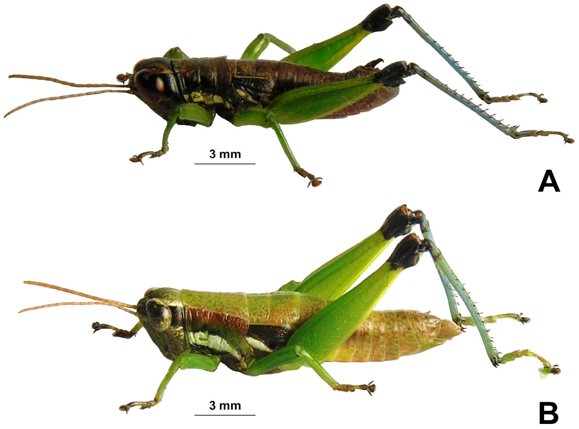
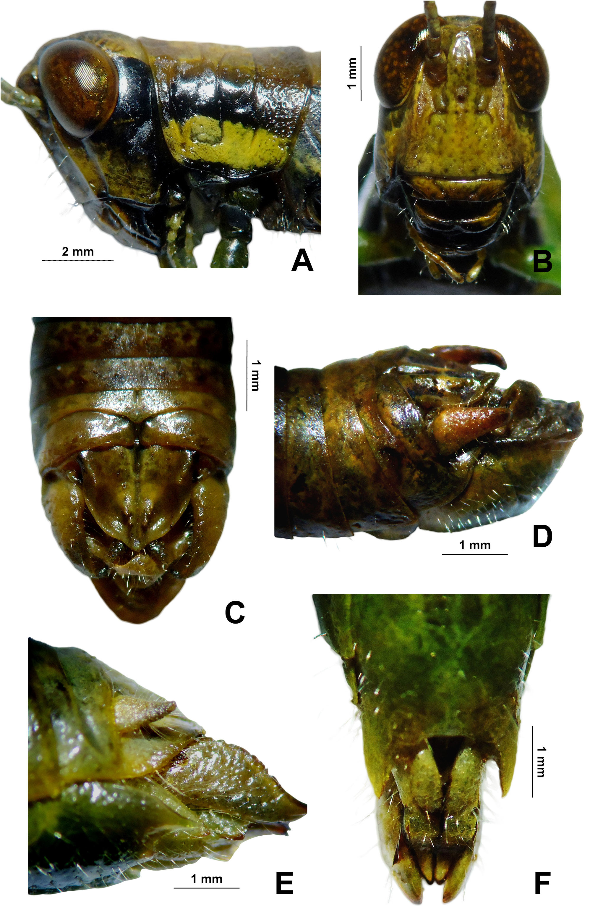
Description. Male Holotype ( Figs. 1–3 View FIGURE 1 View FIGURE 2 View FIGURE 3 ). Body medium in size (18 mm length), moderately robust and deeply pubescent, especially the femorae and tibiae. Braquipterous. Head. Front subvertical, slightly oblique ( Fig. 3A View FIGURE 3 ) and conspicuously larger than usual ( Fig. 3B View FIGURE 3 ). Antennae filiform, with 18 antennomeres surpassing the posterior margin of pronotum. Eyes globular, laterally salient and not exceeding the vertex. Interocular space narrow ( Fig. 3A View FIGURE 3 ). Face little retreating. Frontal costa plan ( Fig.3A View FIGURE 3 ). Occiput smooth ( Fig. 3B View FIGURE 3 ). Fastigium of the vertex subtriangular, shorter, with apex of vertex slightly rounded ( Fig.2A View FIGURE 2 ). Thorax. Pronotum with median carina slightly marked, and lateral absent ( Fig. 2A View FIGURE 2 ); metazona shorter than prozona; pronotum disc crossed by three transverse sulci; pronotum lateral edges parallel, slightly expanding in the metazona; anterior margin straight and the posterior with slight median emargination ( Fig. 2A View FIGURE 2 ). Prosternal process spiniform. Mesosternal lobes rounded. Tegmen short, lobuliform, reaching to the second abdominal segment ( Fig. 2A View FIGURE 2 ). Legs. Hind femur slender, the upper keel smooth, the end of lower knee lobes rounded. Hind tibiae pubescent with 7 outer and 8 inner dorsal spines; absence of hind femur’s terminal spinous projection on the dorsal genicular lobes ( Fig. 2A View FIGURE 2 ). Abdomen. Tympanum large. Abdomen thickened in distal region. Epiproct triangular, longer than wider with deep median sulci in apical portion and five conspicuous protuberances in the median region; edge of the apical region sclerotized and curved apex rounded; furculae divided in two small lobes ( Fig. 3C View FIGURE 3 ). Pallium with small and salient lobes ( Fig. 3C View FIGURE 3 ). Cerci triangular, robust, conic, pubescent, strongly curved, exceeding the epiproct apex; acute and sclerotized apex ( Fig. 3D View FIGURE 3 ). Subgenital plate cupuliform, short, curved up, apex rounded and pubescent ( Fig. 3D View FIGURE 3 ). Phallic complex. Cingulum with long apodemes, arch developed; gonophore with a rectangular ventral projection ( Fig. 4A View FIGURE 4 ); aedeagus long with upper and lower valves of the same size and median longitudinal sulci in the apical valves; sclerites of aedeagus covered by the wide and highly lobulated ectophallic sheath ( Fig. 4A View FIGURE 4 ). Epiphallus with plan bridge and median reentrancy; ancorae larger in size than of the anterior projections; lophi prominent and sclerotized; without internal conical protuberance ( Fig. 4B View FIGURE 4 ).
Body brown light color ( Figs 1A View FIGURE 1 , 2A View FIGURE 2 , 3 View FIGURE 3 A-B). Head mostly yellow color with a black pair of retro-ocular bands that take part of the genae, and continue in the dorsal half of the pronotum lateral lobes, tegmina, pleural and sides of the abdomen. Scape, pedicel, and antennal flagellum brown color. Fastigium of the vertex with five brown spots ( Fig. 2A View FIGURE 2 ). Occiput marked with brown spots. Face, clypeus, and genae yellowish color; mandible and labrum black color; labial and maxillary palpi yellowish color with black tip ( Fig. 3B View FIGURE 3 ). Light brown antennae. Pronotum dorsally with several punctures and brown spots ( Fig. 2A View FIGURE 2 ). Lateral lobes of the pronotum with a yellow ivory band on the lower abdomen ( Fig. 3A View FIGURE 3 ). Tegmen with veins light brown, a narrow ivory band surrounding the dorsal portion of the wing ( Fig. 2A View FIGURE 2 ). Genicular lunules black. Legs I and II green, with pretarsus tip, arolium and claws brown color; femur III yellow-green, with genicular lobe black; tibiae III light-blue, with tarsomeres light green, and tarsal claws and arolium brown. Lateral sclerites of the abdomen yellowed from the second segment. Dorsal and ventral abdominal segments and pallium brown.
Female. Larger in size to male, robust (body length 21mm) ( Fig. 1B View FIGURE 1 , 2B View FIGURE 2 ). Same color as male, but the head more yellowish ( Fig. 2B View FIGURE 2 ); fastigium more prominent and wider than in males and with brown color spots; eyes more prominent. Interocular space wider. Pronotum dorsally light green, with several punctures and brown punctuation spots. Lateral lobes of the pronotum with a reddish, yellow-green and ivory bands, from upper to lower portion ( Fig. 1B View FIGURE 1 ). Abdomen sides with a black spot in between segments 3-6. Ovipositor valves short in lateral view and serrulated margins absent ( Fig. 3E View FIGURE 3 ); dorsal valve of the ovipositor robust and apical tooth of the external ventral valve of ovipositor little prominent. Hind edge of the female subgenital plate forming two large lobes on both sides and without sclerotization ( Fig. 3F View FIGURE 3 ). Legs I and II less robust than in males. Cerci short, conical, not reaching the epiproct tip ( Fig.3E View FIGURE 3 ). Epiproct subtriangular with median sulcus well marked, interrupted transversely in the median region.
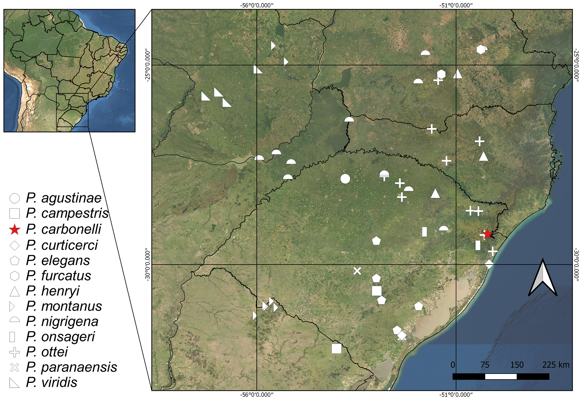
Habitat and Habits. Individuals of P. carbonelli n. sp. were collected mainly during the day, being active on bushes and grasses of the edge of the forest in more open places and with lower canopy cover ( Fig.6 View FIGURE 6 ), usually between 11am to 5pm. The records of the genus Pseudoscopas are between the Pampean and Parana provinces, but P. carbonelli is found only in the southeast of the Parana province. ( Morrone 2001; 2006).
Measurements (mm): Males (n=7): BL= 18.1 (17.4–18.1); PL = 0.3 (0.2–0.3); FL3 = 0.8 (0.7–0.8); TL = 0.3 (0.2–0.3). Females (n = 8): BL = 21.1 (19.2–21.1); PL = 0.5 (0.4–0.5); FL3 = 0.9 (0.8–0.9); TL = 0.4 (0.3 – 0.4).
Type material. Holotype ♂. BRAZIL: Rio Grande do Sul, municipality of São Francisco de Paula , (29°24’44.16”S 50°12’30.44”W), 27-30.i.2020, Zefa, E.; Acosta R.C. & Tim, V., 7♂ and 8♀ ( MCTP) GoogleMaps . Holotype condition: genitalia were extracted and maintained in holotype’s tube.
Chromosomes ( Figs. 5 View FIGURE 5 A-D). Diploid chromosome number of 2n=23, X0 ♂ /24, XX ♀; all chromosomes acrocentrics, grouped in two chromosome pairs large (L 1 –L 2), two pairs medium large ( ML 3 – ML 4), five medium (M 5 –M 9), and two small (S 10 –S 11) ( Fig. 5A View FIGURE 5 ); the X chromosome belongs to the ML group, and the megameric to the M group, both showing positive heteropiknosis until diplotene phase ( Fig. 5C, D View FIGURE 5 ). Metaphase I chiasmata position: L 1, proximal and intersticial; L 2, proximal and intersticial; ML 3, proximal and subterminal; ML 4, subterminal; M 5, terminal; M 6, terminal; M 7, terminal; M 8, intersticial; M 9, intersticial; S 10, terminal; S 11, terminal ( Fig. 5B View FIGURE 5 ). The size relationship between chromosomes can be better seen in ( Fig. 5E View FIGURE 5 ), which includes a metaphase II.
Genus distribution. Brazil (Paraná; Santa Catarina; Rio Grande do Sul); Argentina (Misiones, Concepción); Paraguay (Sapucay, Caaguazú); Uruguay (Caraguatay, Cuchilla Negra, Tacuarembó) ( Cigliano et al. 2021; Ronderos 1987; Mesa et al. 1982; Monné 2018) ( Table 1 View TABLE 1 , Fig. 7 View FIGURE 7 ).


| R |
Departamento de Geologia, Universidad de Chile |
| V |
Royal British Columbia Museum - Herbarium |
| MCTP |
Museu de Ciencias |
| ML |
Musee de Lectoure |
No known copyright restrictions apply. See Agosti, D., Egloff, W., 2009. Taxonomic information exchange and copyright: the Plazi approach. BMC Research Notes 2009, 2:53 for further explanation.
|
Kingdom |
|
|
Phylum |
|
|
Class |
|
|
Order |
|
|
Family |
|
|
Genus |