
Remaneicaris tridactyla, Corgosinho, Paulo Henrique C. & Arbizu, Pedro Martínez, 2007
|
publication ID |
https://doi.org/ 10.5281/zenodo.175898 |
|
DOI |
https://doi.org/10.5281/zenodo.6237261 |
|
persistent identifier |
https://treatment.plazi.org/id/03FE4078-4764-2417-FF6A-B799FE594140 |
|
treatment provided by |
Plazi |
|
scientific name |
Remaneicaris tridactyla |
| status |
sp. nov. |
Remaneicaris tridactyla n. sp.
Type material: Holotype, one dissected male on 7 slides ( INPA 1339a).
Paratypes: One dissected female on 7 slides ( INPA 1339c); 2 undissected males mounted on different slides ( INPA 1339b and INPA 1339d), 1 dissected male mounted on 7 slides ( INPA 1339e), 1 undissected female mounted on one slide ( INPA 1339f).
Etymology: The species name refers to the trifid structure of the modified thumb of the male leg 3.
Type Location: Ribeirão do Ouro river, Sítio do Corgosinho, Florestal, state of Minas Gerais, Brazil. Coordinates: 19°48’19”S, 44°27’45”W.
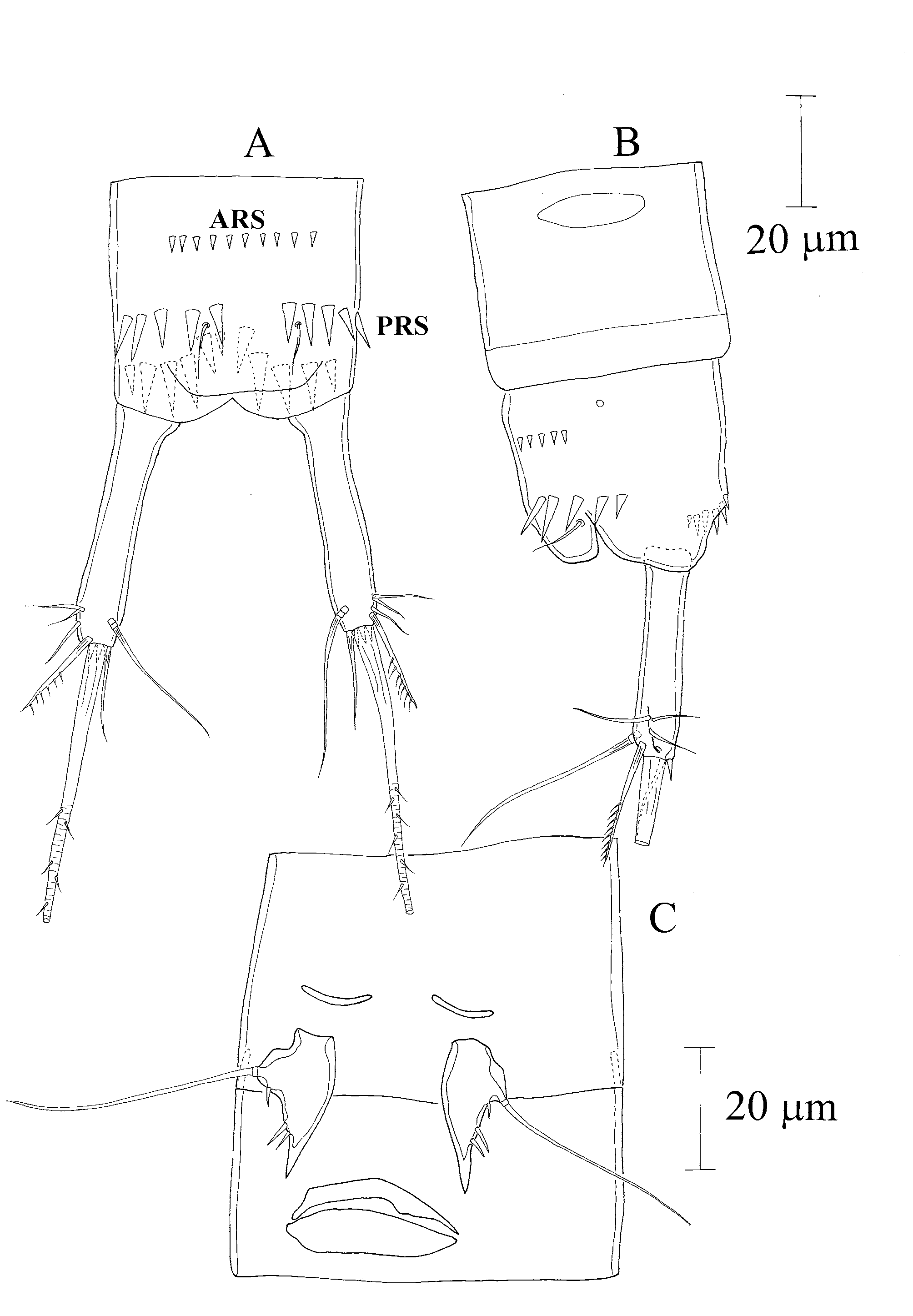
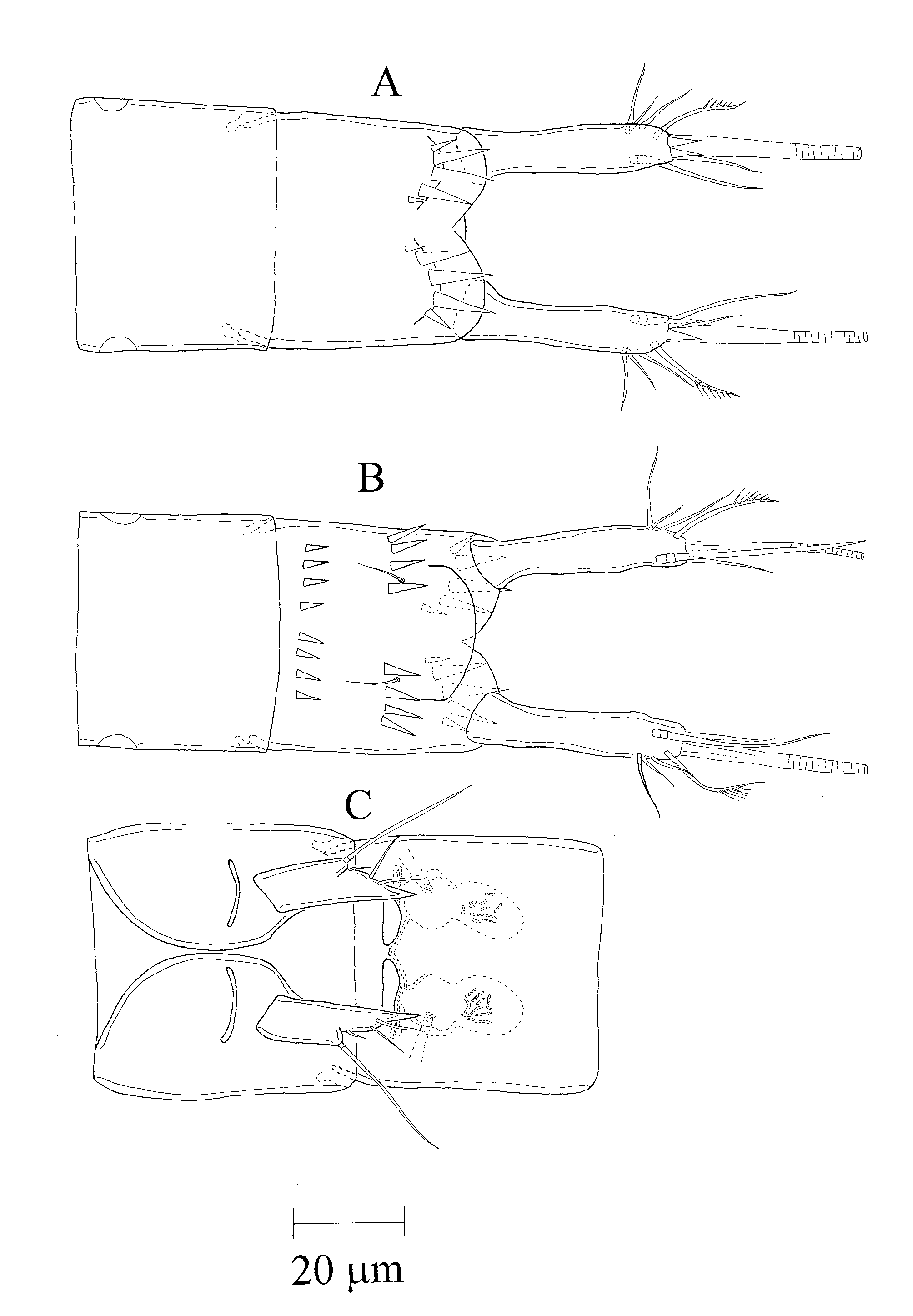
Male ( Fig. 1 View FIGURE 1 A–B). Length 404 µm (measured from the tip of rostrum to the posterior rim of anal operculum). Rostrum not fused to cephalothorax, with a wide base and two sensilla on the tip. Cephalothorax and second urosomite with 1 dorsal integumental window. Urosomite 5 with a pair of lateral integumental windows. Dorsal pores on cephalothorax, thoracic somites 1 and 3 and second urosomite. Telson with 1 pair of lateral pores. For sensilla on tergites see Fig. 1 View FIGURE 1 A–B. Telson with 1 dorsal row of 10 spinules located on the anterior third (ARS), 2 rows of 5 large spinules on the posterior half (PRS), anterior to the sensilla, not reaching the operculum and 2 ventral rows of large spinules on the posterior half, near the insertion of the furca ( Fig. 2 View FIGURE 2 A–B). Anal operculum smooth and quadrate. Furca ( Figs. 1 View FIGURE 1 A–B and 2 A–B) about 6 times as long as wide, with 7 setae. All setae located on the posterior third. A1 ( Fig. 3 View FIGURE 3 A) 9–segmented and prehensile; armature beginning with proximal segment: 0/5/4/2/5+Ae/1/4/2/9+Ae, 1 modified seta on the last segment (indicated by an arrow) and 1 hyaline spinule on the seventh and eighth segments. A2 ( Fig. 3 View FIGURE 3 B) with allobasis; 1- segmented exp with 1 seta, and 1-segmented enp bearing an anterior hyaline frill and 7 setae. Md, Mx1, and Mxp armature as P. h i s p a n i c a Martínez Arbizu 1997; Mx2 with 2 slender setae on the proximal endite and 3 on the distal endite, as in R. analuizae Corgosinho & Martínez Arbizu 2005 . Leg 1 ( Fig. 4 View FIGURE 4 A) coxa without setae or spines, with 2 rows of spinules on the posterior side; basis with outer seta and 1 pore on the anterior side, 3 spinules on the outer margin, and 3 distal spinules anterior to the insertion of the enp; enp and exp of the same size; enp 2-segmented, segment 1 with 1 row of 5 long spinules along the inner margin, 1 row of 4 spinules on the outer margin and a posterior hyaline frill; segment 2 with a posterior hyaline frill and 2 distal setae, 1 of them geniculated; first enp segment of the same size of the first two exopodites; exp 3-segmented, segment 1 with 1 outer spine, segment 2 without setae or spines, segment 3 with 2 outer spines, 2 geniculated setae and a posterior pore. Leg 2 ( Fig. 4 View FIGURE 4 B) coxa without setae or spines, with 1 row of small spinules and 3 outer spinules on the posterior side; basis without outer seta and ornamented with 1 row of spinules on the outer margin, 1 row of small spinules at the level of the enp insertion and 1 pore near the outer margin; exp 3- segmented, the first segment approximately the same length as the remaining exopodites, with a proximal row of spinules on the outer margin, distributed in a “V” shape, a row of spinules anterior to the insertion of the outer spine and an inner hyaline frill, segment 2 without armature, with 2 spinules located medially on the outer margin and with a row of distal spinules, segment 3 with 3 setae, a distal hyaline frill on the inner corner and 2 spinules located medially on the outer margin; enp 1-segmented with 1 distal seta, 2 distal spinules, and 3 spinules along the outer margin. Leg 3 ( Fig. 4 View FIGURE 4 C–D) coxa without setae or spines; basis with an outer seta; enp 1-segmented with 3 subdistal spinules; exp 1-segmented, elongated, bearing 2 rows of spinules along the outer margin and with 1 very modified seta (“thumb”) in a subdistal position. Leg 4 ( Fig. 4 View FIGURE 4 E) coxa without setae or spines, with 3 small spinules on the posterior side; basis with outer seta, 1 pore near the outer margin, a row of spinules on the inner margin ( IBRS) and 1 row of larger spinules near the insertion of the enp ( PERS), in a hyaline area of the basis ( PEHZ); exp 3-segmented, segment 1 almost same length as remaining segments, with an outer spine, a proximal row of spinules distributed in a “V” shape (RVS) on the outer margin, a row of spinules anterior to the insertion of the outer spine and an inner hyaline frill on the distal corner, segment 2 without setae, with a row of spinules on the distal portion, a row of small spinules along the inner margin, conferring on it a serrated shape, and with 2 long spinules located medially on the outer margin, segment 3 with 1 apical and 1 subdistal outer seta, a distal hyaline frill on the inner corner and 3 spinules located medially on the outer margin; enp 1-segmented, leaf-shaped, covered with numerous long spinules (hirsute) along the inner and outer margin and with a distal spine. Leg 5 ( Fig. 2 View FIGURE 2 C) triangular, ending in a spiniform process and with all setae arranged on the outer margin.
Female: Sexually dimorphic in number of body segments, A1, leg 3, leg 4 and genital field.
Habitus ( Fig. 5 View FIGURE 5 A–B). Length 413 µm (measured from the tip of rostrum to the posterior rim of anal operculum). Rostrum as in male. Cephalothorax and genital double-somite with 1 dorsal integumental window. Urosomite 4 with 1 pair of lateral integumental windows. Dorsal pores as in male and 1 pair of lateral pores on the telson. For sensilla on tergites see Fig. 5 View FIGURE 5 A-B. Telson with 2 dorsal rows of 4 spinules on the anterior third (ARS), 2 rows of 4 larger spinules on the posterior half, not reaching the operculum (PRS) and 2 ventral rows of large spinules on the posterior half, near the insertion of the furca ( Fig. 5 View FIGURE 5 A–B and 6 A–B). Anal operculum smooth and convex ( Fig. 6 View FIGURE 6 B). Furca ( Fig. 6 View FIGURE 6 A–B) about 5 times as long as wide, with 7 setae. All setae located on the distal third, as in males. A1 ( Fig. 7 View FIGURE 7 A) 7-segmented, not prehensile; number of setae beginning with proximal segment:
0/4/5/2+Ae/1/2/9+Ae. A2 ( Fig. 7 View FIGURE 7 B) with allobasis, 1-segmented exp with 1 seta, 1-segmented enp bearing 7 setae and a posterior hyaline frill. Buccal parts as in male. Leg 1 ( Fig. 7 View FIGURE 7 C) coxa without setae or spines and with 1 posterior row of spinules; basis with outer seta, 3 outer accessory spinules and 3 distal spinules near the insertion of enp; enp and exp as in males. Leg 2 ( Fig. 7 View FIGURE 7 D) as in males. Leg 3 ( Fig. 7 View FIGURE 7 E) coxa without setae or spines, with 2 rows of spinules on the posterior side; basis with 1 outer seta, and ornamented with 1 row of spinules near the insertion of the enp; exp 2-segmented, segment 1 with an outer spine, a proximal row of spinules and a row of spinules next to the outer spine, segment 2 with 1 spine, 1 seta and 2 long spinules on the outer margin; enp 1-segmented and spiniform, with 2 spinules on the outer margin and 1 spinule on the inner margin. Leg 4 ( Fig. 7 View FIGURE 7 F) as in males, except for the presence of a spiniform enp ornamented with 1 row of 4 spinules on the outer margin and a row of small spinules on the basis, near the insertion of the enp. Leg 5 ( Fig. 6 View FIGURE 6 C) as in male. Genital field as illustrated in Fig. 6 View FIGURE 6 C, with a single, medially located copulatory pore. Gonopore is a transverse slit.


| INPA |
Instituto Nacional de Pesquisas da Amazonia |
No known copyright restrictions apply. See Agosti, D., Egloff, W., 2009. Taxonomic information exchange and copyright: the Plazi approach. BMC Research Notes 2009, 2:53 for further explanation.