
DERMOPHIIDAE, Taylor, 1969
|
publication ID |
https://doi.org/ 10.1111/j.1096-3642.2012.00838.x |
|
persistent identifier |
https://treatment.plazi.org/id/03DB87B7-FFFB-FFB7-FC4A-9189FB316293 |
|
treatment provided by |
Marcus |
|
scientific name |
DERMOPHIIDAE |
| status |
|
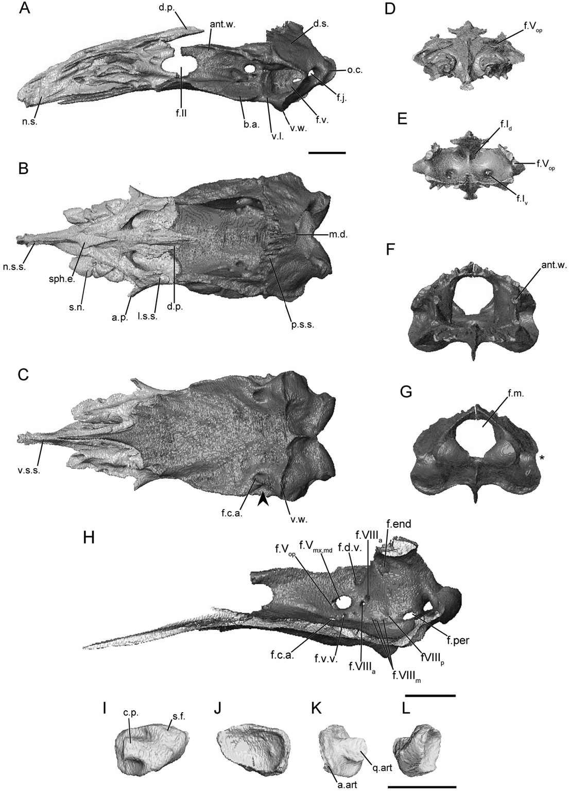
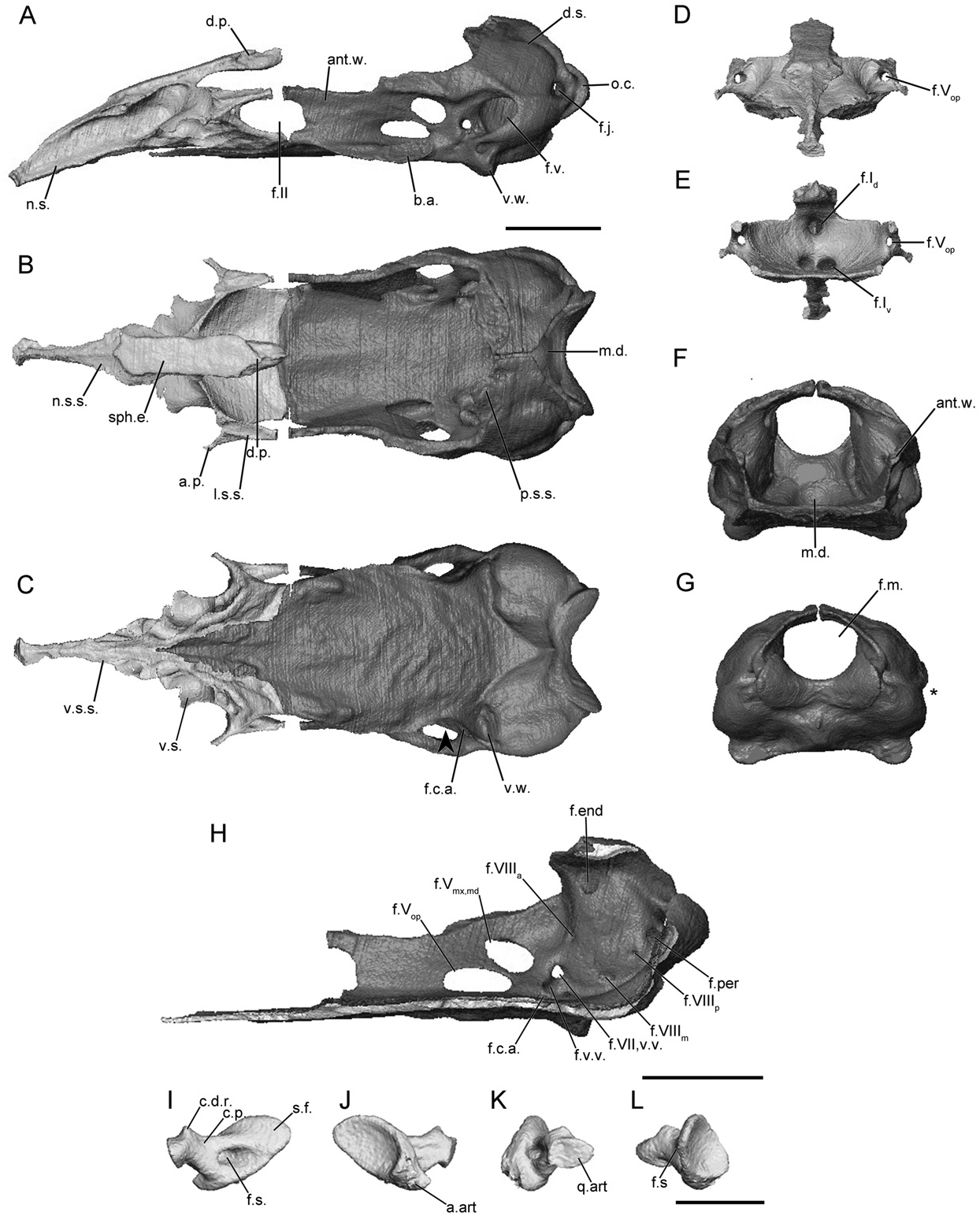
DERMOPHIIDAE View in CoL ( FIGS 3 View Figure 3 , S 4–S View Figure 4 6 View Figure 6 )
In dermophiids the main body of the sphenethmoid accounts for 30% or less of the total length of the sphenethmoid ( Fig. 3A View Figure 3 ). The short lateral wall of its main body is capped by a broad sutural surface (less broad in Geotrypetes seraphini ) that receives the anterolateral lappet of the parietal ( Fig. S5B View Figure 5 ). The anterolateral corner bears an ossified anterolateral process ( Fig. 3B View Figure 3 ), except for Geo. seraphini in which only a small anterolateral expansion is present ( Fig. S5B View Figure 5 ). The dorsomedial process is thick and long, extending well beyond the anterior limit of the lateral walls of the os basale ( Fig. 3A View Figure 3 ). The process is variably exposed dorsally in specimens of Dermophis mexicanus ( Fig. S4B View Figure 4 ), a portion is exposed in Geo. seraphini ( Fig. S5B View Figure 5 ) and Schistometopum thomense ( Fig. S6B View Figure 6 ), and the dorsal exposure of the sphenethmoid in Gymnopis multiplicata corresponds to a region over the nasal septum ( Fig. 3B View Figure 3 ). The posterior margin of the lateral wall of the sphenethmoid is deeply incised by the optic foramen in all dermophiids. The posterior margin of the floor of the main body of the sphenethmoid is deeply incised at the midline, but otherwise the floor is quite broad and only slightly concave ( Fig. 3B View Figure 3 ). Geotrypetes seraphini is distinct amongst dermophiids in the presence of a deeply concave posterior margin of the floor ( Fig. S5B View Figure 5 ).
The ossified portion of the nasal septum is long, and reaches the posterior limit of the external naris. The dorsal surface of the nasal septum is broad near its base (less broad in Geo. seraphini ), and tapers to a thin ridge towards the tip ( Fig. 3B View Figure 3 ). The height of the nasal septum remains roughly uniform throughout its length ( Fig. 3A View Figure 3 ). Long, tubular sola nasi extend anteriorly from the main body of the sphenethmoid ( Fig. 3B, C View Figure 3 ); those in Geo. seraphini are less robust ( Fig. S5C View Figure 5 ) and those in Sc. thomense are connected to the nasal septum via a thin ventral sheet of bone ( Fig. S6C View Figure 6 ).
The anterior wall contains paired dorsal and ventral foramina ( Fig. 3D, E View Figure 3 ). The foramina of the dorsal pair are located close to the midline, and those of the ventral pair are offset laterally from the midline. The dorsal foramina lead to short canals before exiting onto the anterior surface of the sphenethmoid. The ventral pair leads to the tubular sola nasi. A single anterolateral foramen is present in the base of each anterolateral process ( Fig. 3D, E View Figure 3 ) in dermophiids. An additional small foramen is also present in the anterolateral expansion of Geo. seraphini ( Fig. S5D View Figure 5 ).
The antotic wall of the os basale is orientated parallel (angled slightly anteromedially in Geo. seraphini ) to its contralateral wall in dermophiids ( Fig. 3B View Figure 3 ). In anterior view the wall is vertical ( Fig. 3F View Figure 3 ), except for Geo. seraphini in which it is angled slightly dorsolaterally. The dorsal sutural surface is narrow anteriorly and expands dramatically at the level of the antotic foramina into a broad surface in all species, except for Geo. seraphini ( Fig. S5B View Figure 5 ) in which it remains narrow. The surface is continuous with the sutural surface along the anterior margin of the otic capsule. The anterior margin of the antotic wall is deeply incised by the posterior margin of the oval optic foramen; in Geo. seraphini it is posteroventrally obliquely so ( Fig. S5B View Figure 5 ). Five foramina are present in the antotic wall of all dermophiids ( Fig. 3H View Figure 3 ), except for Geo. seraphini in which there are three ( Fig. S5H View Figure 5 ). The difference occurs because the two trunks of the trigeminal and the ventral vein all exit a large common foramen in Geo. seraphini (referred to here as Pattern 3), but discrete foramina are present in the other dermophiid species (referred to here as Pattern 1).
The dorsal surface of the otic-occipital complex of the os basale is posteroventrally tilted, tapered towards the midline ( Fig. 3A, B View Figure 3 ). The anterior portion bears the sutural surface that receives the parietal, which is broad, except for Geo. seraphini . A laterally extending shelf, continuous with the dorsal surface, is present dorsal to the fenestra vestibuli in all dermophiids except Geo. seraphini ( Fig. S5B View Figure 5 ). The fenestra vestibuli is elongate anteroposteriorly. Its ventral margin is thick in D. mexicanus and Gy. multiplicata ( Fig. 3A View Figure 3 ). It opens in the lateral direction and only gently incises the lateral margin of the otic-occipital complex when viewed posteriorly ( Fig. 3G View Figure 3 ), except for D. mexicanus in which it does so very deeply ( Fig. S4G View Figure 4 ). In lateral view the occipital condyle projects far beyond the posterior limit of the otic capsule ( Fig. 3A View Figure 3 ), and the jugular foramen is visible in its base. The foramen magnum is large and subcircular ( Fig. 3G View Figure 3 ).
The medial wall of the otic capsule is perforated by six to eight foramina. Four of these are common to those of all species (endo- and perilymphatic foramina, and anterior and posterior vestibulocochlear nerve foramina; Maddin, 2011). Variably within the family two to four additional foramina transmit portions of the medial branch of the vestibulocochlear nerve ( Fig. 3H View Figure 3 ).
The anterior margin of the floor of the os basale tapers dramatically to a thin, rod-like tip in D. mexicanus ( Fig. S4C View Figure 4 ) and Gy. multiplicata ( Fig. 3C View Figure 3 ), but not the other species ( Figs S5C, S View Figure 5 6C View Figure 6 ). The floor extends far anteriorly, terminating in the anterior half of the nasal septum ( Fig. 3C View Figure 3 ), except for Geo. seraphini in which it just barely underlaps the nasal septum ( Fig. S5C View Figure 5 ). The floor of the os basale is smooth and slightly concave dorsally, and a well-defined median depression is present in the posterior region of the floor ( Fig. 3B View Figure 3 ). A well-developed basicranial articulation, complete with an ovoid sutural surface, is present in all dermophiids ( Fig. 3B View Figure 3 ); however, those in Geo. seraphini are distinctive in their tab-like appearance ( Fig. S5C View Figure 5 ). In ventral view the lateral margin of the floor, just posterior to the basicranial articulation, is slightly constricted (arrowhead; Fig. 3C View Figure 3 ), except in Sc. thomense in which it is strongly constricted (arrowhead; Fig. S6C View Figure 6 ). A well-developed ventral winglike projection is present on the ventral surface of the otic capsule, marking the lateral limit of the ventral muscle attachment site ( Fig. 3A, C View Figure 3 ). In D. mexicanus and Gy. multiplicata the posterior margin of the floor terminates at transverse ridge that does not closely approach the foramen magnum ( Fig. 3C View Figure 3 ). In Geo. seraphini and Sc. thomense the floor terminates at a rounded point, the apex of which closely approaches the foramen magnum ( Fig. S5C View Figure 5 ).
The foramen for the carotid artery that leads to a canal in the floor of the os basale is located just anterior to the otic capsule ( Fig. 3C View Figure 3 ). Its lateral and medial exit points are located ventral to the midpoint of the antotic foramina, on either side of the antotic wall.
The footplate of the stapes is elongate anteroposteriorly ( Fig. 3I, J View Figure 3 ), and is fairly tall in dermophiid species, except for Geo. seraphini ( Fig. S5I, J View Figure 5 ). Its long axis is orientated horizontally to slightly anteroventrally inclined. The anterior margin of the footplate is straight and closely applied to the margin of the fenestra vestibuli, and the rest closely approach its margins, except for Geo. seraphini in which the footplate is somewhat inset from the margin of the fenestra. The medial surface of the footplate is strongly concave. The columellar process is short and robust. It extends anteriorly from the footplate in roughly the horizontal plane. There is no foramen in the base of the columellar process ( Fig. 3L View Figure 3 ).


No known copyright restrictions apply. See Agosti, D., Egloff, W., 2009. Taxonomic information exchange and copyright: the Plazi approach. BMC Research Notes 2009, 2:53 for further explanation.
|
Kingdom |
|
|
Phylum |
|
|
Class |
|
|
Order |
|
|
Family |