
Scorpaena vesperalis, Wibowo & Motomura, 2020
|
publication ID |
https://doi.org/ 10.11646/zootaxa.4852.5.2 |
|
publication LSID |
lsid:zoobank.org:pub:5DE48C24-B9B4-41D3-8AA3-F94C669EE938 |
|
DOI |
https://doi.org/10.5281/zenodo.4504557 |
|
persistent identifier |
https://treatment.plazi.org/id/1B1A0E56-FFC7-F14F-8AFE-C2D8FB15FACF |
|
treatment provided by |
Plazi |
|
scientific name |
Scorpaena vesperalis |
| status |
sp. nov. |
Scorpaena vesperalis n. sp.
New English name: Dwarf Red Scorpionfish
Figures 1 View FIGURE 1 C–D, 2B–C, 3E–F, 4, 6, 7G–L; Tables 1–3
Holotype. WAM P. 28521-003, 58.7 mm SL, Busselton Jetty , Western Australia, 33°39′S, 115°21′E, 8 m, J. Hutchins, 12 Apr. 1885. GoogleMaps
Paratypes. 56 specimens, 16.2–67.6 mm SL, all from the southwestern coast of Western Australia, Australia. AMS I. 20233-014, 6 specimens, 18.5–25.5 mm SL, Canal Rocks, Cape Naturaliste, 33°40′12″S, 115°00′00″E, 1– 15 m, B. Russell, 1 Apr. 1978; GoogleMaps AMS I. 20245-031, 5, 20.2–31.7 mm SL, Horse Shoe Reefs, Rottnest Island , 32°00′S, 115°28′E, 12–15 m, B. Russell, 12 Apr. 1978; GoogleMaps AMS I. 20247-009, 5, 21.1–38.0 mm SL, Kingston Reefs, Rottnest Island , 31°58′48″S, 115°33′00″E, 8 m, B. Russell, 12 Apr. 1978; GoogleMaps AMS I. 20347-001, 67.6 mm SL, off Yallingup, 33°39′00″S, 115°01′12″E, 2 m, N. Coleman, 26 Mar. 1971; GoogleMaps CSIRO A 1696 View Materials , 51.5 mm SL, Strickland Bay , Rottnest Island , 32°00′S, 115°32′E, 21 Jan. 1954; GoogleMaps KAUM–I. 142982, 54.7 mm SL, GoogleMaps WAM P. 25798-011, 2, 38.0– 44.7 mm SL, Eagle Bay , Geographe Bay , 33°33′S, 115°04′E, 0–4 m, G. Allen et al., 21 Oct. 1976; GoogleMaps KAUM–I. 142983, 36.7 mm SL, GoogleMaps KAUM–I. 142985, 35.4 mm SL, GoogleMaps KAUM–I. 142986, 38.1 mm SL, Rocky Bay , Rottnest Island , 32°00′S, 115°28′E, J. Hutchins et al., 6 Jun. 1980; GoogleMaps KAUM–I. 142984, 34.1 mm SL, GoogleMaps WAM P. 28520-009, 4, 28.9–48.9 mm SL, Canal Rocks , 33°39′S, 115°01′E, 3–4 m, J. Hutchins et al., 16 Apr. 1985; GoogleMaps WAM P. 25806-003, 2, 31.9–41.2 mm SL, off Fremantle , Flinders , 32°18′S, 115°30′E, 37 m, N. Sarti et al., 29 Jun. 1977; GoogleMaps WAM P. 26600-007, 28.6 mm SL, Torbay Head , Albany , 35°08′S, 117°38′E, 3 m, J. Hutchins, 12 Apr. 1980; GoogleMaps WAM P. 26608-014, 2, 30.4–30.5 mm SL, Cheyne Beach , east of Albany , 34°53′S, 118°25′E, 12–15 m, J. Hutchins et al., 19 Apr. 1980; GoogleMaps WAM P. 26617- 005, 3, 29.6–34.9 mm SL, Rocky Bay , Rottnest Island , 32°00′S, 115°30′E, 8 m, J. Hutchins, 9 Jun. 1980; GoogleMaps WAM P. 27616-012, 2, 30.1–31.6 mm SL, Fish Hook Bay , Rottnest Island , 32°00′S, 115°30′E, 3–4 m, J. Hutchins et al., 1 Jun. 1982; GoogleMaps WAM P. 27951-009, 45.8 mm SL, Osprey Islet , Jurien Bay , 34°53′S, 118°25′E, 2–5 m, J. Hutchins et al., 10 Apr. 1983; GoogleMaps WAM P. 27988-004, 24.0 mm SL, Hamelin Bay , 34°12′S, 115°01′E, 1 m, N. Sarti, 21 Jan. 1979; GoogleMaps WAM P. 28521-012, 58.8 mm SL, same data as holotype; GoogleMaps WAM P. 29250-001, 2, 33.3–44.5 mm SL, Southern Group , Houtman Abrolhos , 1960; GoogleMaps WAM P. 32040-001, 48.1 mm SL, northwestern coast of Rottnest Island , 183–188 m, J. Hutchins et al. on FV Bluefin, 14 Aug. 1962; GoogleMaps WAM P. 32776-003, 10, 16.2–46.1 mm SL, North Ronsard Rocks , Cervantes, 30°28′42″S, 115°02′48″E, 3–4.7 m, G. Moore, 10 Mar. 2006; GoogleMaps WAM P. 26620-001, 55.9 mm SL, Geordie Bay, 32°00′S, 115°30′E, 5 m, J. Hutchins et al., 14 Jun. 1980 GoogleMaps .
Diagnosis. A new species of Scorpaena distinguished from other members of the S. papillosa complex by the following combination of characters: pectoral-fin rays 14–16 (mode 15); scale rows in longitudinal series 37–41 (40); scale rows above lateral line 4–6 (5), below 9–12 (11); scale rows between 6th dorsal-fin spine base and lateral line 4–6 (5), between last dorsal-fin spine base and lateral line 4–6 (5); body depth 32.3–39.5 (mean 36.0) % of SL; upper-jaw length 19.6–22.5 (20.9) % of SL; maxilla depth 5.7–7.3 (6.4) % of SL; postorbital length 18.2–21.3 (19.6) % of SL; least distance between interorbital ridges 1.4–2.7 (1.9) % of SL; 1st anal-fin spine length 7.2–10.0 (8.7) % of SL; anterior lacrimal spine simple, without additional spinous points on posterior margin; a single united pore behind lower jaw symphysial knob; supraocular tentacle relatively large, its length greater than half pupil diameter; all fins of preserved specimens usually uniformly whitish to translucent; largest recorded specimen 67.6 mm SL.
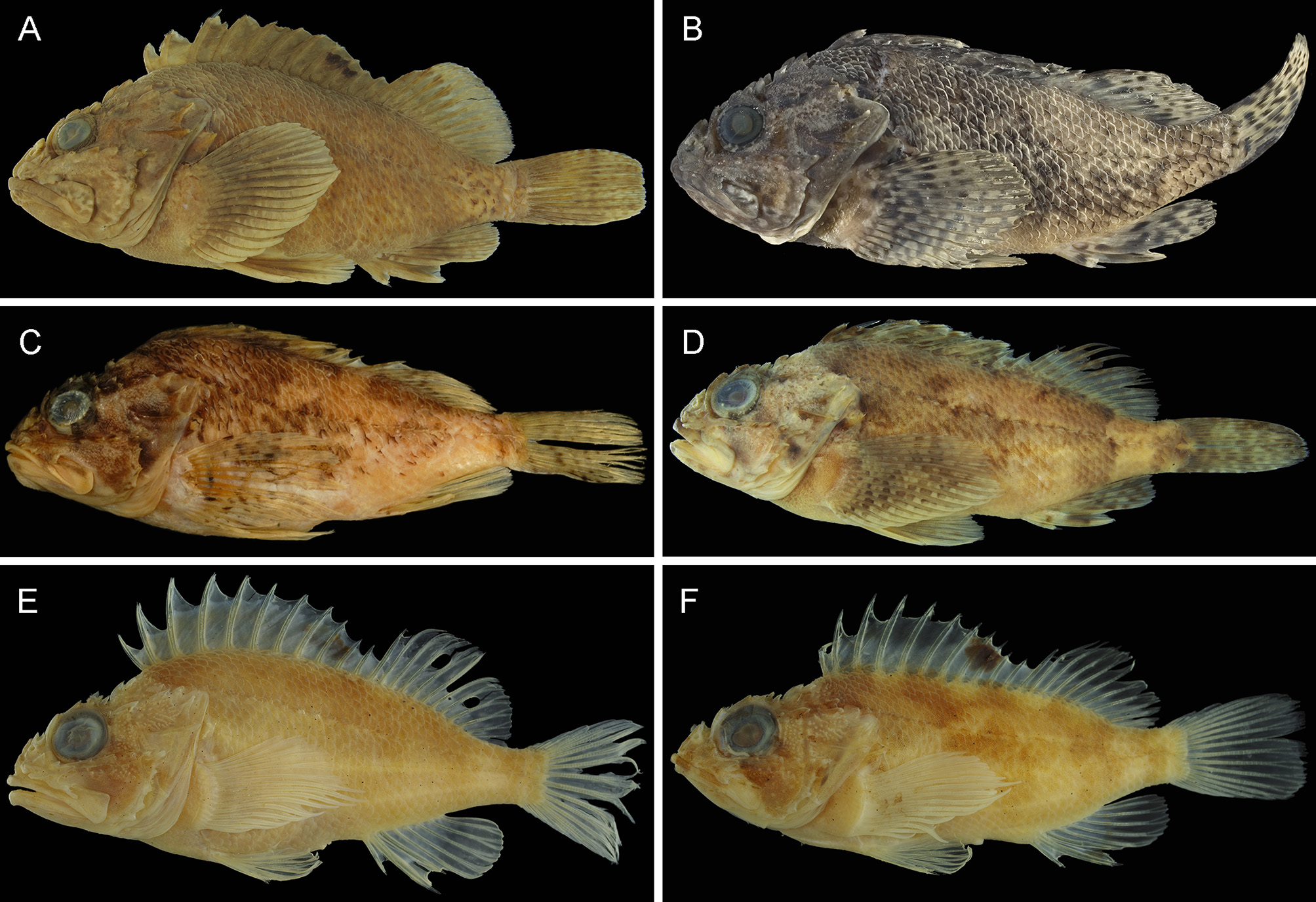
Description. Data for the holotype are given first, followed by paratype data (if different) in parentheses. Head spination illustrated in Fig. 1 View FIGURE 1 C–D. Selected meristics and morphometrics, expressed as percentages of SL, are shown in Tables 1–3. Body moderately compressed anteriorly, width at pectoral-fin base 20.1 (14.2–22.2) % SL, progressively more compressed posteriorly; relatively shallow, depth much less than head length [body depth and head length 39.0 (32.3–39.5) % SL and 41.4 (39.2–45.7) % SL, respectively]. Tiny papillae on snout, interorbital space, occipital pit, dorsal surface of head, outer margin of eye membrane, and behind orbit to an area between upper and lower opercular spines. A single tentacle (its length greater than half pupil diameter) on posterior end of supraocular spine base. Several tiny tentacles on upper part of outer eye margin. A short tentacle, with several short branches along distal margin, associated with anterior nostril, its length approximately equal to nasal spine length. Anterior lacrimal spine associated with tiny fleshy tentacles. Posterior lacrimal spine associated with a small and several tiny fleshy tentacles, its lengths shorter than anterior nostril tentacle, posteriorly continuous with head via fringed skin. Tips of preopercular spines without skin flaps (sometimes tips of 3rd–5th spines with tiny flap). Several (sometimes absent) tentacles occasionally scattered on lateral body surface (associated with pored-lateral line, recognized from left side of body in holotype). Underside of lower jaw smooth, without tentacles. Pectoral-fin axil without skin flaps.
Dorsal fin with 12 spines and 10 (rarely 9, only 4 of all paratypes) soft rays; fourth (sometimes third) spine longest; fifth to eleventh spines becoming progressively shorter; length of last spine about equal to (or slightly greater than) that of second spine; second (sometimes third) soft ray longest; all soft rays branched, posterior branch of last soft ray joined by membrane to caudal peduncle for about four-fifths (two-thirds to four-fifths) its length. Anal fin with 3 spines and 5 soft rays; first spine shortest, located slightly posterior to vertical through last dorsal-fin spine base; second longest, its length greater than 4th dorsal-fin spine length; all soft rays branched; second soft ray longest. Pectoral fins each with 15 (14–16) rays, 1 (1 or 2) upper unbranched rays, 5 (2–5) branched rays, 9 (9–11) lower unbranched rays (usually all rays unbranched in specimens less than 26.3 mm SL); 8th (or 7th) ray longest, slightly elongated; posterior profile of fin pointed (or slightly rounded); tip of fin just reaching (not reaching or slightly beyond) first anal-fin spine base; lower unbranched rays thickened. Pelvic fin with 1 spine and 5 branched soft rays; second soft ray longest; last soft ray joined by membrane to abdomen for about two-thirds its length. Caudal fin with 13 principal rays, posterior margin relatively rounded (or slightly truncate). Gill rakers on upper limb of first gill arch 5 (4–6), lower limb 11 (10–13) [9 (8–10) and 2 (2–4) on ceratohyal and on hypobranchial, respectively], total 16 (14–18), short and spinous, longest raker on first gill arch shorter than gill filaments around angle of gill arch; fourth gill slit closed by membrane. Branchiostegal rays 7. Swimbladder absent.
Area enclosed by opercular margin, and upper and lower opercular spine tips lacking scales. Well exposed ctenoid scales on lateral surface of trunk, becoming cycloid and sometimes embedded in thin skin ventrally. Body scales not extending onto fin rays or membranes, except basal caudal fin. Exposed (sometimes embedded in thin skin) cycloid scales covering pectoral-fin base and ventral body surface, including between pelvic fins. Lateral line above opercle tip and pectoral fin sloping moderately downward. Relatively thick skin with numerous small sensory pores covering predorsal area from posterior edge of occipital pit to anterior margin of predorsal scales, upper and lower suborbital ridges, behind orbit to above upper opercular spine, and between parietal and nuchal and pterotic and lower posttemporal spines. Underside of dentary with three well developed sensory pores, first pore slightly posterior to vertical through anterior lacrimal spine tip, second between anterior and posterior lacrimal spine tips, third on posterior margin of dentary. A single united pore behind lower jaw symphysial knob. A pore on each side of symphysial knob.
Mouth large, slightly oblique, forming an angle of 20 (20–25)º to horizontal axis of head and body. Posterior margin of maxilla just reaching posterior margin of orbit (usually reaching to vertical through or slightly beyond posterior margin of pupil). Upper edge of posterior maxilla swollen laterally, forming a distinct ridge; lateral surface of maxilla smooth, lacking a ridge or tentacles. Symphysial gap separating premaxillary teeth bands narrow, less than width of each band. Upper jaw with a band of short, incurved, conical teeth, about 9 tooth rows anteriorly, band slightly narrowing posteriorly. Lower jaw with a band of short, incurved, conical teeth; most teeth shorter than those of upper jaw, band distinctly narrowing posteriorly. About 6 tooth rows anteriorly on vomer, reducing to 2 rows posteriorly, forming a V-shaped patch. Width of vomerine plate distinctly shorter than length of palatine plate. Two (or 3) tooth rows on palatine plate; plate slightly narrowing posteriorly.
Anterodorsal and lateral surfaces of lacrimal with spines, the former with distinctly larger spine base (anterodorsal lacrimal spine usually indistinct in paratypes less than 26.3 mm SL). Anterior lacrimal spine simple, without small spinous points on its posterior margin, directed forward. Posterior lacrimal spine simple, without small spinous points on its anterior margin, directed ventroposteriorly. Anterior and posterior lacrimal spine sizes about equal. Suborbital ridge with three spines, first below pupil, second extending slightly beyond orbit, third at end of suborbital ridge. Space between ventral margin of eye and suborbital ridge narrow. Suborbital pit absent. Preopercle with 5 pointed spines; uppermost largest with a small supplemental preopercular spine on base; directed posteriorly. Preopercle (between upper end and uppermost preopercular spine) smooth, without serrae or spines. Upper opercular spine simple, with a low median ridge, covered by relatively thick skin with small pores. Lower opercular spine simple, with a distinct median ridge. Space between upper and lower opercular spines smooth, without ridges. Posterior tip of upper opercular spine not reaching opercular margin, posterior tip of lower opercular spine just reaching opercular margin.
Dorsal profile of snout steep, forming 50 (45–50)º angle to horizontal axis of head and body. Nasal spine simple, conical, directed upward. Ascending process of premaxilla short, its posterior margin level with (or slightly beyond) posterior end of nasal spine base. Median interorbital ridge present (or indistinct), extending from just behind posterior margin of ascending process of premaxilla onto midline slightly posterior to posterior end of preocular spine base. Interorbital ridges well developed, separated by moderately deep channel, extending from behind nasal spines and conjoined level with coronal spines. Interorbital ridges divergent anteriorly and posteriorly in dorsal view, narrowest separation level with tip of preocular spine. Preocular spine simple, directed dorsoposteriorly, flattened anteriorly and posteriorly. Supraocular spine simple. Postocular spine simple, its length less than tympanic spine. Coronal spine simple, strongly pointed, directed dorsoposteriorly. Tympanic spine simple, strongly pointed with slightly lateral orientation. Pretympanic spines absent. Occipital pit shallow, center slightly convex with an indistinct transverse ridge at rear between bases of nuchal spines, surrounded laterally by tympanic and parietal spines. Parietal spine simple, its base curving into occipital pit. Nuchal spine simple, base continuous with parietal spine base. Sphenotic with 1 (rarely 0 or 2) small spine(s). Postorbital smooth, usually spineless (rarely with spinous points). Pterotic spine simple, located below parietal and nuchal spines. Upper posttemporal spine small, simple, pointed and directed dorsoposteriorly, its length much shorter than lower posttemporal spine. Lower posttemporal spine simple, its base length slightly less than that of pterotic spine. Supracleithral spines simple, flattened. Cleithral spine flattened, with an indistinct median ridge, strongly pointed.
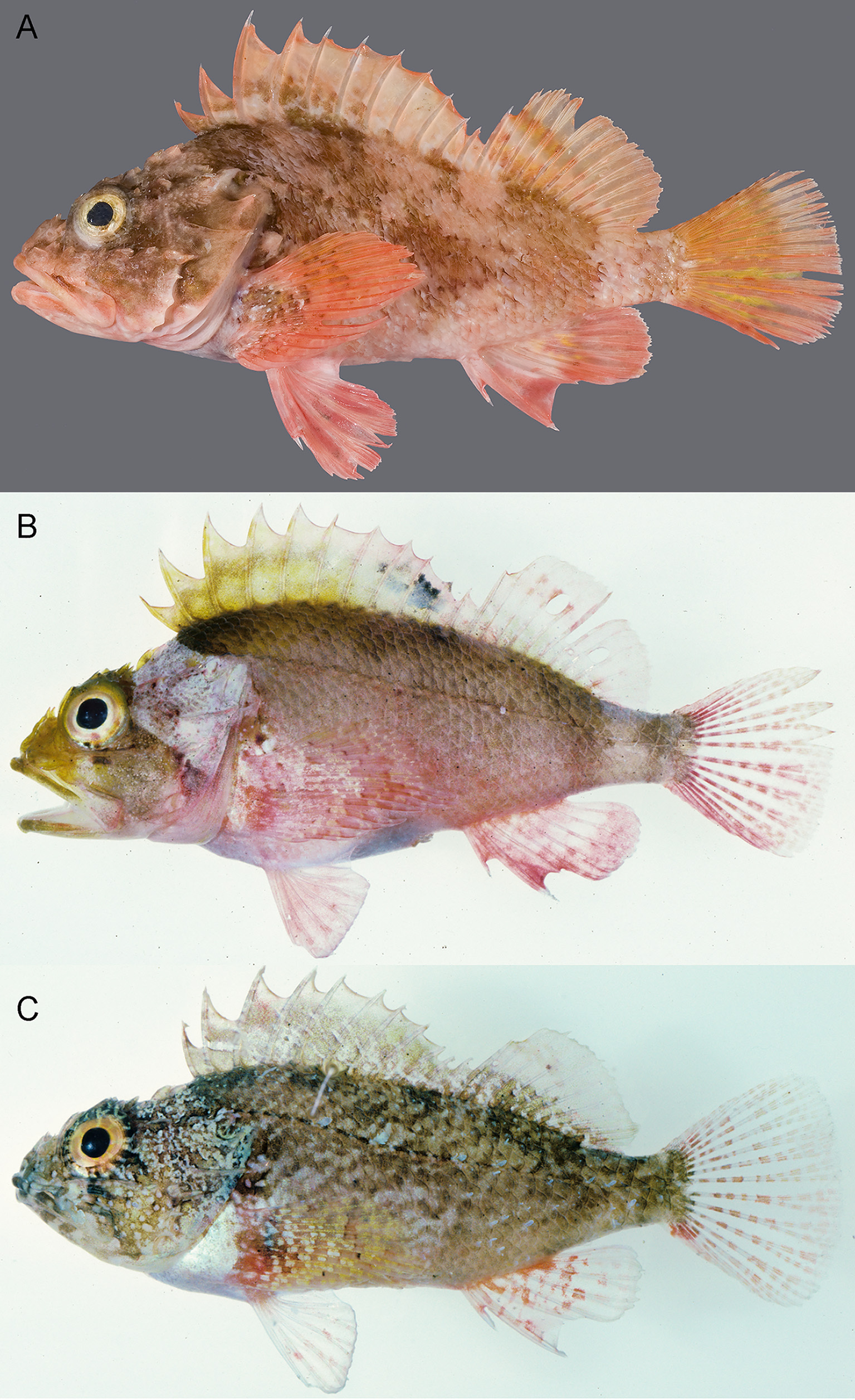
Colour (based on all examined specimens). Fresh specimens vary in coloration from reddish-orange to greenishbrown, suffused with irregular dark brownish to blackish markings, males with a black blotch (absent in females) on posterior portion of spinous dorsal fin between 8th (7th) and 10th spines ( Fig. 2 View FIGURE 2 B–C). Preserved specimens yellowish-brown, persistent dark brownish markings on body and dorsal fin blotch ( Fig. 3 View FIGURE 3 E–F).
Distribution. Distributed off southwestern Australia, ranging from Southern Group, Houtman Abrolhos (28°S) to the Albany coast (35°S) ( Fig. 4 View FIGURE 4 ). Specimens examined in this study were collected mainly from shallow rocky reefs at depths between 0–37 m (a single specimen had been collected from 188 m).
Etymology. The species name from the Latin vesperalis , meaning west, is derived from the type locality of the species (Western Australia), which is also the westernmost occurrence of the S. papillosa complex.


No known copyright restrictions apply. See Agosti, D., Egloff, W., 2009. Taxonomic information exchange and copyright: the Plazi approach. BMC Research Notes 2009, 2:53 for further explanation.
|
Kingdom |
|
|
Phylum |
|
|
Class |
|
|
Order |
|
|
Family |
|
|
Genus |