
Simothraulopsis eurybasis, Do, Jeane M. C., Salles, Frederico F. & Hamada, Neusa, 2017
|
publication ID |
https://doi.org/ 10.5281/zenodo.828100 |
|
publication LSID |
lsid:zoobank.org:pub:5E5CC15A-009D-4E12-9342-315058D35E98 |
|
DOI |
https://doi.org/10.5281/zenodo.6023592 |
|
persistent identifier |
https://treatment.plazi.org/id/03CA6B06-FFBC-6C32-FF0C-FDDEEA3BF846 |
|
treatment provided by |
Plazi |
|
scientific name |
Simothraulopsis eurybasis |
| status |
sp. nov. |
Simothraulopsis eurybasis sp. nov.
( Figs 39–43 View FIGURE 39 View FIGURE 40 View FIGURE 41 View FIGURE 42 View FIGURE 43 )
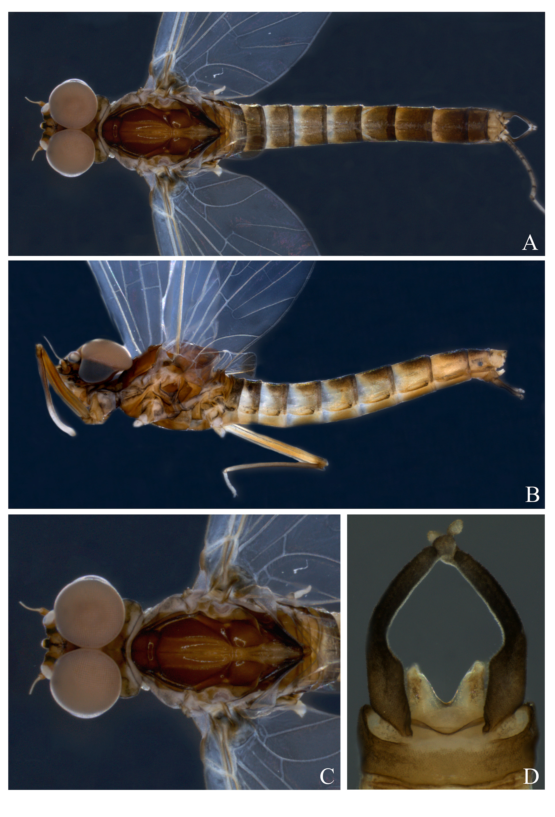
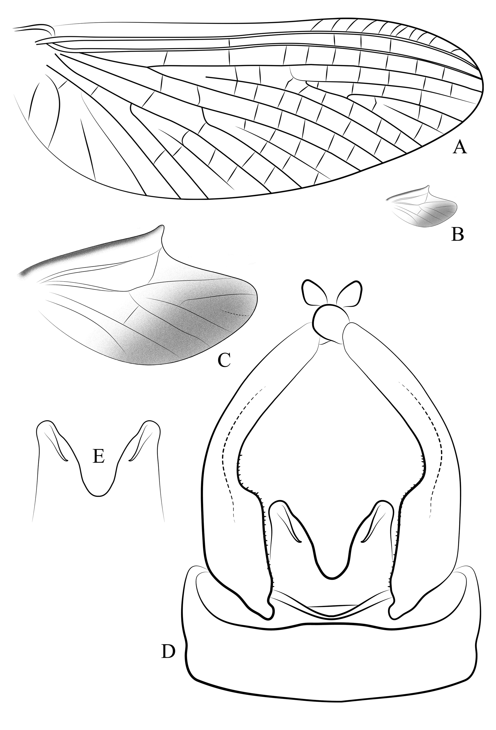
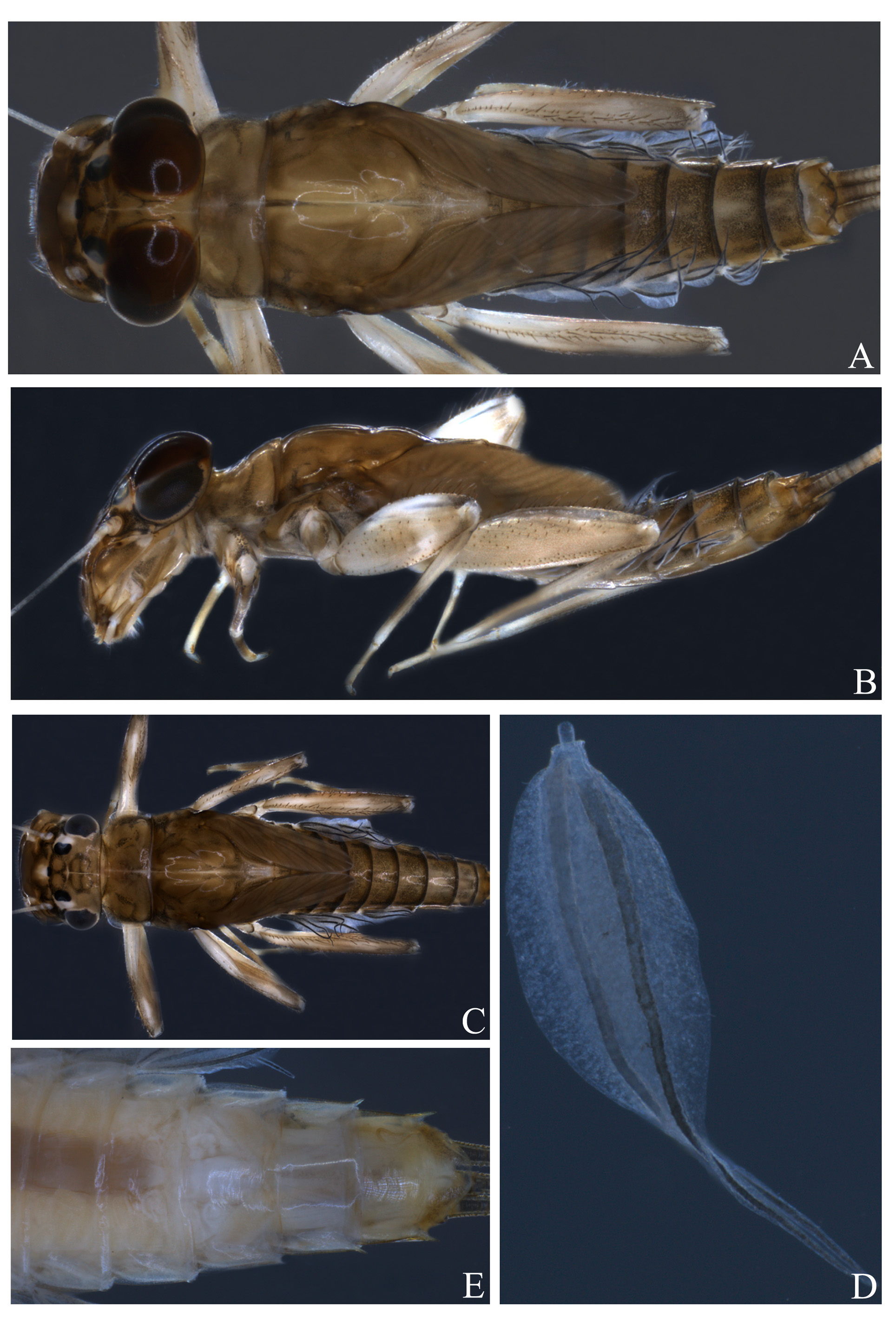
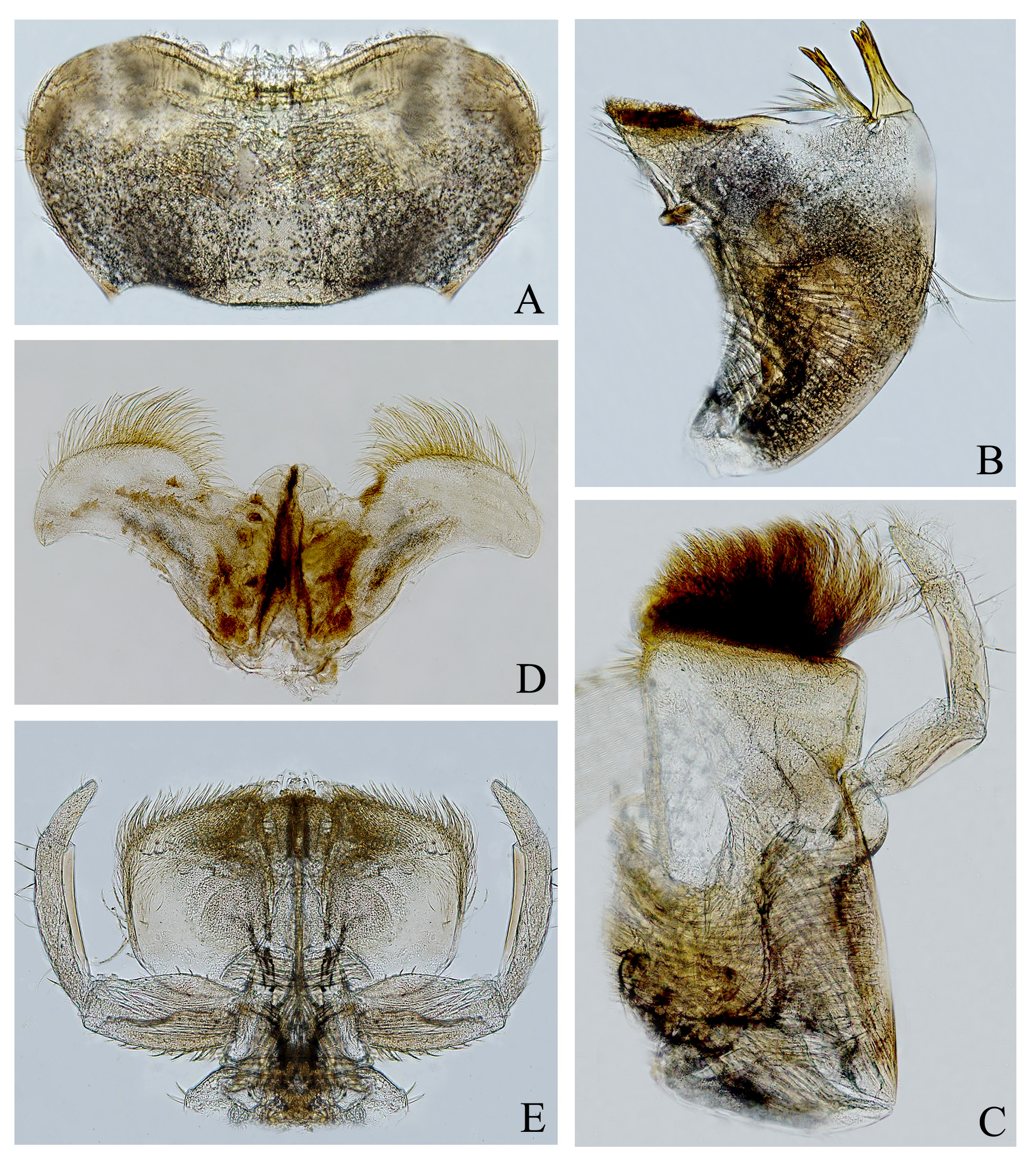
Diagnosis. Male imago: 1) general coloration: thorax orangish brown, abdomen blackish brown ( Figs 39 View FIGURE 39 A–C); 2) hind wing with costal projection almost forming a right angle, located approximately 1/2 distance from base to apex of wing ( Figs 40 View FIGURE 40 B, C); 3) abdominal segment IV almost completely white ( Figs 39 View FIGURE 39 A); 4) penis projection spine-like, short and directed to the midline of the body ( Fig. 40 View FIGURE 40 D, E); 5) penis lobes widely separated, almost underneath by the forceps from ventral view; 6) penis lobes fused on basal 1/4 ( Fig. 40 View FIGURE 40 D); 7) ventral region of penis lobes without a well-marked sclerotized region ( Fig. 39 View FIGURE 39 D). Mature nymph: 1) inner margin of fore tibia with a row of pectinate setae ( Fig. 43 View FIGURE 43 A); 2) femur III slender, about four times longer than wide ( Fig. 43 View FIGURE 43 C); 3) posterolateral projections presents on abdominal segments VIII and IX ( Fig. 41 View FIGURE 41 C); 4) gills hyaline, tracheal region gray ( Fig. 41 View FIGURE 41 D).
Male imago. Length: body: 5.2–5.4 mm; forewing: 6.1–6.3 mm; hind wing: 1.2–1.4 mm.
Head. Dorsal region orangish brown washed with black, with a submedial orangish rounded mark; ventral region whitish, with black marks. Upper portion of compound eyes grayish brown, lower portion black; ocelli whitish, surrounded by black ( Figs 39 View FIGURE 39 A, B). Scape and pedicel of antenna translucent yellow, flagellum translucent white.
Thorax. Pronotum orangish brown, with both lateral and posterior margins darker; submedial and mediolongitudinal stripes black. Mesonotum orangish brown; longitudinal medial suture light brown; anterolateral scutal and lateroparapsidal sutures black; scuto-scutellar impression and scutellum black. Pleura orangish brown; membranous area dark gray. Sterna yellowish brown. Wings membrane hyaline ( Figs 40 View FIGURE 40 A–C). Forewing with longitudinal and cross veins light yellow translucent; costal arms and base of veins C and Sc black; fork of vein MP symmetric ( Fig. 40 View FIGURE 40 A). Hind wing with costal projection well developed, almost forming a right angle, located approximately 1/2 distance from base to apex of wing; longitudinal veins brown, except base of vein C and Sc grayish brown; cross veins translucent; costal region dark gray, almost the entire lower portion of hind wing blackish brown ( Figs 40 View FIGURE 40 B, C). Legs. Coxae and trochanters whitish yellow, washed with gray. Leg. I: femur orangish brown with a whitish apical band; tibia with a dark gray basal band, remainder whitish; tarsi whitish. Legs II and III: femora orangish brown with basal and apical bands whitish; tibiae gray with subapical region whitish; tarsi whitish with distal region of tarsomeres light brown.
Abdomen. Terga I–III blackish brown, except posterior margin black and anterolateral region of terga II and III translucent white; tergum IV almost completely white, except medial region washed with grayish brown and posterior margin black; terga V–IX black, except anterior region of terga V and VI whitish and anterior region of tergum VII yellowish. Tergum X yellowish brown, with margins dark brown and both submedial and medial stripes brown ( Figs 39 View FIGURE 39 A, B). Sterna I–IV translucent white, lateral region of sterna V and VI with a brown mark; other sterna brown with lateral region blackish brown. Genitalia ( Figs 39 View FIGURE 39 D; 40D, E). Styliger plate yellowish brown, posterolateral angle and lateral margin blackish brown. Forceps segment I blackish brown, segments II and III whitish, except apical region of segment II brown. Segment II 0.1 length of segment I, 1.2 length of segment III. Penis lobes yellowish, lobes with a small black mark located near outer margin; penis lobes widely separated, almost underneath by the forceps from ventral view and fused on basal 1/4; inner margins divergent; each lobe rounded apically, with a short (less than the half of total length of penis lobes), spine-like projection, directed to the midline of the body; ventral region of penis lobes without a well-marked sclerotized region ( Fig. 39 View FIGURE 39 D). Caudal filaments broken and lost.
Female subimago. Length: body: 5.4–5.7 mm; forewing: 6.3–6.5 mm; hind wing: 1.3–1.5 mm. Color pattern similar to male imago, except by coloration more yellowish ( Fig. 39 View FIGURE 39 C). Sternum IX with lateral margins light brown.
Mature nymph. Length: body, 4.4–4.9 mm; antenna, 2.3–2.4 mm; cerci, 4.5–4.9 mm; caudal filament, 5.1–5.3 mm. General coloration brown washed with black ( Figs 41 View FIGURE 41 ).
Head. Brown, with blackish gray marks. Upper portion of male compound eyes reddish brown, lower portion black. Eyes of female black. Ocelli dark gray. Scape of antenna dark brown, pedicel and flagellum translucent white. Maximum width of labrum 1.2 times maximum width of clypeus. Labrum ( Fig. 42 View FIGURE 42 A) yellowish brown washed with gray, with a submedial rounded dark gray mark; anteromedial emargination broad, with four denticles of equal size. Mandible ( Fig. 42 View FIGURE 42 B) brown, translucent on apical region (except incisors and molar area orangish yellow) and near basal articulation; with a translucent yellow, rounded mark medially located and scattered gray marks; outer margin slightly curved, with 2–4 filiform, long, thick setae medially located. Maxilla ( Fig. 42 View FIGURE 42 C) yellowish translucent, outer margin grayish brown, region near basal articulation gray. Segment II of maxillary palpi 1.3 length of segment I, 2.0 length of segment III. Hypopharynx as in figure 42D. Labium as in figure 42E.
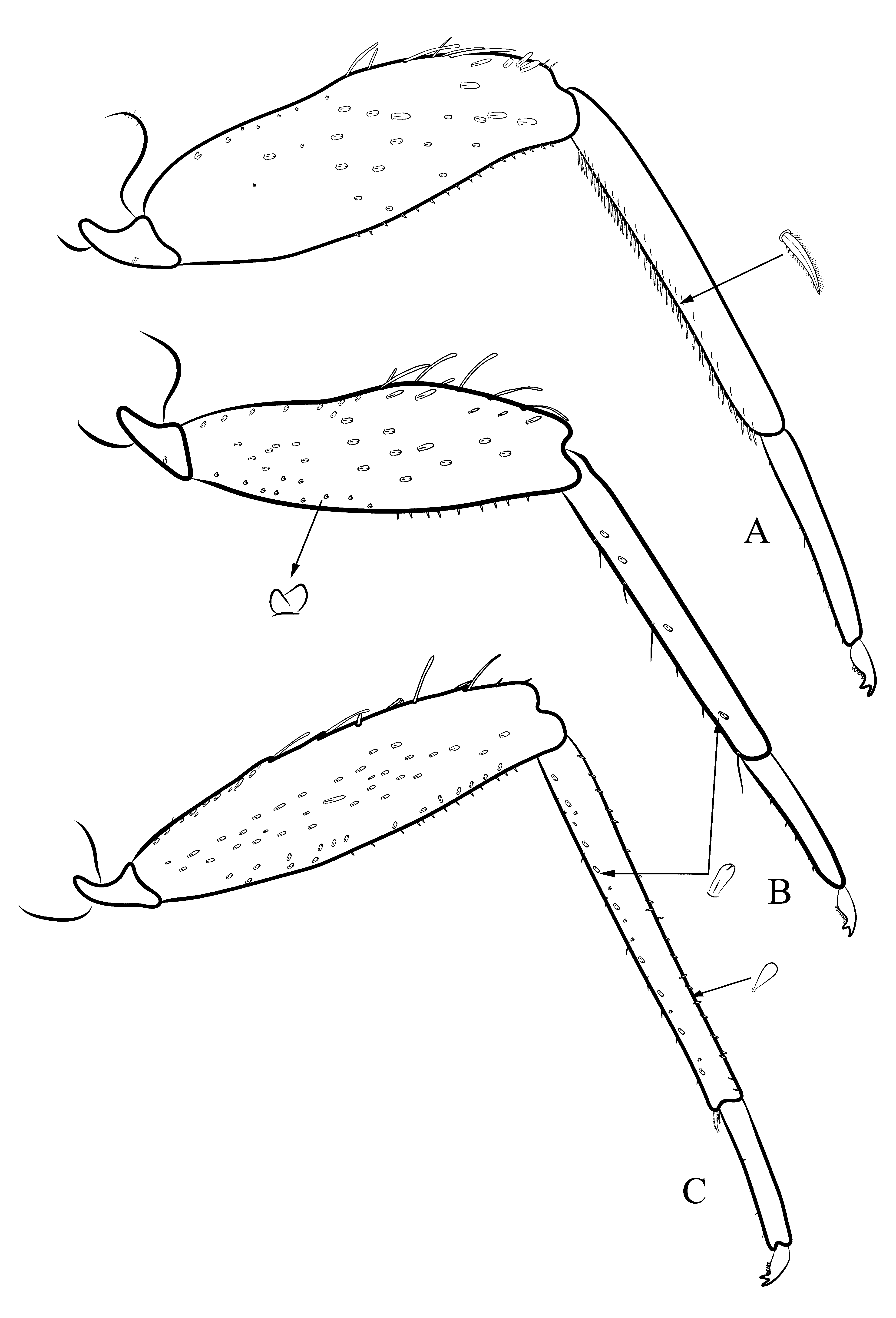
Thorax. Terga brown, irregularly washed with blackish gray, central region yellowish; anterior margin of pronotum with 1–2 spines; pleura brown, irregularly washed with gray; sterna whitish. Legs. General coloration whitish; coxae with dorsal region dark brown, femora with a light gray transverse line. Femora I and II almost completely whitish, except a mediolongitudinal light brown band and subapical dark gray band; femur III almost completely brown, without well-marked bands; with a subapical dark gray brown mark and apical whitish mark. Tibiae and tarsi whitish; tibiae with basal and submedial brown bands; tarsi with a basal brown band. Femur III slender, about four times longer than wide ( Fig. 43 View FIGURE 43 C). Leg I. ( Fig. 43 View FIGURE 43 A). Coxa with few filiform setae located near dorsal margin. Trochanter with few lanceolate setae. Femur: outer margin with apical region with long, stout setae; apical half of inner margin with short stout setae; dorsal surface with: few spatulate setae, spatulate setae with bifid apex and robust, short, bifid setae. Tibia: inner margin with pectinate setae and with a longitudinal dorsal row of filiform, thick setae near inner margin. Tarsus with filiform thick setae along inner margin. Leg II ( Fig. 43 View FIGURE 43 B). Coxa bare. Trochanter with few filiform setae. Femur: apical region of outer margin with long stout setae; inner margin with apical half with short stout setae; dorsal surface with spatulate setae with bifid apex and with robust, short, bifid setae. Tibia: inner margin with few stout setae; dorsal surface near inner margin with spatulate setae with bifid apex. Tarsus with few stout setae along inner margin. Leg III ( Fig. 43 View FIGURE 43 C). Coxa and trochanter bare. Femur: outer margin with apical half with long, stout setae; inner margin with apical half with short stout setae; dorsal surface with simple spatulate setae and spatulate setae with bifid apex. Tibia: inner margin with few stout setae and with spatulate setae with bifid apex near inner margin; outer margin with a row of teardrop shaped setae. Tarsus with few stout setae along to outer margin.
Abdomen. Terga blackish brown with posterior margins black and lateral regions yellowish brown, except tergum II almost completely yellowish brown. Sterna yellowish, lateral region washed with gray. Gill translucent, tracheal region gray ( Fig. 41 View FIGURE 41 D). Posterolateral projections present on abdominal segments VIII and IX ( Fig. 41 View FIGURE 41 C). Caudal filaments yellowish brown, with basal area light dark brown.
Life cycle association. Male subimago extracted from nymph and compared with the imagos.
Etymology. From the Greek word eurys, wide; and basis, base. In reference to the wide distance between the apices of penis lobes.
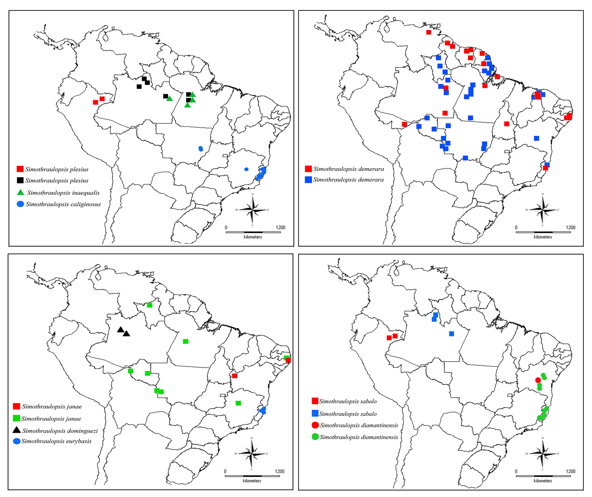
Comments. Simothraulopsis eurybasis sp. nov. has a distribution restricted to the north of the Espírito Santo state ( Fig. 49 View FIGURE 49 ). Imagos were collected mostly by light traps from large rivers. The two collected nymphs were found in submerged stones in a rock bottomed stream.
Male imagos of S. eurybasis sp. nov. and S. diamantinensis have similar penis lobes, with short, spine-like, penis projection ( Figs 20 View FIGURE 20 D, E; 40D, E). The new species, however, can be distinguished from other species of Simothraulopsis by the distance between the penis lobes (they are so widely separated, that are almost covered by the forceps in ventral view). Besides that, coloration of hind wings and general coloration of the body may also prove helpful with identification: in S. eurybasis sp. nov. the pigmented area below vein R is wider and the abdominal coloration is darker ( Figs 19 View FIGURE 19 A, B; 20C; 39A, B; 40C). Nymphs of these two species can be separated from each other by the general coloration: in S. eurybasis sp. nov. the thorax is more marked and the abdomen is darker ( Figs 21 View FIGURE 21 A, E; 41A, E).
Material examined. Holotype: Male imago (light trap), BRAZIL, Espírito Santo State, Sooretama, São José river (S 19°07'33.1"; W 40°14'26.1"; 24m) 19/v/2011, Salles FF & Nascimento JMC, cols. ( INPA) GoogleMaps . Paratypes: five male imagos, six female imagos and two nymphs (same data as holotype) (two male imagos, two female imagos, one nymph at IBN; two male imagos, two female imagos and one nymph at CZNC; two male imagos, two female imagos and one nymph at DZRJ) GoogleMaps ; two male imagos, Nova Venécia, Patrimônio do Bis (S 18°33'27.5"; W 40°20'6.5"; 188m) 25-26/vii/2012, Salles FF & Angeli K cols ( INPA) GoogleMaps ; one male imago, Nova Venécia, Santa Rita do Pipi Nuck (S 18°39'51.4''; W 40°30'44.9''; 80m) 18-19/iv/2012, Salles FF & Angeli K cols ( CZNC) GoogleMaps .


| INPA |
Instituto Nacional de Pesquisas da Amazonia |
No known copyright restrictions apply. See Agosti, D., Egloff, W., 2009. Taxonomic information exchange and copyright: the Plazi approach. BMC Research Notes 2009, 2:53 for further explanation.
|
Kingdom |
|
|
Phylum |
|
|
Class |
|
|
Order |
|
|
Family |
|
|
Genus |