
Stenorhamphus phuphan, Smith & Hwang & Weirauch, 2019
|
publication ID |
https://doi.org/ 10.26107/RBZ-2019-0011 |
|
DOI |
https://doi.org/10.5281/zenodo.3681397 |
|
persistent identifier |
https://treatment.plazi.org/id/DC5787F2-FFD5-FA76-FC3F-8013FB8CF894 |
|
treatment provided by |
Carolina |
|
scientific name |
Stenorhamphus phuphan |
| status |
sp. nov. |
Stenorhamphus phuphan , new species
( Figs. 1–3 View Fig View Fig View Fig , 5B View Fig , 6 View Fig E–H, 7C, D, H, I, 8)
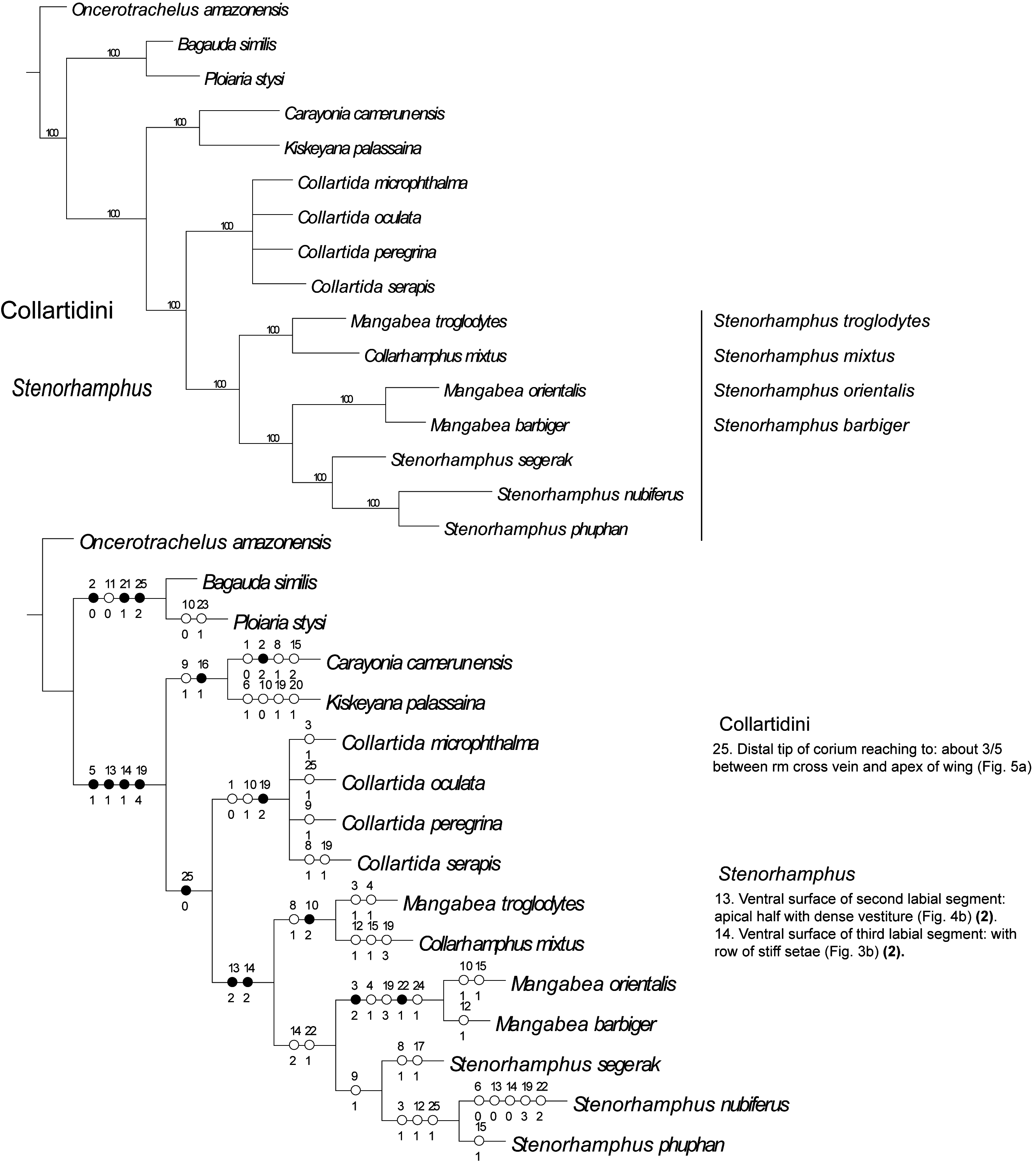
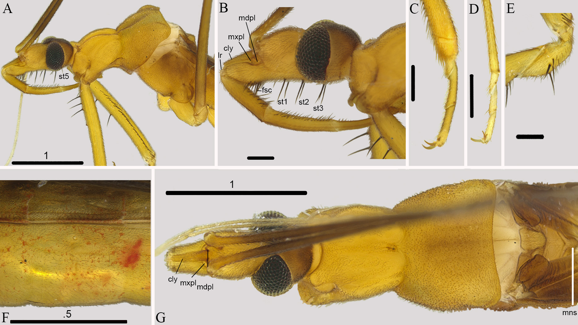
Diagnosis. Recognised within Stenorhamphus by the total length approximately 6.9 mm, one pair of setae posterior to each eye, setae along apex of second labial segment, along entire third segment, and basally along the fourth segment, four spines on the trochanter, fascicle of stout setae on the anterior area of the gena, postocular region long, not globulose, posterior lobe of pronotum approximately equal to anterior lobe, legs long, mid and hind coxae longer than length of abdomen, without spines on posterior lobe of pronotum.
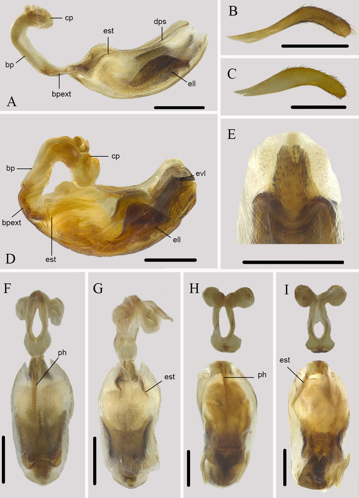
Description. Male: small (total length, holotype: 6.89 mm) COLOURATION: general colouration yellow, with base of wings and posterior portion of head brown ( Figs. 2C, D View Fig ). Head: postocular region brown, anterior anteocular region lighter ( Fig. 3B View Fig ). Antenna: brown, flagellomeres light brown. Labium: light brown to yellow. Thorax: anterior pronotum yellow, posterior dark yellow to brown. Legs: coxae light brown to yellow, trochanters, femora, tibiae, and tarsi pale brown. Wings: basally brown, rest hyaline. Abdomen: tergites yellow; mediosternites pale brown, laterosternites somewhat darker; pygophore brown. VESTITURE: as in genus description with the following additions: Head: ventral surface with three to four pairs of long, stout setae located posterior to antennifer, at anterior and posterior margins of eye, lateral surface of head withone long, stout setae posterior to eye (four ventral and one lateral setae visible on right side, three setae on left) ( Figs. 3A, B View Fig ) fascicle of eight stout setae on gena ventrad of apex of maxillary plate ( Figs. 3A, B View Fig ); second labial segment (first visible) with medium-length stout setae on ventral surface in apical half of segment ( Figs. 3A, B View Fig ). Legs: forecoxa, in addition to short vestiture, with posterodorsal series and four stout, long setae anteroventral ( Figs. 3A View Fig ), foretrochanter with four stout setae on anterior surface ( Fig. 3E View Fig ), ventral surface of forefemur with about 16 medium stout setae in basal 2/3, interspersed with short setae ( Fig. 2C View Fig ). STRUCTURE: Head ( Figs. 2C, D View Fig , 3A, B, G View Fig ): postocular region long and slender, anteocular region less globulose than in S. segerak , new species. Thorax ( Figs. 2C, D View Fig , 3A, G View Fig ): posterior lobe wider than long, distinctly wider than anterior lobe, slightly depressed medially and with distinct, raised, lateral areas in posterior half of lobe, posterior margin slightly concave ( Figs. 2C, D View Fig , 3A, G View Fig ). Raised portion of mesoscutellum tongue shaped ( Fig. 3G View Fig ). Legs ( Figs. 2C, D View Fig , 3A, C, D, E View Fig ). Wings ( Figs. 2D View Fig , 5B View Fig ): elongate, surpassing apex of abdomen, rmcu cross vein not present ( Fig. 5B View Fig ). Abdomen ( Figs. 2C, D View Fig , 3F View Fig ). Genitalia ( Figs. 6E, F, G, H View Fig , 7C, D, H, I View Fig ): segment 8 well developed; pygophore elongate ovoid, with spine-like medial process, transverse bridge present ( Figs. 6E, F, G, H View Fig ); parameres slender, curved, apex pointed ( Figs. 6E, F View Fig , 7C View Fig ); aedeagus ( Figs. 7D, H, I View Fig ) with basal plates stout and strongly curved, ponticulus basilaris very slender to nonexistent, basal plate extension relatively short, stout ( Fig. 7D View Fig ), dorsal phallothecal sclerite heavily sclerotised posteriorly ( Fig. 7A View Fig ), endosoma with ventral and lateral, heavily sclerotised lobes, lateral lobes as tall as wide ( Figs. 7D, H, I View Fig ).
Measurements. See Table 1 View Table 1 .
Female. Unknown.
Etymology. Named after the locality of the holotype, Phu Phan National Park in Thailand; a noun in apposition.
Distribution. Only known from the type locality in Thailand.
Biology. Collected in lowland dry dipterocarp forest with deciduous trees and high canopy cover.
Type material. Holotype: male, THAILAND: Sakon Nakhon: Phu Phan National Park, behind forest protection unit at Huay Wien Prai, 17.1143°N, 104.0054°E, 387m, 25 Feb – 3 March 2007 Malaise trap, Sailom Tongboonchai ( RCW 4869), type deposited in the Queen Sirikit Botanic Garden, Chang Mai ( Thailand) ( QSBG) GoogleMaps .
Discussion. Most similar to S. nubiferus due to the following shared characters: the distance from the posterior margin of the head to the anterior margin of the eye is approximately 1/3 the total length of the head (3:1), the third labial segment is the longest (12:1), and the pterostigma reaches 4/5 between rm cross vein and apex of wing (25:1). However, it is separated from S. nubiferus by the length of the posterior lobe of the pronotum in dorsal view being approximately equal to the anterior lobe (15:1), the pair of ventral setae in position 2 present (6:1), and four ventral spine-like setae on forecoxa (19:4).


| RCW |
RCW |
| QSBG |
Thailand, Chaing Mai, Queen Sirikit Botanic Garden |
No known copyright restrictions apply. See Agosti, D., Egloff, W., 2009. Taxonomic information exchange and copyright: the Plazi approach. BMC Research Notes 2009, 2:53 for further explanation.
|
Kingdom |
|
|
Phylum |
|
|
Class |
|
|
Order |
|
|
Family |
|
|
Genus |